
Parodontophora paramicroseta, Li, Yongxiang & Guo, Yuqing, 2016
|
publication ID |
https://doi.org/10.11646/zootaxa.4109.4.1 |
|
publication LSID |
lsid:zoobank.org:pub:8A6824E2-428F-4C5A-9CC3-2E009FA8BBF3 |
|
DOI |
https://doi.org/10.5281/zenodo.6057442 |
|
persistent identifier |
https://treatment.plazi.org/id/03DA0D64-FF80-FF97-FF56-95F5FF1F8C28 |
|
treatment provided by |
Plazi (2016-05-10 06:08:46, last updated 2024-11-27 18:31:00) |
|
scientific name |
Parodontophora paramicroseta |
| status |
sp. nov. |
Parodontophora paramicroseta sp. nov.
( Figure 10 View FIGURE 10 , Figure 11 View FIGURE 11 , Table 9 View TABLE 9 )
Type material. ♂1 and ♀1 were collected from Station NDWU in January 2013, and ♂2 and ♀2 were collected respectively from Station ZZZJ in July 2012 and October 2012.
Holotype: ♂1 on slide number NDWW 201310 H2104. Paratypes: ♂2 on slide number ZZZJ 201207 M2114, ♀1 on slide number NDWW 201310 H2110, ♀2 on slide number ZZZJ 201210 L1104. The holotype and paratypes are deposited in the Third Institute of Oceanography, State Oceanic Administration, Xiamen.
Type locality and habitat. All specimens were collected from mangrove forests in Wanwu mangrove forest in Ningde City, East China Sea, and Zhang River estuary in Zhangzhou City, East China Sea. Station NAFA: 119.86°E, 26.85°N. Station ZZZJ: 117.35°E, 23.96°N.
Etymology. This species is named paramicroseta (english: para-, like) because its morphological characters are similar to those of P. microseta sp. nov..
Measurements. Table 9 View TABLE 9 .
Description. Body narrows gradually in the anterior region. Cuticle with distinct outer striation discernible in the lateral field. Lip region more or less rounded, with six outer labial papillae. Four cephalic setae 3–4 µm, 20–29% of head diameter, 3–4 µm from the anterior end. The head behind the cephalic setae has a distinct constriction. Opisthocephalic setae 1 µm, arranged as two single subdorsal setae and two single subventral setae, i.e. (1D- 1V)2. Somatic setae length 2–3 µm, arranged as two groups of two longitudinally arranged setae respectively located on both sides of the amphid, extended from the posterior part of buccal cavity to the conical portion of the tail. Buccal cavity 32–34 µm long and 6–7 µm wide, cylindrical with conspicuous sclerotized parallel walls and conoid posterior parts. Six bifurcate teeth at the tip of the buccal cavity with the anterior tip narrower than the posterior one. Armilliths absent. Amphid 3–4 µm from the anterior end, crook-shaped with the shorter dorsal branch and parallel much longer ventral branch extending past the base of buccal cavity. Length of the dorsal branch equals 0.19–0.21 of ventral branch length and the amphid length is 1.35–1.5 times the buccal cavity length. Pharynx starts at the base of buccal cavity, muscular, and gradually broadens to the base and forms a bulb. Cardia small, rounded-conoid, surrounded with intestinal tissue. Renette cell slightly behind the base of pharynx, elongated oval or rectangular, 53–70 µm, 33–37% of the pharynx length. Nerve ring at 57–67% of pharynx length. Excretory pore located at 1 µm from the anterior end of body. Tail length 164–212 µm, conical anteriorly and cylindrical posteriorly, pointed terminal end without terminal setae. Three caudal glands open to the spinneret.
Male: Five pairs of subventral setae and several irregular setae on the conical portion of the tail, length 3 µm. Reproductive system diorchic. Testes paired, opposed and outstretched. Anterior testis to the right and posterior testis to the left of the intestine. Vas deferens well developed. Spicules length 44–48 µm, paired, equal and arcuate, several slight thickened septa respectively closes to dorsal sides and ventral sides, circle aperture in distal end and enlarged thickened proximal end with a front, ventral and dorsal constriction. Gubernaculum with dorsal-caudally directed apophysis 14–15 µm long, thickened rib-like edge extended into two distinct points. A precloacal seta present, length 3 µm, 11–12 fibriform precloacal supplements in front of it, they extend 290–489 µm from cloaca to the anterior end, 27–28% of body length.
Female: Morphologically similar to the male. Unpaired setae on tail. Reproductive system ampdidelphic, ovaries outstretched. Anterior gonad to the left and posterior gonad to the right of the intestine. Vulva is at 50–52% of the total body length. Short vagina with thick walls is perpendicular to the longitudinal body axis. Vulval glands are present anterior and posterior to the vagina. No spermathecae and spermatozoa are found in uteris.
Diagnosis and relationships. Parodontophora paramicroseta sp. nov. is characterized by relatively short cephalic setae (0.2–0.29 h. d.); the positon of the posterior end of the amphid extending past the base of the buccal cavity (1.35–1.5 times the length of the buccal cavity); opisthocephalic setae arranged as (1D- 1V)2; regular arranged somatic setae respectively located on both sides of the amphid from the posterior part of buccal cavity to the conical portion of the tail; excretory pore almost at the anterior end of body; armilliths absent; renette cell occupying 33–37% of the pharynx length; 11–12 fibriform precloacal supplements present and extend 290–489 µm anteriad; anterior gonad to the left and posterior gonad to the right of the intestine.
P. paramicroseta sp. nov. is almost similar to P. microseta sp. nov. than others species in which the positon of the posterior end of the amphid extending past the base of the buccal cavity. Both P. paramicroseta sp. nov. and P. microseta sp. nov. as new species of mangrove forest habitats in different regions of Fujian Province have relatively high morphological similarity, whereas it still differs from that species in the ventral branch length of the amphid (45–48 µm vs. 55–69 µm), the ratio of ventral branch length of the amphid to the buccal cavity length (1.35–1.5 vs. 1.71–2.1), female gonads arrangement (anterior gonad to the left and posterior gonad to the right of the intestine vs. anterior gonad to the right and posterior gonad to the left of the intestine) and the number of supplements (11–12 ind vs. 13–15 ind), which shows in Table 8 View TABLE 8 . In morphological difference of the spicule, P. microseta sp. nov. has a posterior-ventral rib next to circle aperture in distal end, while P. paramicroseta sp. nov. does not.
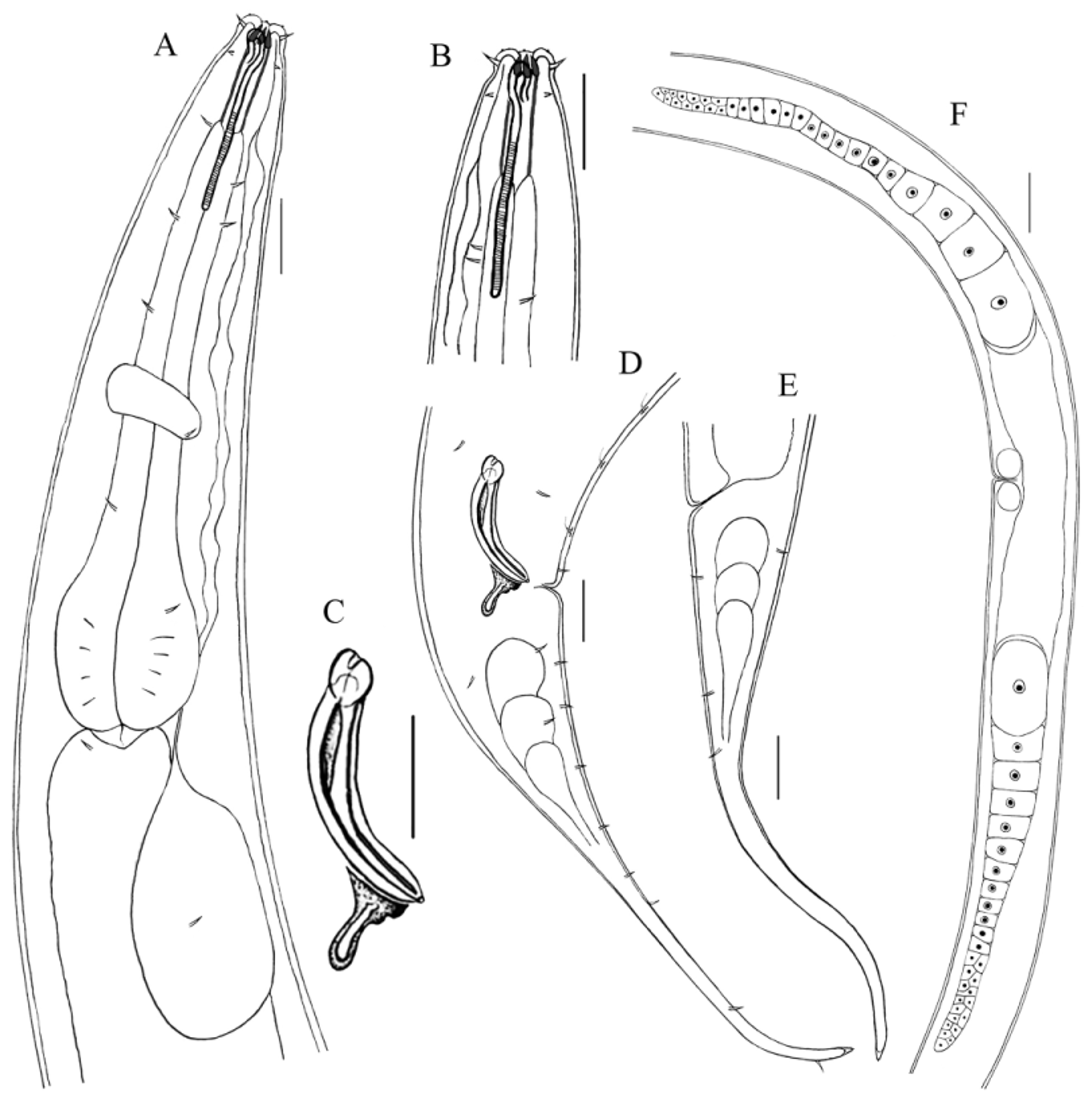
FIGURE 10. Parodontophora paramicroseta sp. nov. A: lateral view of male anterior part; B: lateral view of female head; C: lateral view of copulatory apparatus; D: lateral view of male tail and spicule; E: lateral view of female tail; F: lateral view of female reproductive system. Scale bar: A, B, C, D, E = 20 Μm; F = 40 Μm.
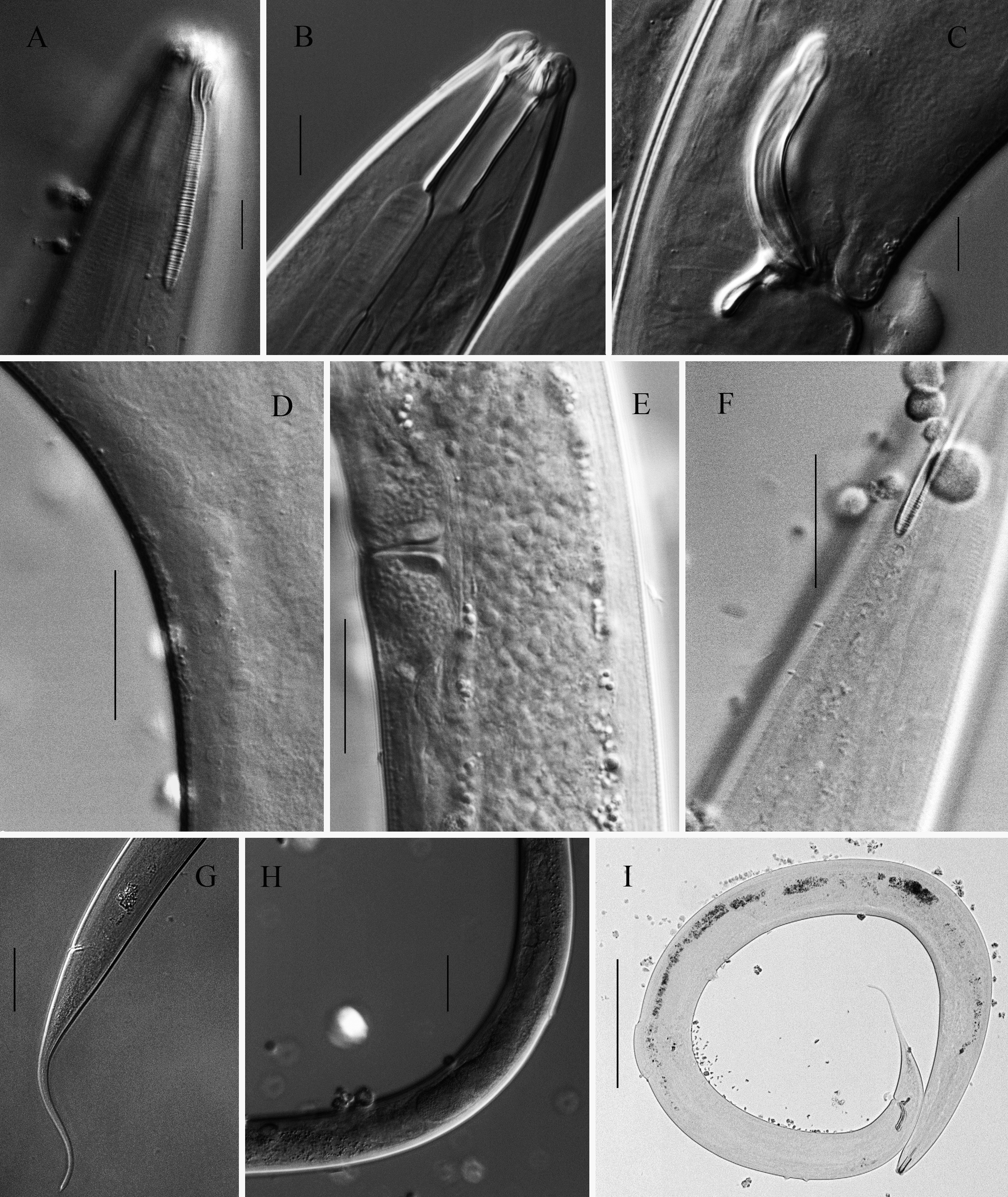
FIGURE 11. Light micrographs of Parodontophora paramicroseta sp. nov. A: anterior body end of female, showing amphid; B: anterior body end of male, showing buccal cavity; C: lateral view of male body part, showing copulatory apparatus; D: supplements in male; E: vulva region; F: longitudinally arranged somatic setae on the ventral side of the amphid; G: posterior body end of female; H: ovary region; I: full view of male body. Scale bars: A, B, C, D = 10 Μm; E, F = 25 Μm; G, H = 50 Μm; I = 200 Μm.
TABLE 9. Individual measurement of Parodontophora paramicroseta sp. nov. (in µm).
| Characteristics | Holotype | Paratypes | ||
|---|---|---|---|---|
| ♂1 | ♂2 | ♀1 | ♀2 | |
| Total body length | 1723 | 1054 | 1586 | 1315 |
| Head diameter | 15 | 14 | 15 | 14 |
| Cephalic setae | 3 | 3 | 4 | 4 |
| Opisthocephalic setae | 1 | 1 | 1 | 1 |
| Buccal cavity length | 33 | 32 | 33 | 34 |
| Buccal cavity diameter | 6 | 7 | 7 | 6 |
| Amphid from anterior end | 3 | 3 | 4 | 3 |
| Amphid c. b. d. | 30 | 26 | 26 | 28 |
| Amphid dorsal branch length | 10 | 10 | 9 | 9 |
| Amphid ventral branch length | 48 | 48 | 48 | 46 |
| Excretory pore from anterior end | 1 | 1 | 1 | 1 |
| Excretory pore c. b. d. | 10 | 10 | 11 | 10 |
| Nerve ring from anterior end | 117 | 108 | 116 | 115 |
| Nerve ring c. b. d. | 47 | 36 | 43 | 41 |
| Pharynx length | 204 | 161 | 195 | 183 |
| Pharynx c. b. d. | 69 | 39 | 51 | 46 |
| Renette gland length | 68 | 53 | 70 | 67 |
| Maximum body diameter | 91 | 44 | 62 | 66 |
| a. b. d. | 47 | 28 | 34 | 39 |
| Tail length | 212 | 164 | 197 | 168 |
| c' | 4.5 | 5.9 | 5.8 | 4.3 |
| Spicule length as chord | 42 | 37 | - | - |
| Spicule length as arc | 48 | 44 | - | - |
| Gubernaculum length | 12 | 11 | - | - |
| Apophysis length | 14 | 15 | - | - |
| Vulva c. b. d. | - | - | 56 | 57 |
| Vulva, % | - | - | 50.1 | 52.0 |
| a | 18.9 | 24.0 | 25.6 | 19.9 |
| b | 8.4 | 6.5 | 8.1 | 7.2 |
| c | 8.1 | 6.4 | 8.1 | 7.8 |
No known copyright restrictions apply. See Agosti, D., Egloff, W., 2009. Taxonomic information exchange and copyright: the Plazi approach. BMC Research Notes 2009, 2:53 for further explanation.
|
Kingdom |
|
|
Phylum |
|
|
Class |
|
|
Order |
|
|
Family |
|
|
Genus |
1 (by plazi, 2016-05-10 06:08:46)
2 (by ImsDioSync, 2016-05-10 06:48:56)
3 (by ImsDioSync, 2017-02-01 15:06:28)
4 (by ImsDioSync, 2017-02-01 15:07:40)
5 (by ImsDioSync, 2017-02-08 01:42:13)
6 (by ImsDioSync, 2017-06-25 17:55:27)
7 (by ExternalLinkService, 2019-09-26 09:39:48)
8 (by ExternalLinkService, 2022-01-29 21:17:40)
9 (by ExternalLinkService, 2022-02-13 02:27:26)
10 (by plazi, 2023-10-30 00:55:44)