
Parodontophora huoshanensis, Li, Yongxiang & Guo, Yuqing, 2016
|
publication ID |
https://doi.org/10.11646/zootaxa.4109.4.1 |
|
publication LSID |
lsid:zoobank.org:pub:8A6824E2-428F-4C5A-9CC3-2E009FA8BBF3 |
|
DOI |
https://doi.org/10.5281/zenodo.6057438 |
|
persistent identifier |
https://treatment.plazi.org/id/03DA0D64-FF98-FF8F-FF56-95FCFD388E7D |
|
treatment provided by |
Plazi (2016-05-10 06:08:46, last updated 2024-11-27 18:31:00) |
|
scientific name |
Parodontophora huoshanensis |
| status |
sp. nov. |
Parodontophora huoshanensis sp. nov.
( Figure 6 View FIGURE 6 , Figure 7 View FIGURE 7 , Table 5 View TABLE 5 , Table 6 View TABLE 6 )
Type material. Two males and two females were collected from Station ZZHS in September 2012.
Holotype: ♂1 on slide number ZZHS 201209 L3113. Paratypes: ♂2 on slide number ZZHS 201209 L3112, ♀1 on slide number ZZHS 201209 L3111, ♀2 on slide number ZZHS 201209 L3102. The holotype and paratypes are deposited in the Third Institute of Oceanography, State Oceanic Administration, Xiamen.
Type locality and habitat. All specimens were collected from intertidal sandy sediment at Huoshan Island, Zhangzhou City, East China Sea. Station ZZHS: 118.025°E, 24.213°N.
Etymology. This species is named for the type locality, intertidal sediments on Huoshan Island.
Measurements. Table 5 View TABLE 5 .
Description. Body narrows gradually in the anterior region. Cuticle with faint outer striation discernible in the lateral field. Lip region more or less rounded, with six outer labial papillae. Four cephalic setae 3–4 µm, 27–33% of head diameter, 3 µm from the anterior end. Opisthocephalic setae 2–3 µm, arranged as two subdorsal groups of two longitudinally arranged setae and two single subventral setae, i.e. (2D- 1V)2. Somatic setae scattered, length 2–3 µm. Buccal cavity 26–29 µm long and 4–6 µm wide, cylindrical with conspicuous sclerotized parallel walls. Six bifurcate teeth at the tip of the buccal cavity with the anterior tip narrower than the posterior one. Armilliths absent. Amphid 3 µm from the anterior end, crook-shaped with the shorter dorsal branch and parallel much longer ventral branch. Posterior end of the amphid near the base of the buccal cavity. Length of the dorsal branch equals 0.54–0.65 of ventral branch length and the amphid length is 0.77–0.83 times the buccal cavity length. Pharynx starts at the base of buccal cavity, muscular, and gradually broadens to the base. Cardia small, rounded-conoid, surrounded with intestinal tissue. Renette cell slightly behind the base of pharynx, elongated oval, 56–91 µm, 34–60% of the pharynx length. Nerve ring at 63–65% of pharynx length. Excretory pore indistinct near cephalic setae at the anterior part of buccal cavity. Tail length 130–146 µm, conical anteriorly and cylindrical posteriorly, pointed terminal end without terminal setae. Three caudal glands open to the spinneret.
Male: Several irregular setae on the conical portion of the tail, length 3–4 µm. Reproductive system diorchic. Testes paired, opposed and outstretched. Anterior testis to the left and posterior testis to the right of the intestine. Vas deferens well developed. Spicules length 34–35 µm, paired, equal and arcuate, several slight thickened septa closes to dorsal sides, circle aperture in distal end and enlarged thickened proximal end with a front and dorsal constriction. Gubernaculum with dorsal-caudally directed apophysis 11–13 µm long, thickened edge extended into two small points. 6 fibriform precloacal supplements present, they extend 218–223 µm from cloaca to the anterior end, 16–18% of body length.
Female: Morphologically similar to the male. Unpaired setae on tail. Reproductive system ampdidelphic, ovaries outstretched. Anterior gonad to the right and posterior gonad to the left of the intestine. Vulva is at 50% of the total body length. Short vagina with thick walls is perpendicular to the longitudinal body axis. Vulval glands are present anterior and posterior to the vagina. Two spermathecae and no spermatozoa are found in uteris.
Diagnosis and relationships. Parodontophora huoshanensis sp. nov. is characterized by relatively short cephalic setae (0.27–0.33 h. d.); the positon of the posterior end of the amphid far from the base of the buccal cavity (0.77–0.83 times the length of the buccal cavity); opisthocephalic setae arranged as (2D- 1V)2; excretory pore near cephalic setae at the anterior part of buccal cavity; armilliths absent; renette cell occupying 34–60% of the pharynx length; 6 fibriform precloacal supplements present and extend 218–223 µm anteriad.
P. huoshanensis sp. nov. comes close to P. brevamphida Timm, 1952 , P. probata Smolyanko & Belogurov, 1995 , P. limnophila Wu, 2000 , P. amoyensis Zou, 2000 and P. fluviatilis Gagarin & Thanh, 2008 in the ratio of dorsal branch length of the amphid to the ventral branch length, the ratio of amphid ventral branch length to buccal cavity length, the size of renette cell and the positon of excretory pore. However, the new species can be separated from P. probata by armilliths absent, while P. probata has the large, roundedly heart-shaped in the buccal cavity. It also can differ from P. brevamphida , P. limnophila , P. amoyensis and P. fluviatilis in males have supplements and different opisthocephalic setae arrangement ( Table 6 View TABLE 6 ).
continued.
Notes: Anterior, at the anterior part of buccal cavity; Middle, at the middle of buccal cavity; 1, excretory pore about at the level of cephalic setae; 2, excretory pore almost at the anterior end of body; *, the data were calculated from original data or published pictures; -, no data; Y, present; N, absent.
Gagarin, V. G. & Thanh, N. V. (2008) Four new species of free-living nematodes of family Axonolaimidae (Nematoda, Araeolaimida) from mangrove of Mekong River Delta, Vietnam. International Journal of Nematology, 18 (2), 133 - 143.
Smolyanko, O. I. & Belogurov, O. I. (1995) On the morphology of four species of free-living marine nematodes of the genus Paradontophora (Araeolaimida, Axonolaimidae). Hydrobiological Journal, 31 (2), 94 - 108.
Timm, R. W. (1952) A survey of the marine nematodes of Chesapeake Bay, Maryland. Chesapeake Biological Laboratory, Solomons Island, MD, 70 pp.
Wu, J. H., Somerfield, P. J., Austen, M. C. & Liang Y. L. (2000) The freeliving nematode genus Parodontophora Timm 1963 (Nematoda: Axonolaimidae) is not exclusively marine: Parodontophora limnophila sp. nov. from freshwater in China. Hydrobiologia, 431 (2 - 3), 205 - 210. http: // dx. doi. org / 10.1023 / A: 1004052531906
Zou, C. Z. (2000) Research on the Free-living Marine Nematodes near the Xiamen Island II - Description of two species of the family Axonolaimidae (Filipjev, 1918). Journal of Xiamen University (Natural Science), 39 (6), 862 - 868. [in Chinese] http: // dx. doi. org / 10.3321 / j. issn: 0438 - 0479.2000.06.024

FIGURE 6. Parodontophora huoshanensis sp. nov. A: lateral view of male anterior part; B: lateral view of female head; C: lateral view of copulatory apparatus; D: lateral view of male tail and spicule; E: lateral view of female tail; F: lateral view of female reproductive system. Scale bar: A, B, C, D, E = 20 Μm; F = 40 Μm.
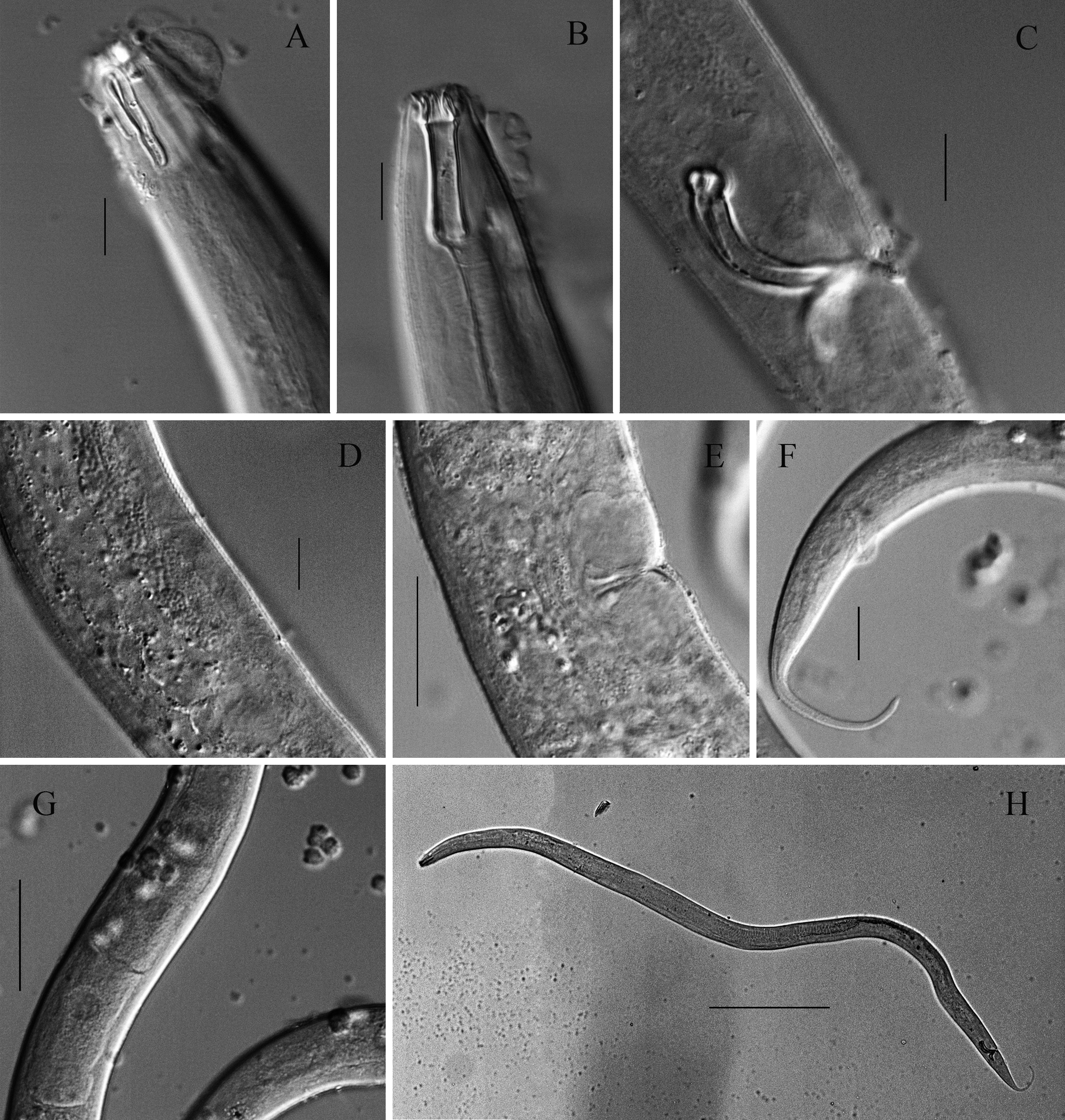
FIGURE 7. Light micrographs of Parodontophora huoshanensis sp. nov. A: anterior body end of male, showing amphid; B: anterior body end of female, showing buccal cavity; C: lateral view of male body part, showing copulatory apparatus; D: supplements in male; E: vulva region; F: posterior body end of female; G: ovary region; H: full view of male body. Scale bars: A, B, C, D = 10 Μm; E, F = 25 Μm; G = 50 Μm; H = 200 Μm.
TABLE 5. Individual measurement of Parodontophora huoshanensis sp. nov. (in µm).
| Characteristics | Holotype | Paratypes |
|---|---|---|
| ♂1 | ♂2 ♀1 ♀2 | |
| Total body length | 1235 | 1377 1408 1325 |
| Head diameter | 12 | 11 12 15 |
| Cephalic setae | 4 | 3 4 4 |
| Opisthocephalic setae | 2 | 2 2 2 |
| Buccal cavity length | 26 | 27 27 29 |
| Buccal cavity diameter | 4 | 4 4 6 |
| Amphid from anterior end | 3 | 3 3 3 |
| Amphid c. b. d. | 17 | 18 17 24 |
| Amphid dorsal branch length | 13 | 13 13 13 |
| Amphid ventral branch length | 20 | 21 21 24 |
| Nerve ring from anterior end | 98 | 102 95 101 |
| Nerve ring c. b. d. | 30 | 28 34 43 |
| Pharynx length | 150 | 161 151 164 |
| Pharynx c. b. d. | 33 | 35 37 55 |
| Renette gland length | 80 | 75 91 56 |
| Maximum body diameter | 46 | 44 42 72 |
| a. b. d. | 27 | 30 25 34 |
| Tail length | 132 | 130 142 146 |
| c' | 4.9 | 4.3 5.7 4.3 |
| Spicule length as chord | 30 | 28 - - |
| Spicule length as arc | 35 | 34 - - |
| Gubernaculum length | 10 | 9 - - |
| Apophysis length | 11 | 13 - - |
| Vulva c. b. d. | - | - 41 65 |
| Vulva, % | - | - 49.5 50.1 |
| a | 26.8 | 31.3 33.5 18.4 |
| b | 8.2 | 8.6 9.3 8.1 |
| c | 9.4 | 10.6 9.9 9.1 |
TABLE 6. Comparison of Parodontophora huoshanensis sp. nov. with allied species.
| Species | P. amoyensis | P. brevamphida | P. fluviatilis |
|---|---|---|---|
| Buccal cavity length (Μm) | 39 | 18–20 | 19–21 |
| Cephalic setae length/head diameter | 0.51–0.54 | 0.7 | 0.41–0.48 |
| Amphidael dorsal branch length/amphidael ventral branch(%) | 66* | 50* | 50 |
| Amphidael dorsal branch length/buccal cavity length | 0.7* | 0.8 | 0.7–0.8 |
| Opisthocephalic setae arrangement | ♂(2D-2V)2,♀(3D-2V)2 | irregular | (3D-1V)2 |
| Renette gland length/pharynx length (%) | 48–54 | 40 | 48–59 |
| Position of excretory pore | Anterior | Anterior2 | Anterior1 |
| Spicule length as arc (Μm) | 35–36 | - | 19–21 |
| Supplements | N | N | N |
| Armilliths | N | N | N |
| Reference | Zou 2000 | Timm 1952 | Gagarin & Thanh 2008 |
No known copyright restrictions apply. See Agosti, D., Egloff, W., 2009. Taxonomic information exchange and copyright: the Plazi approach. BMC Research Notes 2009, 2:53 for further explanation.
|
Kingdom |
|
|
Phylum |
|
|
Class |
|
|
Order |
|
|
Family |
|
|
Genus |
Parodontophora huoshanensis
| Li, Yongxiang & Guo, Yuqing 2016 |
P. fluviatilis
| Gagarin & Thanh 2008 |
P. limnophila
| Wu 2000 |
P. amoyensis
| Zou 2000 |
P. probata
| Smolyanko & Belogurov 1995 |
P. brevamphida
| Timm 1952 |
1 (by plazi, 2016-05-10 06:08:46)
2 (by ImsDioSync, 2017-02-01 15:06:28)
3 (by ImsDioSync, 2017-02-01 15:07:40)
4 (by ImsDioSync, 2017-02-08 01:42:13)
5 (by ImsDioSync, 2017-06-25 17:55:27)
6 (by ExternalLinkService, 2019-09-26 09:39:48)
7 (by ExternalLinkService, 2022-01-29 21:17:40)
8 (by ExternalLinkService, 2022-02-13 02:27:26)
9 (by plazi, 2023-10-30 00:55:44)