
Dysdera naouelae Bellvert & Dimitrov, 2024
|
publication ID |
https://doi.org/ 10.5852/ejt.2024.921.2429 |
|
publication LSID |
lsid:zoobank.org:pub:02633F29-4CDF-4027-BEBF-07AD2F925B42 |
|
DOI |
https://doi.org/10.5281/zenodo.10671228 |
|
persistent identifier |
https://treatment.plazi.org/id/65B05A67-B976-49F1-A9E3-110A537035BE |
|
taxon LSID |
lsid:zoobank.org:act:65B05A67-B976-49F1-A9E3-110A537035BE |
|
treatment provided by |
Plazi |
|
scientific name |
Dysdera naouelae Bellvert & Dimitrov |
| status |
sp. nov. |
Dysdera naouelae Bellvert & Dimitrov sp. nov.
urn:lsid:zoobank.org:act:65B05A67-B976-49F1-A9E3-110A537035BE
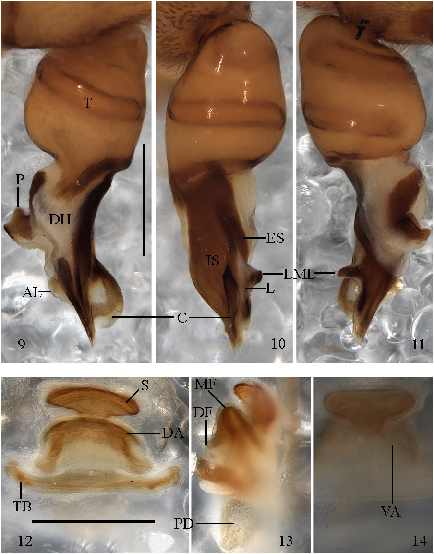
Figs 9–14 View Figs 9–14. 9–11
Dysdera tartarica View in CoL – Lazarov 2009: 104, figs 1–5 [misidentification].
Diagnosis
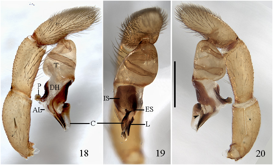
The new species is morphologically most similar to Dysdera kourosh sp. nov., Dysdera mikhailovi Fomichev & Marusik, 2021 , and Dysdera sagartia Zamani, Marusik & Szűts, 2023 by the well-developed, wide and rounded crest ( Figs 9–11 View Figs 9–14. 9–11 ), internal sclerite significantly wider than the exterior one, and the characteristic lateral margin of the lateral sheet ( Fig. 10 View Figs 9–14. 9–11 ). The male differs by (1) the thin, well sclerotized processus-like lateral margin of the lateral sheet, almost perpendicular to the embolic division tip ( Fig. 11 View Figs 9–14. 9–11 ), vs pointed backward in D. kourosh (arrowed in Fig. 20 View Figs 18–20 ) and significantly wider in D. mikhailovi ( Fomichev & Marusik 2021: fig. 46) and D. sagartia ( Zamani et al. 2023b: fig. 19a), (2) the convex crest, with wider apical part ( Fig. 9 View Figs 9–14. 9–11 ), vs concave at the middle in D. kourosh ( Fig. 18 View Figs 18–20 ) and wider in the basal part than in the apical part in D. mikhailovi ( Fomichev & Marusik 2021: fig. 44), and (3) proximal border of the additional lateral sheet not fused with distal haematodocha ( Figs 9, 11 View Figs 9–14. 9–11 ), vs fused in the other 2 species. Female differs by the curved spermatheca in dorsal view ( Fig. 12 View Figs 9–14. 9–11 ), vs straight in D. mikhailovi ( Fomichev & Marusik 2021: fig. 54) and the presence of a sclerotized neck in the spermatheca of D. naouelae sp. nov. ( Fig. 14 View Figs 9–14. 9–11 ).
Etymology
The new species is named after Naouel El Jaarani Oualkadi, partner of the first author, for all her patience and support.
Type material
Holotype
“ TURKESTAN ” • 1 ♂; precise country, collection date and collector unknown; SMF.
Paratype
“ TURKESTAN ” • 1 ♀; precise country, collection date and collector unknown; SMF .
Comparative material examined
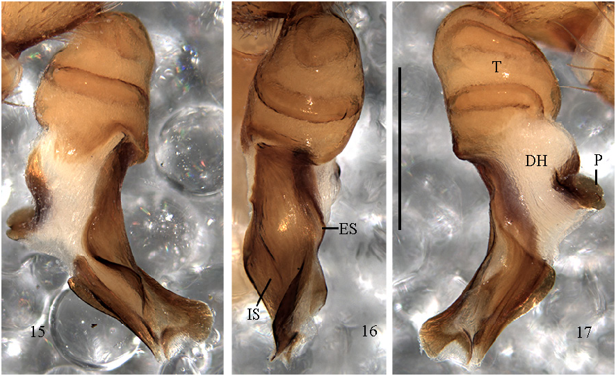
Dysdera tartarica Kroneberg, 1875 View in CoL ( Figs 15–17 View Figs 15–17 ) KYRGYZYSTAN • 1 ♂; Jalal-Abad Region , Ferganski range, Arslanbob; 12 Jun. 1983; S.L. Zonstein leg.; NHMW .
Description
Male ( Figs 9–11 View Figs 9–14. 9–11 )
PROSOMA. 3.55 long; maximum width 2.63; minimum width 1.63. Uniformly orange-red; smooth with some small black grains mainly at front. Frontal border roughly triangular, about ½ of carapace length; anterior lateral borders convergent; rounded at maximum dorsal width, back lateral borders rounded; posterior margin narrow, straight. Eye diameters: AME 0.18; PLE 0.14; PME 0.11; AMEs on edge of the frontal border, separated from one another by about ⅔ diameter, close to PLEs; PMEs very close to one another, about ⅓ of PME diameter from PLEs. Labium trapezoid-shaped, base wider than the distal part; longer than wide at base; semi-circular groove at the tip. Sternum orange, frontally darker, becoming lighter towards the back; wrinkled; covered with setae mainly on margin.
CHELICERAE. 1.36 long, about ⅖ of carapace length in dorsal view; fang medium-sized, 0.99 long; paturon dorsal and ventral side completely covered with piligerous granulations. Cheliceral inner groove short, about ⅓ of cheliceral length; armed with three teeth and lamina at base; B> D= M; D triangular, located roughly at the centre of the groove; B close to basal lamina; M close to B.
LEGS. Front legs orange, back legs yellow. Lengths of leg segments: fe1 2.85; pa1 1.81; ti1 2.40; me1 2.65; ta1 0.67; total 10.38; fe2 2.62; pa2 1.65; ti2 2.17; me2 2.48; ta2 0.61; total 9.52; fe3 2.03; pa3 1.12; ti3 1.30; me3 1.98; ta3 0.52; total 6.94; fe4 2.74; pa4 1.40; ti4 1.93; me4 2.57; ta4 0.66; total 9.30; fe Pdp 1.67; pa Pdp 0.94; ti Pdp 0.81; ta Pdp 0.93; total 4.35; leg formula: 1>2>4>3. Leg 1 has two terminal spines on the forward margin; leg 2 two terminal spines on the forward margin. Fe3d spines in two rows; forward 2; backward 1; pa3 spineless; tb3d spines arranged in two bands; proximal 1.0.1; medial-proximal 0; medial-distal 0; distal 1.0.1; tb3v spines arranged in four bands; proximal 0.1.0; medial-proximal 1.1.0; medial-distal 0.1.0; distal 1.0.0; with two terminal spines. Fe4d spines in two rows, forward 2; backward 6-5; pa4 spineless; tb4d spines arranged in two bands; proximal 1.0.1; medial-proximal 0; medial-distal 0; distal 1.0.1; tb4v spines arranged in four bands; proximal 1.2.0; medial-proximal 1.1.0; medial-distal 0.1.0; distal 1.0.0; with two terminal spines. Dorsal side of the frontal legs smooth; ventral side of the pedipalp covered with small piligerous grains. Claws with 8 teeth or less; hardly larger than claw width.
OPISTHOSOMA. 3.76 long; cream-colored; cylindrical. Abdominal dorsal setae 0.02 long; thick, roughly straight, blunt, tip not enlarged; uniformly and thickly distributed.
PALP ( Figs 9–11 View Figs 9–14. 9–11 ). T slightly shorter than ED, external distal border sloped forwards, internal one sloped backward. ED not bent, same T axis in lateral view, internal distal border not expanded. IS wider than ES, both more or less parallel; IS continuous to tip ( Fig. 10 View Figs 9–14. 9–11 ). ED tip sloped towards back in lateral view. C present, long; distal end beside ED internal tip; distal border rounded, smooth, markedly expanded, perpendicular to ED ( Fig. 9 View Figs 9–14. 9–11 ). AC absent. LF absent. L well developed, with a sclerotized processus-like basal lateral apophysis. LA absent. F absent. AL present, well developed; proximal border in posterior view smooth, not fused with distal haematodocha. P not fused to T ( Fig. 9 View Figs 9–14. 9–11 ); lateral length from ⅔ to as long as T width; ridge present, parallel to T; not expanded, upper margin smooth; distally ridge-like expanded; posterior margin not folded.
Female ( Figs 12–14 View Figs 9–14. 9–11 ) PROSOMA. 3.77 long; maximum width 2.73; minimum width 1.75. Orange, anteriorly darker, becoming lighter towards posterior. Anterior border almost round. Eye diameters: AME 0.19; PLE 0.11; PME 0.15, separated from one another by about 1 diameter and about ½ PME diameter from PLEs. Sternum uniformly orange.
CHELICERAE. 1.53 long; 1.18. Cheliceral inner groove short, about ⅓ of cheliceral length; armed with three teeth and lamina at base; B> D> M.
LEGS. Lengths of leg segments: fe1 2.74; pa1 1.81; ti1 2.17; me1 2.26; ta1 0.61; total 9.61; fe2 2.51; pa2 1.69; ti2 2.02; me2 2.19; ta2 0.55; total 8.96; fe3 1.96; pa3 1.13; ti3 1.32; me3 1.87; ta3 0.49; total 6.78; fe4 2.61; pa4 1.38; ti4 1.93; me4 2.53; ta4 0.56; total 9.01; fe Pdp 1.66; pa Pdp 0.80; ti Pdp 0.77; ta Pdp 0.94; total 4.18; leg formula 1>4> 2>3. Leg 1 one terminal spine on forward margin. Fe3d spines in one row; 3; tb3v spines arranged in three bands; proximal 1.1.1; medial-proximal 1.1.1; medial-distal 0; distal 1.0.0; with two terminal spines. Fe4d spines in two rows; forward 3-2; backward 6-5; tb4v spines arranged in four bands; proximal 1.1.1-0; medial-proximal 1.1.0; medial-distal 0.0-1.0-1; distal 1.1-0.0; with two terminal spines.
OPISTHOSOMA. 4.41 long. Abdominal dorsal hairs 0.08 long. All other somatic characters as in male.
VULVA ( Figs 12–14 View Figs 9–14. 9–11 ). DA wider than long, fused to VA ( Fig. 13 View Figs 9–14. 9–11 ); DF wide in dorsal view. MF margins fused, sheet-like, well developed, and completely sclerotized. VA rectangular, transparent ( Fig. 14 View Figs 9–14. 9–11 ); frontal region completely sclerotized; posterior region sclerotized except for most internal area; AVD absent. S attachment projected under VA; arms as long as DA ( Fig. 12 View Figs 9–14. 9–11 ), distinctly curved; tips not projected; neck as wide as arms. Laterals of TB directed forward.
Distribution
Central Asia (“Turkestan”).
Remarks
This species belongs to the aculeata group (sensu Deeleman-Reinhold & Deeleman 1988). It was erroneously recorded as Dysdera tartarica Kroneberg, 1875 by Lazarov (2009). We examined the male specimen of D. tartarica , described and depicted by Deeleman-Reinhold & Deeleman (1988), which fits the original description and the drawing of Charitonov (1956: 22, fig. 2). As clearly seen from its photographs ( Figs 15–17 View Figs 15–17 ), it is not conspecific with D. naouelae sp. nov. ( Figs 9–14 View Figs 9–14. 9–11 ).
No known copyright restrictions apply. See Agosti, D., Egloff, W., 2009. Taxonomic information exchange and copyright: the Plazi approach. BMC Research Notes 2009, 2:53 for further explanation.
|
Kingdom |
|
|
Phylum |
|
|
Class |
|
|
Order |
|
|
Family |
|
|
Genus |
Dysdera naouelae Bellvert & Dimitrov
| Bellvert, Adrià, Dimitrov, Dragomir, Zamani, Alireza & Arnedo, Miquel A. 2024 |
Dysdera tartarica
| Lazarov S. 2009: 104 |