
Trichrysis sinica Rosa, Nguyen & Wiśniowski, 2022
|
publication ID |
https://doi.org/10.11646/zootaxa.5194.1.8 |
|
publication LSID |
lsid:zoobank.org:pub:FA4ACDB7-064A-4BD8-9E7C-DC548A916E55 |
|
DOI |
https://doi.org/10.5281/zenodo.10548386 |
|
persistent identifier |
https://treatment.plazi.org/id/3F7C60AA-D301-485F-816B-B5BB52816566 |
|
taxon LSID |
lsid:zoobank.org:act:3F7C60AA-D301-485F-816B-B5BB52816566 |
|
treatment provided by |
Plazi |
|
scientific name |
Trichrysis sinica Rosa, Nguyen & Wiśniowski |
| status |
sp. nov. |
Trichrysis sinica Rosa, Nguyen & Wiśniowski , sp. nov.
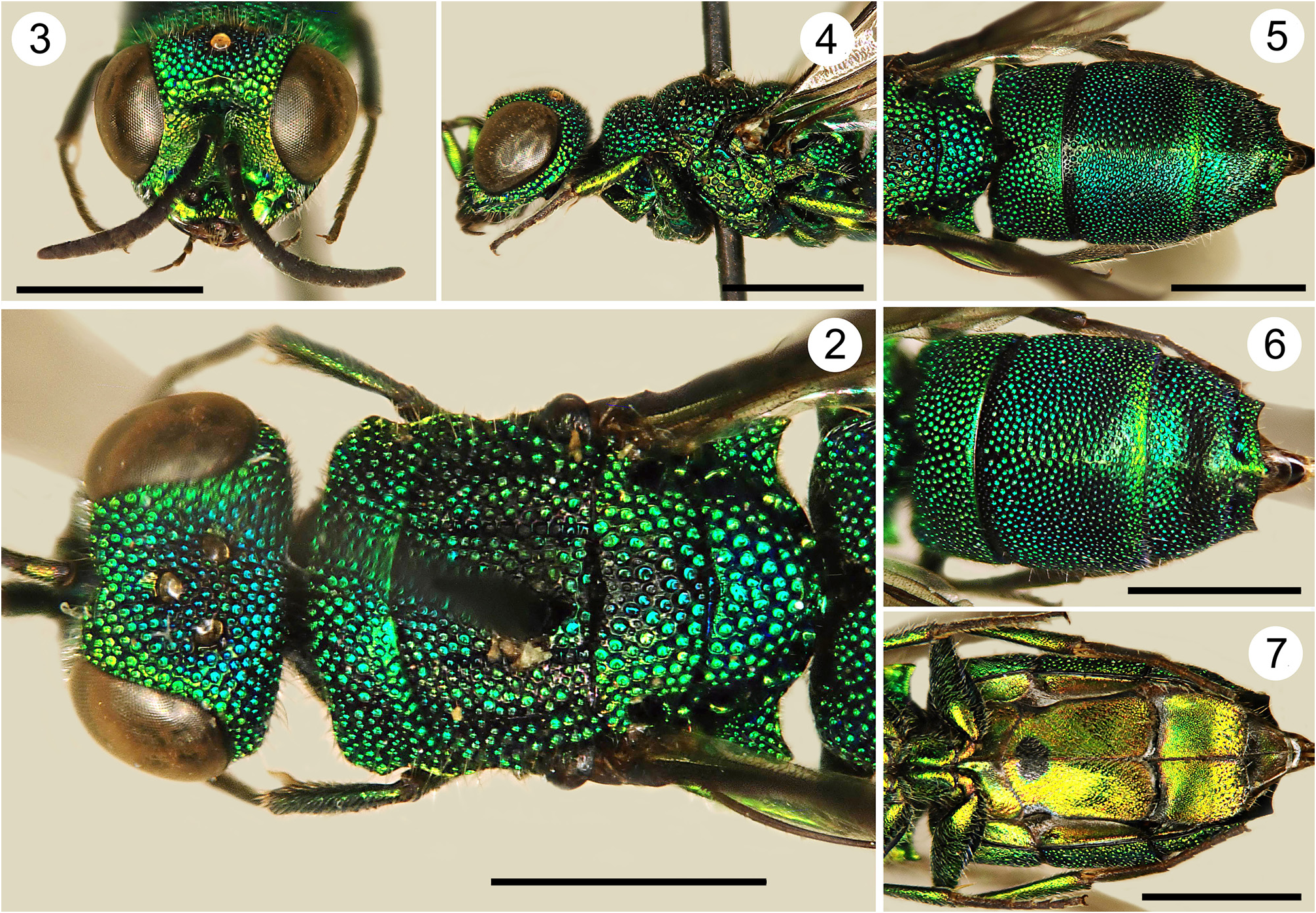
( Figs 2–7 View FIGURES 2–7 )
urn:lsid:zoobank.org:act:
Material examined. Holotype, 1♀, CHINA; / S China —10.– 14.7.1990 / Jinghong pr. YUNNAN / lgt. S. Bečvář ( NMLU).
Diagnosis. Middle-sized species with robust, densely punctate body and golden-green colouration with olivegreen to brown colour on interstices of vertex and mesosoma ( Fig. 2 View FIGURES 2–7 ); scape and pedicel fully metallic green; tegula brown with indistinguishable metallic reflection. TFC weak, slightly raised and angled medially. Sublateral carina fully developed. T2 with weak median carina. Median tooth on apex of T3 sharp and pointed ( Fig. 6 View FIGURES 2–7 ). S2 black spots ogival, fused medially, longer than wider ( Fig. 7 View FIGURES 2–7 ).
Description. Female. Holotype, body length 5.2 mm.
Head. Scapal basin deep, punctate laterally and striate medially ( Fig. 3 View FIGURES 2–7 ). TFC single, short, slightly inverted V-shaped. Area between TFC and scapal basin slightly raised. Relative length of P:F1:F2:F3=1.0:1.6:0.7:0.6; F1 l/w=2.9; OOL=2.0 MOD; BOL=1.7 MOD; POL=1.8 MOD; MS=1.0 MOD; clypeal apex slightly concave.
Mesosoma. Median pronotal line deep, extending to 3/4 length of pronotum; sublateral carina distinct and complete ( Figs 2, 4 View FIGURES 2–7 ), lateral margin of pronotum concave medially. Mesoscutellum antero-medially with wide interstices; punctation contiguous on metanotum, with punctures larger than on mesonotum. Episternal sulcus and scrobal sulcus with large areolate punctures ( Fig. 4 View FIGURES 2–7 ).
Metasoma. Punctures on T2 geminate, intersticespunctate ( Fig. 5 View FIGURES 2–7 ); punctuation decreasing in diameter toward posterior margin on T2. T2 and T3 with weak median carina. T3 prepit bulged medially convex; pit row with small and round separated pits. Apex of T3 with three pointed teeth similar in size, with interval between median tooth and lateral tooth straight ( Figs 5, 6 View FIGURES 2–7 ). S2 black spots ogival and medially fused ( Fig. 7 View FIGURES 2–7 ).
Colouration. Body metallic green with olive colour on interstices of the punctation of the fore body, with golden reflection on face and sternites. Scape and pedicel metallic green, flagellum black. Tegula brown, with weak metallic reflections. Legs metallic green, with tarsi black. Wings slightly infuscate.
Male. Unknown.
Distribution. China ( Yunnan).
Remarks. After examination of more material, we can state that the Chinese specimen photographed and identified by Rosa et al. (2016) as Trichrysis tonkinensis actually belongs to an undescribed species. At that time, we examined several specimens preserved in the Linsenmaier collection (NMLU) from China, India, Indonesia and Malaysia and identified by the Swiss author as T. tonkinensis . They all showed a certain variability, which was difficult to evaluate in a few specimens collected in such a wide area. Therefore, we considered all these specimens to belong to the same taxon, with regional variability. At least the Chinese species photographed by Rosa et al. (2016) can be now considered separated from the Vietnamese T. tonkinensis . We include in the type series only the specimen photographed in Rosa et al. (2016) from Rosa’s collection, that will be deposited in NMLU. Specimens listed in Rosa et al. (2016) from other Oriental localities must be re-evaluated in the light of recent studies ( Rosa et al. 2021, 2022; Wiśniowski et al. 2020).
Trichrysis sinica sp. nov. is very similar to T. excisifrons for the habitus and ogival shape of the black spots on S2 (see pictures of the type in Rosa et al. 2021); it is separated by the weak TFC, slightly arched (vs. raised and inverted V-shaped in T. excisifrons ); brown tegulae, with a weak green reflection (vs. fully metallic blue); body colour green based (vs. blue); the colour of tegulae is often considered as a diagnostic feature in some Chrysidini genera ( Linsenmaier 1959; Rosa et al. 2016). Trichrysis sinica is similar to T. tonkinensis and T. striata Nguyen & Wiśniowski , sp. nov. for habitus and colour, but is separated by the weak, medially acute TFC (vs. raised, straight, and only medially slightly angled, with endings laterally bent under and close to eye margin); ogival shape of the black spots on S2 (vs. triangular, wider than long); T3 sparsely punctate before prepit bulge (vs. densely punctate).
No known copyright restrictions apply. See Agosti, D., Egloff, W., 2009. Taxonomic information exchange and copyright: the Plazi approach. BMC Research Notes 2009, 2:53 for further explanation.
|
Kingdom |
|
|
Phylum |
|
|
Class |
|
|
Order |
|
|
Family |
|
|
Genus |