
Spintherobolus ankoseion,, 1999
|
publication ID |
https://doi.org/ 10.5281/zenodo.10881992 |
|
DOI |
https://doi.org/10.5281/zenodo.10881811 |
|
persistent identifier |
https://treatment.plazi.org/id/03DA87B2-2A2A-FFF6-FE83-F65A4A99F481 |
|
treatment provided by |
Juliana |
|
scientific name |
Spintherobolus ankoseion, |
| status |
sp. nov. |
Spintherobolus ankoseion, View in CoL new species
( Figs. 29-31 View Fig View Fig View Fig )
Holotype. MZUSP 35430 , male, 23.1 mm SL; creek in the forest, near Barra do Sai , between Barra do Sai and Itapema, northern Santa Catarina, Brazil; 22 Dec 1985, W. Costa .
Paratypes. MZUSP 51024 , 2 males, 22.5-23.0 mm SL, 9 females, 22.6-26.0 mm SL; MCP 19253 , 1 male, 20.6 mm SL, 3 females, 24.4-27.9 mm, same data as holotype . The following paratypes are all from Parana: Brazil: MZUSP 51025 , 1, male, 21.1 mm SL; praia de Guaratuba , 5 km north of Matinhas; 4 Dec 1975, P. S. Santos Filho. - USNM 297935 , 1 male, c&s, 23.1 mm SL, 1 female, c&s, 23.3 mm SL, 1 female, 23.6 mm SL, 1 juv., 13.0 mm SL; rio da Praia , near Guaratuba; 18 Feb 1988, R. M. C. Castro. - USNM 297936 , 2 males, 18.8- 21.6 mm SL, 2 females, 20.1 -21.1 mm SL; unnamed blackwater stream crossing road “ Elisio Pereira Alves Filho ” about 15 km from road BR-277, SW of Paranaguá; 20 Feb 1988, R. M. C. Castro .
Diagnosis. This species is distinguished by the low vertebral count 32-33, x = 32.2, versus 32-34, x = 33.0 for S. broccae and 32-34, x = 32.9 for S. leptoura ( Fig. 21 View Fig ); a larger preanal distance in males, 61.3-63.9 % SL, x = 62.6, versus for S. broc cae 55.9-61.5, x = 59.7, and S. leptoura 57.3-61.9, x = 58.9. Although some overlap occurs in these ranges, Mann-Whitney rank sum tests presented in caption of Figure 21 View Fig indicate significant differ ences at the 0.05 level of probability. Other differ ences among these species are expressed in the diagnoses of the other species.
Description. Morphometric data are summarized in Table 3 View Table 3 . Body compressed, moderately elongate. Predorsal body profile convex with concavity at nape. Body profile along dorsal-fin base posteroventrally inclined, becoming straight or slightly concave between dorsal and caudal fins. Ventral body profile gently convex between tip of mandible and pelvic fins. Belly profile convex between pelvic and anal fins in females and slightly concave in males. Body profile along anal-fin base slightly concave in females and slightly convex in anterior lobe in males and concave in their posterior lobe. Ventral profile of caudal peduncle concave in females, with a keel formed by prominent ventral procurrent caudal-fin rays.
Snout shorter than eye diameter. Mouth ter minal, angled posteroventrally; maxilla short, not reaching line drawn vertically through anterior margin of eye.
Dorsal-fin rays ii,9 (branched rays 8-9, x = 8.9). Dorsal-fin origin near midbody. Adipose fin absent.
Anal-fin rays iv, 13 (anterior unbranched rays iii-iv, x = 3.2, n = 22; branched rays 11-13, x = 12.6, n = 22; Fig. 20 View Fig ). Profile of anterior lobe of anal fin of males large and rounded ( Fig. 9 View Fig ). Distal tip of largest rays not reaching posteriorly beyond distal tip of posterior anal-fin rays; distal border of posterior anal-fin lobe concave. Anal-fin profile of females with distal border deeply concave, each ray decreasing in length from posterior unbranched ray to 6th branched ray. Anal-fin origin at vertical line drawn through dorsal-fin posterior basal termination. Three of six males examined have hooks on anal-fin rays. Hooks extremely reduced in size, occurring on posterior face of 2 to 6 segments of posterior branch of first and sometimes first and second branched anal-fin rays. Each segment bears two to three hooks on each side. Branched anal-fin rays 1-5 of males enlarged; branched rays two to four even more developed, reaching more than five times wider than more posterior rays. These rays with a posterior process on anteroproximal face of each lepidotrich. These processes are extensions of rays two to four, each inserted between lepidotrichia of next anterior ray. Each bony ray segment of posterior branch of branched rays 2 to 4, and sometimes also rays 1 and 5, not squarish like other branches of same ray, but with the posteroventral angle acute and elongated to distal tip of that fin ray. Segments ofbranched fin rays 2 to 4 are progressively fused to each other.
Pectoral-fin rays i,8 (branched rays 7- 11, x = 8.9). Posterior tip of longest pectoral-fin ray ex tends posterior to pelvic-fin origin in both males and females. Pelvic-fin rays i,5. Tip of largest ray reaching posteriorly beyond anal-fin origin in males, but not reaching that point in females. Pelvic fin of males bearing hooks on ventromedial face of unbranched ray and three (rarely two) following branched rays. Hooks present along their entire length, except for third branched ray which has hooks, when present, along only half this ray’ s length. Two hooks present on each ray segment, only on medialmost branch.
Principal caudal-fin ray count 10/ 9. Dorsal procurrent caudal-fin rays 11 (9- 11, x = 10.0, n = 22). Ventral procurrent caudal-fin rays, 12 (12-16, x = 14.0, n = 22). Ventral procurrent caudal-fin rays of males large, with theirproximal tips fused, slab shaped and expanded in sagittal plane. Four to five terminal caudal (preural) vertebrae (including posterior half centrum) support caudal-fin rays and ventral procurrent rays, with enlarged and modified hemal spines. Those ventral procurrent rays anterior to hemal spine of antepenultimate vertebra fused to each other and with lateral bony processes between muscles and skin. Anteriormost ventral procurrent rays with proximal tips absent.
Scales cycloid. Lateral line with 4 perforated scales (4-5, x = 4, n = 22). Scales in lateral series, 33 (31 -33, x = 31.8, n = 19; Fig. 22 View Fig ). Scale rows between dorsal-fin origin and lateral-line series 5 (5-6, x = 5.5, n = 21), and between lateral line and pelvic-fin origin 6 (4-6, x = 5.3, n = 21). Scale rows around caudal peduncle 14 (n = 19). No modified scales on caudal fin. Scale sheath along anal-fin base absent.
Teeth. All jaw teeth with a basal pedicle and somewhat laterally expanded distal conical cusp. Anterior dentary teeth tricuspid, but each with single cusp laterally and posteriorly. Premaxillary and maxillary teeth all conical. Premaxillary teeth 8, maxillary teeth 5-6 and dentary teeth 12- 13 in two c&s specimens.
Vertebrae. 32 (32-33, x = 32.2, n = 19; Fig. 21 View Fig ). Branchiostegal rays 4 in two cleared and stained specimens. Arranged as in S. papilliferus.
Color in alcohol. See Figures 29-30 View Fig View Fig for the preserved color patterns adult males and females which are approximately the same and very close to that of S. broccae . A discrete lateral body stripe extends from just dorsal to the dorsalmost part of the gill opening to the caudal peduncle where it continues onto the caudal fin as a solid dark wedge with its apex covering the two middle caudal-fin rays. In the photographs of a male and female, there appears to be a dark humeral spot just posterior to the more dorsal region of the opercular flap. This is the area of the anterior and posterior pseudotympanums and the black pigment coating the swimbladder can be seen through the skin. The dark chromatophores of the lateral stripe occur in this area but they are not increased in size or number and do not form a humeral spot.
The scale borders are covered by a double or, in some areas a triple row of dark chromatophores above the lateral stripe, giving the dorsal part of the body a reticulate pattern. The median scale row along the back from the nape to the caudal fin is very dark, nearly black, and the scales do not have pale centers as they do in most of the dorsal body region. The abdomen and sides ventral to the lateral stripe are pale cream color or yellowish (the color of preserved muscle tissue) and the scale borders of one or two horizontal scale rows just ventral to the lateral stripe have dark chromatophores in one or two rows. The posterior termination of the caudal peduncle, both dorsal and ventral to the lateral stripe, is white, without dark pigment of any kind.
The pectoral and pelvic fins of both sexes are hyaline with a few scattered dark chromatophores, especially on the anterior undivided ray of both fins. The dorsal fin is mostly hyaline except for its anterior dorsal border where the anterior undivided rays and the first branched ray have numerous dark chromatophores. The rays posterior to these dark rays progressively lose the dark chromatophores. The caudal fin is hyaline with a thin scattering of small dark chromatophores over the rays and membranes, especially those of the dorsal parts of the dorsal lobe of the male. A second dark, relatively broad stripe covers some of the muscular mid-base of the fin and extends posteriorly to completely cover the short anal-fin rays that follow the large, elongate, anterior anal-fin lobe. The anterior lobe of the fin, distal to its muscular base, is hyaline except for the unbranched rays.
The head is pale brown around the mouth with a scattering of dark chromatophores over the ventral surface of the snout, jaws, and oper cular region. In the photographs ( Figs. 29-30 View Fig View Fig ), the snout region, especially the jaws, is somewhat overexposed and the dark chromatophores do not show well. The dorsal surface of the snout and the area between the eyes are pale brown, while the dorsum of the cranium and the nape are dark brown. The head area posterior to the eye and extending ventrally from the parietal region to the preopercle and branchiostegal rays is white. The circumorbital region ventral to the eye is dark, with a series of dark chromatophores, giving producing dark spot ventral to the eye. The opercular region is dark dorsally and pale brown to white ventrally in both sexes.
Color in life. See Figure 31 View Fig of a male photographed immediately after capture. Life colors are very much like those in preservative except that the dark stripes are black, the abdominal region bluish white and the two white spots at the posterior termination of the caudal peduncle are intense white with very pale orange just anterior to them. The distal half of the caudal-fin rays are greenish brown to almost pale orange. The dorsal fin bears the same colors. The pelvic and pectoral fins are hyaline except for the anterior unbranched rays which are darkened with the black chromatophores. The procurrent rays are black, at least in the male. The dorsal surface of the snout and head is dark brown to black, except for the nares which are translucent much like most of the body lacking black pigment.
Sexual dimorphism. Males and females can be externally recognized by the anal-fin shape; compare Figures 29 View Fig and 30 View Fig . The distal profile of this fin is deeply concave with the anterior lobe comparatively acutely angled in females. In males the anterior lobe is rounded in profile and the distal border of this lobe extends further posteriorly than in females. Not all males have hooks on the anal fin but all have hooks on the pelvic-fin rays and large branched anal-fin rays 1 to 5. Males have a keel along ventral profile of caudal peduncle, formed by the ventral procurrent caudal-fin rays.
Etymology. The name ankoseion is from the Greek ankos, a mountain valley or glen and eïon, a beach and is given in reference to the fact that this species lives between the coastal mountains and the sea. A noun in apposition.
Fig. 32. Spintherobolus broccae, USNM 287324, adult male 18.9 mm SL, Brazil, Rio de Janeiro, Cachoeira de Macacu.
Ecological notes. This species, like the others in subgroup b, lives in the lower elevations of coastal blackwater streams that drain the surrounding forests.
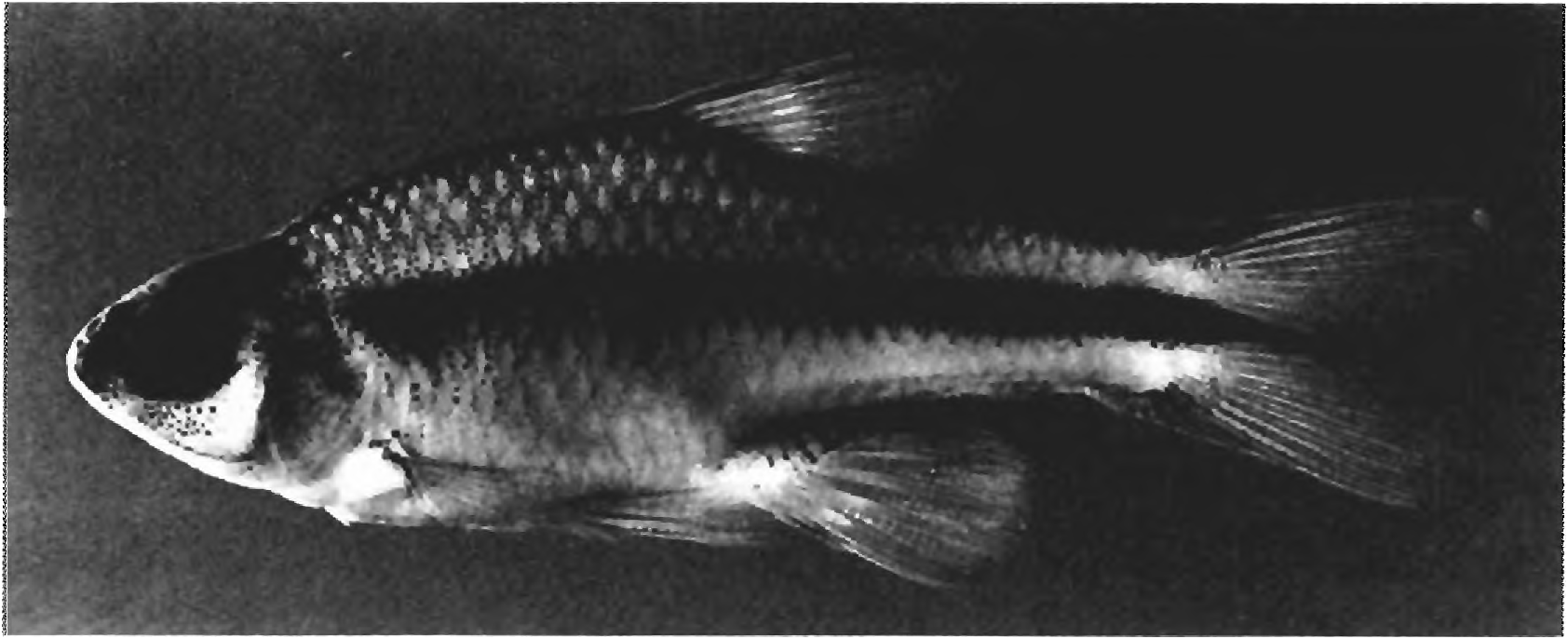
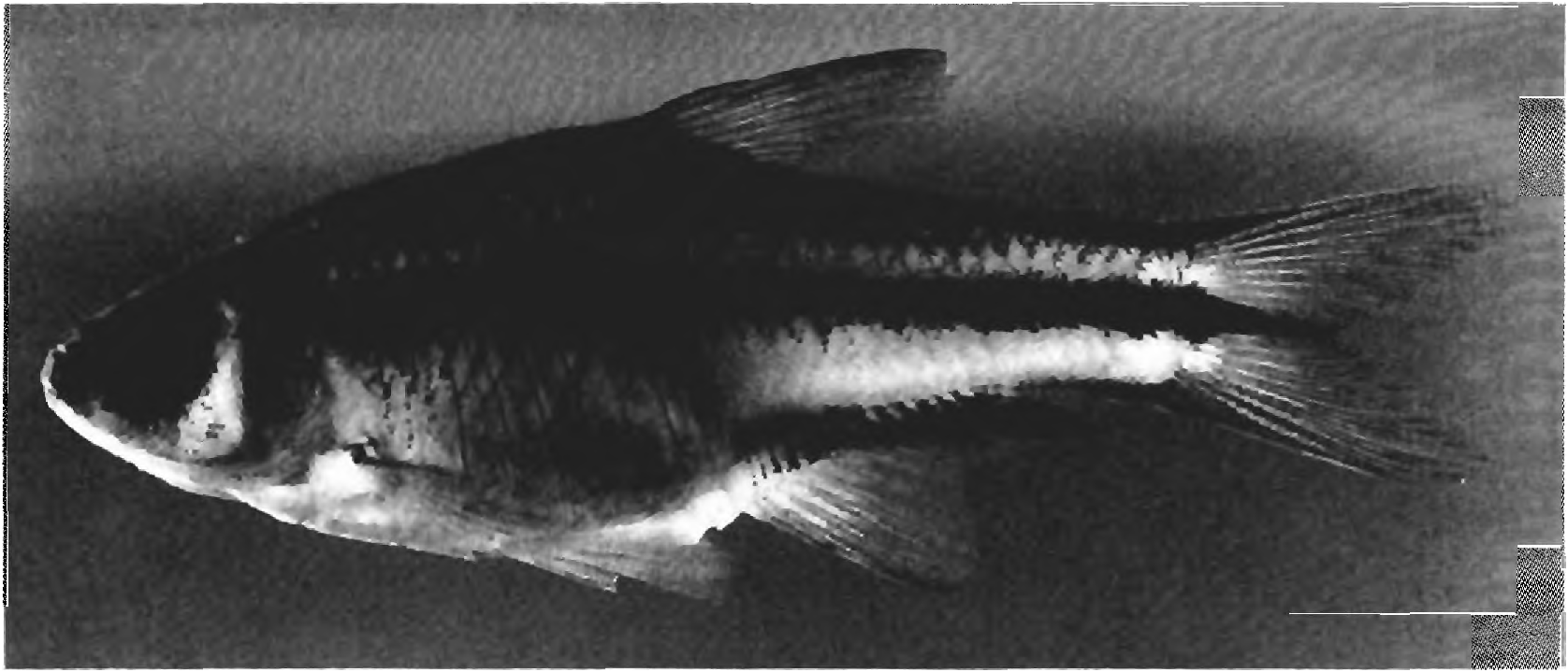
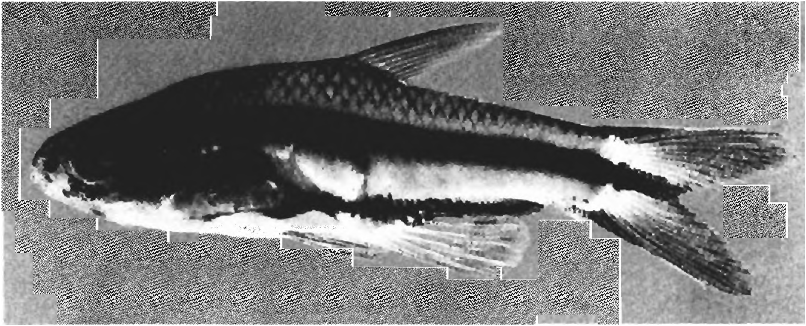
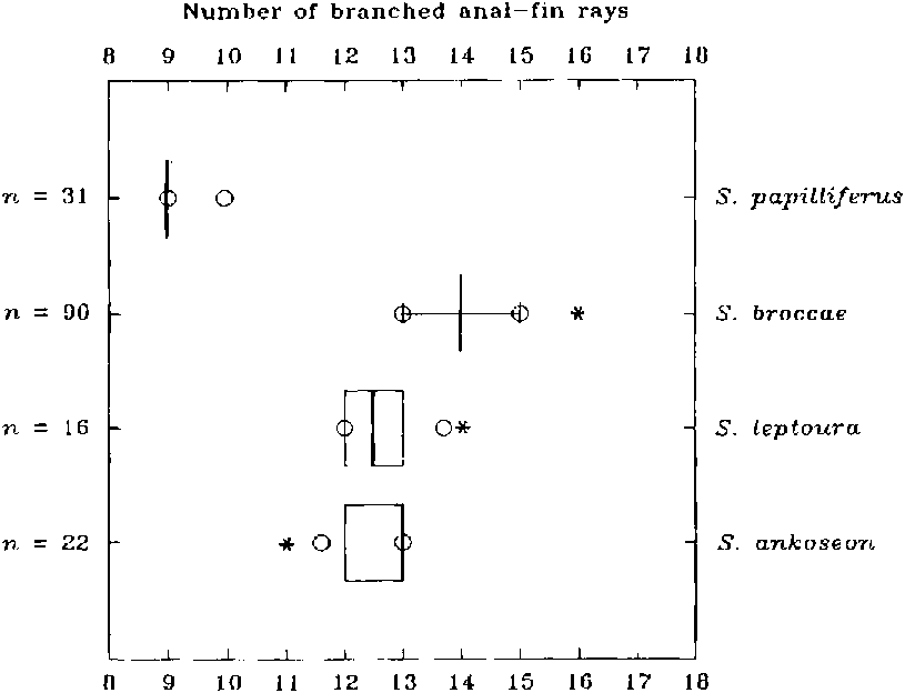
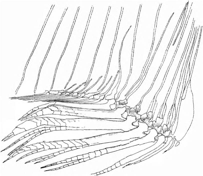
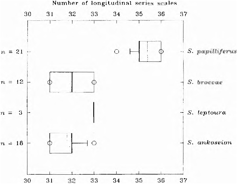
Table 3. Morphometries of Spintherobolus ankoseion. Table includes measurements of all type specimens. Values for the holotype are given separately.
| holotype | males | females | |||||||
|---|---|---|---|---|---|---|---|---|---|
| n | low | high | mean | n | low | high | mean | ||
| Standard length [mm] | 23.1 | 7 | 18.8 | 23.1 | 21.5 | 16 | 13.0 | 27.9 | 23.4 |
| Percentage of standard length | |||||||||
| Snout to anal-fin origin | 62.3 | 7 | 61.3 | 63.9 | 62.6 | 16 | 60.0 | 63.5 | 62.0 |
| Snout to dorsal-fin origin | 56.3 | 7 | 53.2 | 57.8 | 55.6 | 16 | 53.2 | 59.0 | 55.8 |
| Snout to pelvic-fin origin | 48.9 | 7 | 41.3 | 48.9 | 44.6 | 16 | 40.4 | 45.4 | 42.9 |
| Dorsal-fin base length | 9.5 | 7 | 9.0 | 12.6 | 10.8 | 16 | 10.2 | 13.6 | 11.4 |
| Anal-fin base length | 21.7 | 7 | 21.3 | 23.8 | 22.0 | 16 | 20.7 | 26.5 | 22.9 |
| Caudal peduncle length | 15.1 | 7 | 16.5 | 19.1 | 17.2 | 16 | 13.4 | 17.7 | 16.3 |
| Caudal peduncle depth | 12.9 | 7 | 12.8 | 15.6 | 14.8 | 16 | 11.9 | 16.5 | 14.7 |
| Depth at dorsal-fin origin | 35.5 | 7 | 29.3 | 37.9 | 34.2 | 16 | 29.2 | 38.5 | 35.4 |
| Dorsal-fin length | 28.6 | 7 | 26.6 | 30.8 | 29.0 | 16 | 27.0 | 30.3 | 28.5 |
| Pelvic-fin length | 19.1 | 7 | 18.5 | 21.8 | 19.9 | 16 | 13.4 | 19.9 | 17.8 |
| Pectoral-fin length | 20.8 | 6 | 18.2 | 20.8 | 19.0 | 16 | 13.9 | 19.7 | 17.5 |
| Bony head length | 28.6 | 6 | 28.4 | 30.1 | 29.1 | 16 | 27.2 | 31.5 | 28.8 |
| Percentage of head length | |||||||||
| Snout length | 19.7 | 6 | 19.0 | 24.2 | 21.0 | 16 | 17.5 | 23.4 | 21.1 |
| Upper jaw length | 15.5 | 6 | 15.2 | 20.0 | 18.4 | 16 | 14.8 | 21.9 | 18.5 |
| Horizontal eye diameter | 5.8 | 6 | 24.6 | 27.4 | 26.2 | 16 | 25.0 | 31.0 | 28.2 |
| Least interorbital width | 28.8 | 6 | 27.0 | 30.0 | 28.3 | 16 | 24.7 | 29.9 | 26.7 |
No known copyright restrictions apply. See Agosti, D., Egloff, W., 2009. Taxonomic information exchange and copyright: the Plazi approach. BMC Research Notes 2009, 2:53 for further explanation.
|
Kingdom |
|
|
Phylum |
|
|
Order |
|
|
Family |
|
|
SubFamily |
Cheirodontinae |
|
Tribe |
Cheirodontini |
|
Genus |