
Nemesia maltensis, Cassar & Mifsud & Decae, 2022
|
publication ID |
https://doi.org/10.5852/ejt.2022.806.1705 |
|
publication LSID |
lsid:zoobank.org:pub:4991BA30-038F-4B86-983B-E4E738C56759 |
|
DOI |
https://doi.org/10.5281/zenodo.6384567 |
|
persistent identifier |
https://treatment.plazi.org/id/BB791B03-F14E-4D15-9338-EE2E02244C4A |
|
taxon LSID |
lsid:zoobank.org:act:BB791B03-F14E-4D15-9338-EE2E02244C4A |
|
treatment provided by |
Felipe |
|
scientific name |
Nemesia maltensis |
| status |
sp. nov. |
Nemesia maltensis View in CoL sp. nov.
urn:lsid:zoobank.org:act:
Figs 54–73 View Figs 54–63 View Figs 64–69 View Figs 70–73
Diagnosis
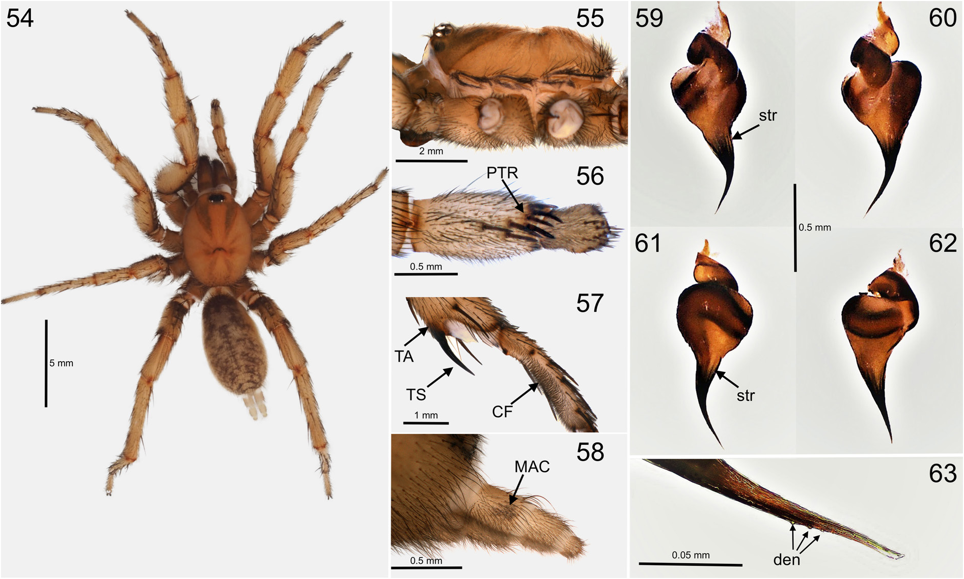
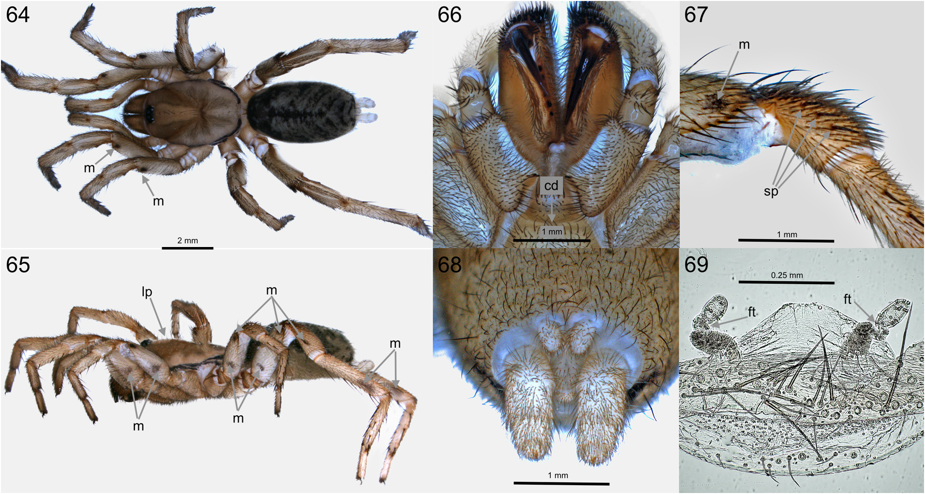
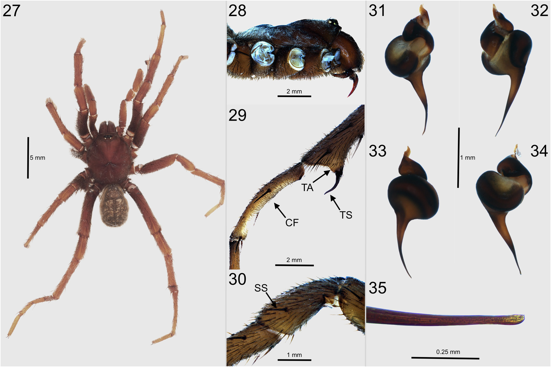
Nemesia maltensis groups out with a species-complex that is common in the central and eastern Mediterranean Basin ( Decae 2012). In this species-complex the palpal organ of males has striae (fine ribs) on the proximal embolus ( Figs 59–62 View Figs 54–63 ), and females have tube shaped, tripartite spermathecae in which the median part is folded and twisted ( Fig. 69 View Figs 64–69 ). Within this species-complex, N. maltensis fits in a central Mediterranean subgroup that has informally been named the maculatipes group ( Decae et al. 2015). Species in this subgroup, such as N. maculatipes Ausserer, 1871 ( Sardinia) , N. meridionalis (Costa, 1835) (southern Italy), N. sanzoi Fage, 1917 ( Sicily) and N. pannonica Herman, 1879 ( Serbia) have conspicuous dark coloured patches (maculae) on legs and/or the external proximal article of the PLS ( Figs 58 View Figs 54–63 , 64–65, 67 View Figs 64–69 ). Within the maculatipes group N. maltensis keys out with N. meridionalis on grounds of a similar morphology of the distal embolus in males being ornamented with a short row of tiny denticles ( Fig. 63 View Figs 54–63 cf. Isaia & Decae 2012: fig. 4). This character is not known from any other Nemesia species. Males of N. maltensis can be distinguished from those of N. meridionalis by the absence of an abrupt narrowing of the distal embolus ( Figs 59–62 View Figs 54–63 cf. Isaia & Decae 2012: fig. 3), females of N. maltensis can be distinguished from those of N. meridionalis by the relatively flat profile of the carapace (lateral view Fig. 65 View Figs 64–69 ), lack of sharply defined light coloured spigot fields on the PMS and PLS ( Fig. 68 View Figs 64–69 cf. Isaia & Decae 2012: fig. 5), and absence of a strong colour contrast between the labium and the sternum and the indistinct central division of the labial furrow ( Fig. 66 View Figs 64–69 cf. Isaia & Decae 2012: figs 7–8).
Etymology
The name refers to Malta, the largest island in the Maltese Archipelago and the only Mediterranean island where the species is currently known to occur.
Type material
Holotype MALTA • ♂; Buskett ; 8–13 Sep. 2020; 35.856° N, 14.396° E; T. Cassar leg.; (no. TC.004); NHMR. GoogleMaps
Paratypes MALTA • 4 ♂♂; same collection data as for holotype; (no. TC.005 to TC.008); NHMR GoogleMaps • 2 ♂♂; same collection data as for holotype; (no. TC.009, TC.010); NHMW GoogleMaps • 2 ♀♀; same collection data as for holotype; 14 Mar. 2021; (no. TC.012, TC.013); NHMR GoogleMaps .
Description
Male holotype (no. TC-004, NHMR)
PRESERVATION AND CONDITION. Specimen 6 months preserved in 70% ethanol, in good condition ( Fig. 54 View Figs 54–63 ), right bulb detached for study ( Figs 59–63 View Figs 54–63 ).
GENERAL COLORATION. Carapace yellowish with dark flanks of cephalic part and dark margin ( Fig. 55 View Figs 54–63 ), chelicerae brown, darker than carapace with dorsal lighter coloured patch, opisthosoma dorsal anterior dark greyish-brown, further light coloured with dark, irregular chevrons and a dark cardiac line ( Fig. 54 View Figs 54–63 ), opisthosoma ventral uniform creamy white, palps and legs lighter coloured than body in dorsal view and ventrally lighter coloured than dorsally, sternum and ventral coxae creamy white, labium conspicuously darker than other ventral parts.
CARAPACE. Longer than wide (CW/CL 0.8), covered with very fine silvery pubescence, curved bristles concentrated along the margins and posterior on the thoracic part, longitudinal row on the crest of the cephalic part and few forwardly projecting bristles on clypeus. Cephalic not elevated, thoracic part bulging slightly up from the fovea before sloping down to the posterior margin ( Fig. 55 View Figs 54–63 ).
EYES. Ocular-tubercle dome-shaped, clypeus narrow sloping down from eyes, eye-group rectangular, twice as wide as long (EL/PR 0.51), AME less than their diameter apart (dis.AME/dia.AME 0.87), dis. ALE–PLE less than ½ dia.ALE (ALE–PLE/ALE 0.44).
CHELICERAE. Rastellum with few strong teeth on the apical prolateral corner of chelicerae, prolateral row of furrow teeth, fangs sharp, bent, ventral un-serrated ridge.
VENTRAL PROSOMA. Maxillae rounded distal lobe, few spiky cuspules on proximo-anterior margin. Labium: wider than long (LW/LL 0.7), cuspules absent, labial furrow narrow. Sternum: longer than wide (SW/SL 0.8) three pairs round sigilla, posteriors sub-marginal, evenly covered with bristles.
PALPS. Cymbium with distal group of spines, tibia with PTR strongly develop ( Fig. 56 View Figs 54–63 ), proximally inflated (TibW/PTib 0.6), patella spineless, femur longer than tibia (PFem/PTib 1.6) with group of slender spines dorsally on distal half of the article. Palpal organ, proximal bulbous part pyriform extending distally into a slender curved embolus. Embolus curved, distally gradually tapering to a sharp, bevelled tip. Striae on proximal embolus, few tiny denticles just proximal of the embolus tip ( Figs 59–63 View Figs 54–63 ).
LEGS. All tarsi with fine ventral scopulae. Leg: I: metatarsus and tibia modified, metatarsus curved, lateral longitudinal rows of spines over the length of the article, CF with dense brush of short bristles, tibia distally widened, TA reduced ( Fig. 57 View Figs 54–63 cf Fig. 29 View Figs 27–35 ), TS smoothly upward curved ( Fig. 57 View Figs 54–63 ), spines present on all articles proximal of tarsus, patella with two prolateral spines. Legs I–III; tarsi without spines, other articles with numerous spines, maculae vague, most prominent on prolateral femur (apical) and patella (central). Leg IV, tibia> femur> metatarsus (Fem4/Met4 = 1.02, Tib4/Met4 = 1.07). Leg formula: 4123. PTC on all tarsi with ventrally two parallel combs of small teeth (genus character), ATC smooth.
OPISTHOSOMA. Ovoid, anterior narrowing with numerous forward directed bristles, dorsal bristles are backward directed, Spinnerets: PMS knob-shaped, spigots apically concentrated, PLS proximal article with external macula ( Fig. 58 View Figs 54–63 ), as long as medial + distal articles, spigots spread over ventral proximal and medial articles and on apical distal article.
MEASUREMENTS. TBL = 12.3; CL = 5.0; CW = 3.8; CP = 3.0; AR = 0.83; PR = 0.83; EL = 0.42; dia.ALE = 0.25; dia.PLE 0.15; dia.AME = 0.15; dia.PME = 0.12; dis.AME–AME = 0.13; dis.ALE–PLE = 0.11; SL = 2.6; SW = 1.9; LL = 0.6; LW = 0.8; Palp = 4.9 (0.7 + 1.2 + 1.1 + 1.9); Leg I = 13.2 (2.0 + 2.6 + 2.6 + 2.4 + 3.6); Leg II = 12.5 (2.0 + 2.5 + 2.4 + 2.1 + 3.5); Leg III = 11.9 (2.0 + 3.0 + 2.1 + 1.7 + 3.1); Leg IV = 16.8 (2.3 + 3.9 + 4.2 + 2.4 + 4.0); BuL = 1.12; BuW = 0.39; EmL = 0.51.
VARIATION MALES (n = 7). TBL = 8.7–12.3 (av. 10.1, sd. 1.2); CL = 3.8–5.0 (av. 4.4, sd. 0.4); CW = 2.8–3.8 (av. 3.3, sd. 0.3); CP = 2.1–3.0 (av. 2.5, sd. 0.3); AR = 0.67–0.83 (av. 0.74, sd. 0.05); PR = 0.69– 0.83 (av. 0.76, sd. 0.05); EL = 0.36–0.44 (av. 0.40, sd. 0.03); dia.ALE = 0.20–0.25 (av. 0.22, sd. 0.02); dia.PLE = 0.14–0.18 (av. 0.17, sd. 0.02); dia.AME = 0.13–0.15 (av. 0.14, sd. 0.01); dia.PME = 0.11– 0.14 (av. 0.13, sd. 0.01); dis.AME–AME = 0.08–0.14 (av. 0.11, sd. 0.02); dis.ALE–PLE = 0.04–0.11 (av. 0.07, sd. 0.02); SL = 1.9–2.6 (av. 2.2, sd. 0.2); SW = 1.5–1.9 (av. 1.7, sd. 0.1); LL = 0.3–0.6 (av. 0.4, sd. 0.1); LW = 0.6–0.8 (av. 0.7, sd. 0.1); Palp = 4.0–4.9 (av. 4.6, sd. 0.3); Leg I = 10.4–13.0 (av. 11.7, sd. 0.9); Leg II = 9.5–12.5 (av. 11.0, sd. 1.0); Leg III = 9.0–11.8 (av. 10.3, sd. 0.9); Leg IV = 13.5–16.8 (av. 15.1, sd. 1.2); Bul = 0.96–1.12 (av. 1.06, sd. 0.06); BuW = 0.31–0.39 (av. 0.36, sd. 0.03); EmL = 0.47–0.52 (av. 0.52, sd. 0.02).
Female paratype (no. TC-012, NHMR)
Note. The females available for description are small and might not be reproductive adults, the spermathecae of the here described paratype are however sufficiently developed for study. Specimens, 6 months preserved in 70% ethanol, in perfect condition.
GENERAL COLORATION. Carapace cephalic part has wide light brown crest-zone and is generally darker in colour than thoracic part, black margin prominent ( Fig. 64 View Figs 64–69 ), chelicerae bicoloured brown, slightly darker than carapace, abdomen dorsal as in male, palps and legs generally as in male, but dorsal femora lighter coloured and with prominent maculae ( Figs 64–65, 67 View Figs 64–69 ), sternum and labium slightly darker coloured than ventral coxae.
CARAPACE. Longer than wide (CW/CL 0.7), sparsely covered with very fine black pubescence, bristles centrally in longitudinal row on crest-zone. Cephalic part only slightly elevated and gradually sloping down over the thoracic part to the posterior margin ( Fig. 65 View Figs 64–69 ).
EYES. Ocular-tubercle as in male, eye-group rectangular, twice as wide as long (EL/PR 0.53), AME less than their diameter apart (dis.AME/dia.AME 0.92), dis.ALE–PLE less than ½ dia.ALE (ALE–PLE/ ALE 0.42).
CHELICERAE. Stronger developed than in male with distinct rastellum, prolateral row of furrow teeth with six conical teeth, fangs sharp, bent, ventral un-serrated ridge.
VENTRAL PROSOMA. Maxillae small rounded distal lobe, few knobby cuspules on proximo-anterior margin. Labium: twice as wide as long (LW/LL 2.3), cuspules absent, labial furrow centrally divided ( Fig. 66 View Figs 64–69 ). Sternum: longer than wide (SW/SL 0.7), sigilla not distinguished.
PALPS. Femur and patella spineless, tibia ventral and prolateral sharp distally pointing spines and dorsal long parallel rows of trichobothria, tarsus, distally pointed, ventral half fully scopulate with group of sharp distally pointing spines, dorsal row trichobothria in central part of article, palpal claw with short proximal row of teeth.
LEGS. All femora with external maculae ( Figs 64–65 View Figs 64–69 ). Anterior legs with ventro-prolateral scopulae extending from tip of tarsus to distal patella, ventral spines on tibia and metatarsus. Posterior legs with dense groups of spiny bristles prolateral on distal femur and dorsal patella. Leg III, patella with three prolateral spines ( Fig. 67 View Figs 64–69 ) and one retrolateral spine. Leg IV patella spineless, tibia> femur> metatarsus (Fem4/Met4 1,1, Tib4/Met4 1,3). Leg formula: 4123. PTC and ATC as in male.
OPISTHOSOMA. Generally as in male, anterior bristle group less distinct, spinnerets without sharply defined spigot-fields ( Fig. 68 View Figs 64–69 ). Spermathecae, tripartite, tube shaped, proximal part with dense concentration of pigmented cells, proximally widest, median part folded and twisted ( Fig. 69 View Figs 64–69 ), distal part digitiform with few pigmented cells.
MEASUREMENTS. TBL = 9.7; CL = 3.9; CW = 2.9; CP = 2.3; AR = 0.71; PR = 0.74; EL = 0.39; dia.ALE = 0.18; dia.PLE 0.13; dia.AME = 0.10; dia.PME = 0.10; dis.AME–AME = 0.10; dis.ALE–PLE = 0.05; SL = 1.7; SW = 1.3; LL = 0.3; LW = 0.6; Palp = 5.1 (1.1 + 1.1 + 1.1 + 1.8); Leg I = 8.3 (1.0 + 1.3 + 1.6 + 1.8 + 2.6); Leg II = 7.4 (1.0 + 1.3 + 1.4 + 1.5 + 2.2); Leg III = 7.1 (1.1 + 1.6 + 1.1 + 1.3 + 2.0); Leg IV = 11.3 (1.1 + 2.4 + 3.1 + 2.0 + 2.7).
VARIATION FEMALE (n = 2). TBL = 9.2, 9.7; CL = 3.4, 3.9; CW = 2.3, 2.9; CP = 2.0, 2.3; AR = 0.62, 0.71; PR = 0.61, 0.74; EL = 0.33, 0.39; dia.ALE = 0.18, 0.19; dia.PLE = 0.13, 0.16; dia.AME = 0.10, 0.12; dia.PME = 0.10; dis.AME–AME = 0.10, 0.11; dis.ALE–PLE = 0.05, 0.08; SL = 1.7, 2.1; SW = 1.3, 1.5; LL = 0.3, 0.4; LW = 0.6, 0.8; Palp = 4.2, 5.1; Leg I = 6.7, 8.3; Leg II = 6.3, 7.4; Leg III = 5.9, 7.1; Leg IV = 9.6, 11.3.
Observations
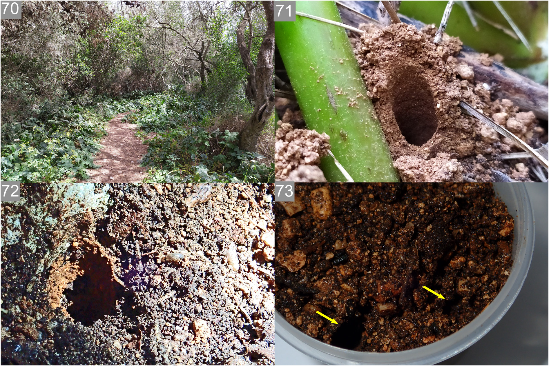
In the field ( Fig. 70 View Figs 70–73 ), the burrows of this species appeared to be completely lidless, with the burrow entrance opening at the ground surface directly ( Fig. 72 View Figs 70–73 ). The wall of the burrow is lined with almost imperceptibly thin strands of silk from the inside; but the burrow walls derive most of their structural integrity from the compaction of the soil around them ( Fig. 71 View Figs 70–73 ). In captivity, once soil was moistened, a female constructed a bifurcated burrow with two shafts opening at the soil surface with no lids ( Fig. 73 View Figs 70–73 ), joining together into one shaft deeper into the soil. Females were collected from burrows some 15 cm deep in the soil, congregated at the edge of a small boulder embedded in deep soil in a wooded area populated by trees of Laurus nobilis L., Olea europaea , Rhamnus alaternus L. and Pinus halepensis Mill. Burrow entrances always appeared at the soil surface at the edge of the boulder or, in one instance, at the base of the thick stems of an Acanthus mollis L. plant nearby. Males were collected in pitfall traps after several heavy rainfall events which occurred in September – the so called “first rains” which bring an end to the dry season.
No known copyright restrictions apply. See Agosti, D., Egloff, W., 2009. Taxonomic information exchange and copyright: the Plazi approach. BMC Research Notes 2009, 2:53 for further explanation.
|
Kingdom |
|
|
Phylum |
|
|
Class |
|
|
Order |
|
|
SubOrder |
Mygalomorphae |
|
Family |
|
|
Genus |