
Pleurotrocha sigmoidea Skorikov, 1896 (Notommatidae)
|
publication ID |
https://doi.org/ 10.5281/zenodo.188361 |
|
DOI |
https://doi.org/10.5281/zenodo.5694442 |
|
persistent identifier |
https://treatment.plazi.org/id/03DAC45D-D010-FFC1-FF68-F885A526FF6D |
|
treatment provided by |
Plazi |
|
scientific name |
Pleurotrocha sigmoidea Skorikov, 1896 (Notommatidae) |
| status |
|
Pleurotrocha sigmoidea Skorikov, 1896 (Notommatidae) View in CoL
Pleurotrocha macropoda Zavadowsky, 1926 View in CoL Proales sigmoidea Fadeew, 1927
Diagnosis. Species with bulbous body; toes short and conical; cerebral ganglion with small retrocerebral organ (less than half of head length), apical field of corona with palp-like ciliary tufts; pedal glands as long as foot; virgate trophi; triangular rami with pointed alulae; manubria with a small, lateral, knob-like protuberance; crescentic unci with 2 large and several minor uncinal teeth; fulcrum dorsoventrally expanded and terminally broadening.
General body organization of parthenogenetic female
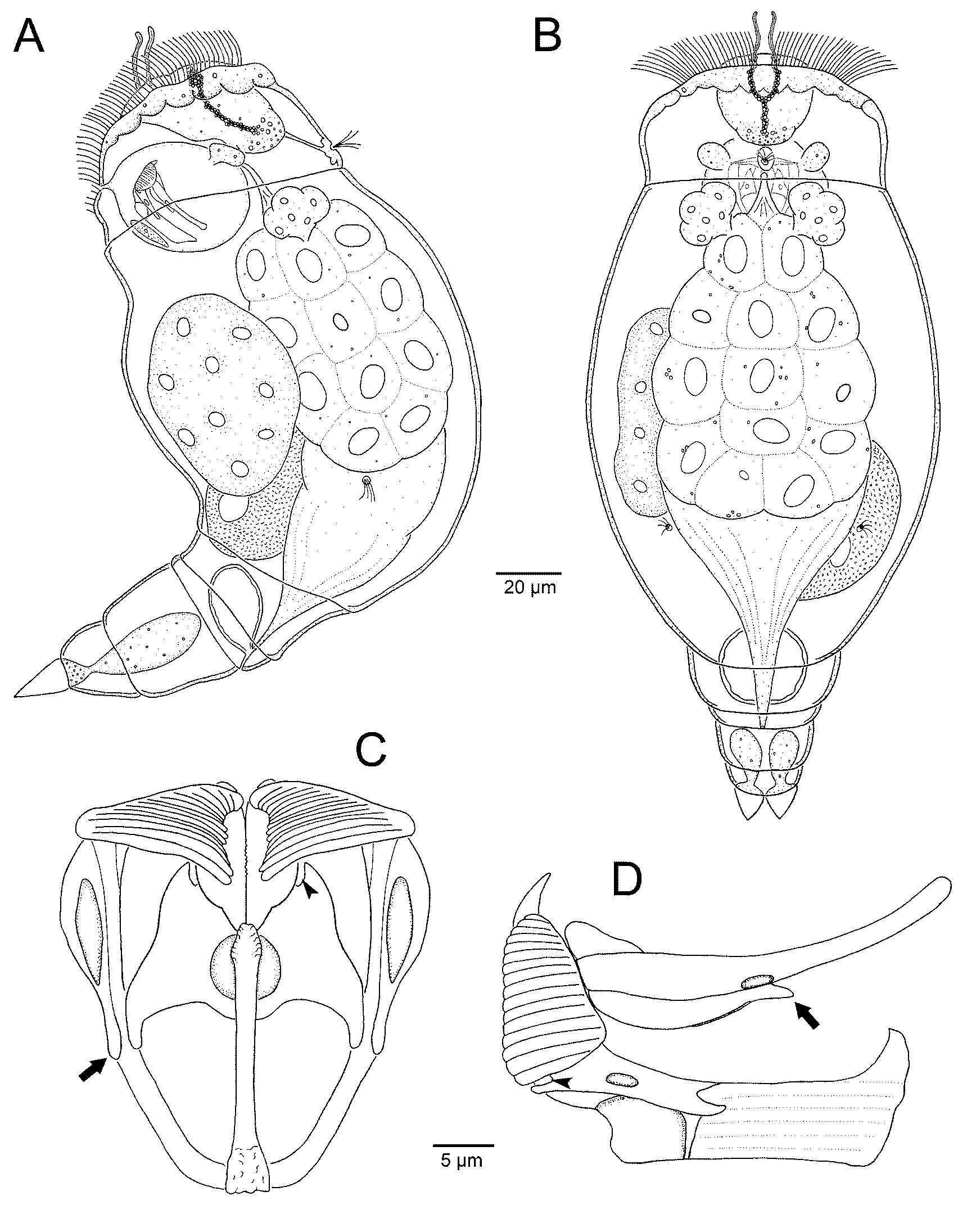
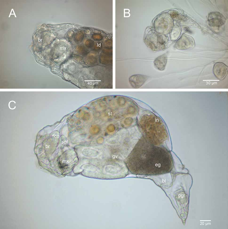
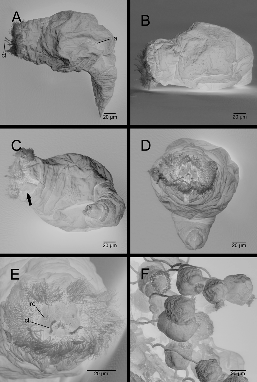
Habitus ( Figs 1 View FIGURE 1 A, B; 2A–C, 3A–E). Body bulbous and hyaline, divided into 3 distinct regions: head, trunk and foot with toes. Epidermis hardly stiffened, with smooth surface. Head short, sloped, contractile and offset from trunk by neck fold. Corona coursing from apical to ventral head region, characterized by a dense pseudotrochus composed of locomotory cilia. Pseudotrochus interrupted dorsally ( Fig. 3 View FIGURE 3 E), merging ventrally with cilia field leading to mouth opening. A pair of lobe-like epidermal projections ( Fig. 3 View FIGURE 3 C) limits mouth opening post-orally. Locomotory cilia enclosing an apical field that bears a pair of long, palp-like tufts composed of fused cilia, with a pair of shorter ciliary tufts above openings of retrocerebral organ ( Fig. 3 View FIGURE 3 E). Center of apical field shows additional short cilia arranged along an inverted, U-shaped fold ( Fig. 3 View FIGURE 3 E). Trunk bulbous, arched dorsally, composed of 3 pseudosegments and forms ovate outline in dorsal view. First pseudosegment comprises most of trunk; it is followed by a narrower, shorter lumbar pseudosegment and finally by a preanal pseudosegment ( Figs 1 View FIGURE 1 A, B). Foot massive, tapering gradually, divided into 2 almost equal-sized pseudosegments with 2 short, pointed, conical toes distally. During swimming, foot is caudally directed. Foot and toes are retractile.
Digestive system ( Figs 1 View FIGURE 1 A, B; 2A, C). Mouth opening in ventral part of corona leads to short, slender, ciliated buccal tube that discharges ventrally to spherical mastax. Oesophagus diverges dorsally from first third of mastax ( Fig. 1 View FIGURE 1 A). Stomach multicellular, with large, yellow-brownish, egg-shaped droplets, and occupies one-third of trunk ( Fig. 1 View FIGURE 1 A, 2A). Ciliated intestine offset from the stomach ( Fig. 2 View FIGURE 2 C). Anus discharges below preanal pseudosegment.
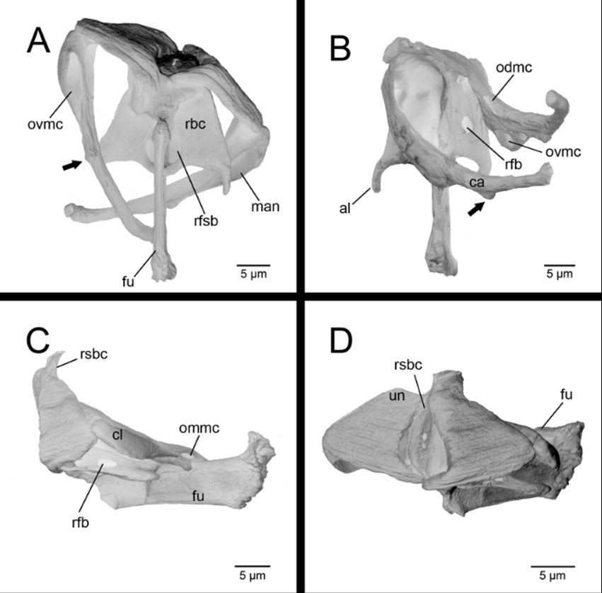
Trophi ( Figs 1 View FIGURE 1 C, D, 4A–D). Virgate type, bilaterally symmetrical with fulcrum residing in the longitudinal axis of the body. Trophi elements appear as follows:
Rami. Stout and triangular in ventral view, curved upwards anteriorly in a sickle-shaped bend, inner margins provided with small denticles ( Fig. 1 View FIGURE 1 C). Ramus subbasal and basal chambers exhibit distinct openings with the large, oval ramus foramen subbasalis directed ventrally ( Figs 1 View FIGURE 1 C, D, 4A) and circular ramus foramen basalis pointing caudodorsally ( Figs. 1 View FIGURE 1 D, 4B). Basal rami chambers provided with slender, acuminate alulae distally; space between rami forms a convex bowl.
Fulcrum. Long, rod-shaped and slender in ventral view ( Fig. 4 View FIGURE 4 A). In lateral view high, with longitudinal striae, expanding distally into a stamp-like plate characterized by a bunchy surface, margins arched upwards and a diagonal orientation to the longitudinal fulcrum axis ( Fig. 4 View FIGURE 4 C).
Unci. Bent, crescent-shaped plates, with 2 distinct ventral teeth and 3 very small median teeth. Some additional reduced teeth recognizable only by series of denticles and jugal lines ( Fig. 4 View FIGURE 4 D).
Manubria. Taper gradually from broad basal clava to distal end of long, rod-shaped cauda. Clava composed of 3 chambers that are not clearly distinguishable in detail but show distinct openings. Ventral manubrial chamber shows elongate, ventrally directed opening. It features a small, knob-like projection ( Figs 1 View FIGURE 1 C, D, 4A, B) situated ventrolateral to longitudinal middle of manubrium. Median manubrial chamber constitutes largest part of clava and also forms long cauda. Its opening lies above projection of ventral manubrial chamber and is ventrolaterally directed ( Figs 1 View FIGURE 1 D, 4C). Small dorsal manubrial chamber opens dorsolaterally ( Fig. 4 View FIGURE 4 B).
Nervous system and sensory organs. Cerebral ganglion rounded, very small ( Figs 1 View FIGURE 1 A, 2A). Dorsal antenna consists of several cilia encircled by flat, rounded collar; located on head in front of neck fold ( Fig. 3 View FIGURE 3 B). Lateral antennae lying dorsolaterally in posterior third of trunk; cilia surrounded by flat, rounded collar. Two pairs of ciliary tufts with possible sensory function arranged apically on head ( Fig. 3 View FIGURE 3 E).
Glandular system ( Figs 1 View FIGURE 1 A, B, 2A). Retrocerebral organ a dark Y-shaped duct located posterior to brain ( Fig. 1 View FIGURE 1 B); in some specimens, only the duct is visible with light microscopy. A pair of large, kidney-shaped salivary glands resides in mastax complex. Both gastric glands possess 5 nuclei, are cauliflower-shaped and diverge anteriorly from stomach with short stalks. Pedal glands as long as foot, with terminally constricted mucus reservoir above toes.
Protonephridial system. Paired protonephridia with distinct terminal organs (exact number and position not documented), opening into contractile bladder ( Figs 1 View FIGURE 1 A, B, 2C) that in turn empties into terminal part of the intestine (cloaca).
Reproductive system ( Figs 1 View FIGURE 1 A, B, 2A, B): Germovitellarium unpaired, eight-nucleated, in ventral region of trunk; a large, dark egg often present.
Measurements. Total length 300–310 µm, maximum dorsoventral extent 108–121 µm, maximum width 124–132 µm, foot length 60 µm, toe length 20 µm, trophi length 35 µm, ramus length 22 µm, manubrium length 32 µm, manubrium width 8 µm, fulcrum length 19–23 µm.
Ecology. The species is limnosaprobic and uncommon, occurring in stagnant or running fresh waters in colder seasons. It has been reported from Germany ( Koste 1968), France ( De Beauchamp 1948), Russia ( Skorikov 1896), Canada ( De Smet 1996) and England ( Reiss & Schmid-Araya 2008). It lives solitarily among degraded macrophytes or between Lemna on colonies of large sessile ciliates (especially Vorticella ) ( Fig. 3 View FIGURE 3 F) that it feeds on. The ciliates are relatively large in comparison to the small mouth opening of the rotifers (compare Figs 3 View FIGURE 3 E and 3F) and are eaten in parts. De Beauchamp (1948) also reported the species living on Campanella umbellaria . It appears that P. sigmoidea has a preference for larger ciliates and does not switch to other prey. Because of its rare appearance, however, and uncommon field observations, P. sigmoidea may have a broader prey spectrum.
No known copyright restrictions apply. See Agosti, D., Egloff, W., 2009. Taxonomic information exchange and copyright: the Plazi approach. BMC Research Notes 2009, 2:53 for further explanation.
|
Kingdom |
|
|
Phylum |
|
|
Class |
|
|
Order |
|
|
Family |
|
|
Genus |
Pleurotrocha sigmoidea Skorikov, 1896 (Notommatidae)
| Wilts, Eike F., Bininda-Emonds, Olaf R. P. & Ahlrichs, Wilko H. 2009 |
Proales sigmoidea
| Fadeew 1927 |
Pleurotrocha macropoda
| Zavadowsky 1926 |