
Pleurotrocha petromyzon ( Ehrenberg, 1830 ) (Notommatidae)
|
publication ID |
https://doi.org/ 10.5281/zenodo.188361 |
|
DOI |
https://doi.org/10.5281/zenodo.5694444 |
|
persistent identifier |
https://treatment.plazi.org/id/03DAC45D-D017-FFC8-FF68-FA12A69EFEB4 |
|
treatment provided by |
Plazi |
|
scientific name |
Pleurotrocha petromyzon ( Ehrenberg, 1830 ) (Notommatidae) |
| status |
|
Pleurotrocha petromyzon ( Ehrenberg, 1830) (Notommatidae) View in CoL
Notommata petromyzon Ehrenberg, 1830 Notommata gibba Ehrenberg, 1832
Notommata petromyzon: Ehrenberg 1838 Proales petromyzon: Hudson & Gosse 1886 Notops laurentinus Jennings, 1894
Proales laurentius Jennings, 1896
Pleurotrocha laurentina: Harring 1913
Diagnosis. Species with fusiform, stout body; toes short and conical; retrocerebral organ with posterior sphere followed by dark red eye; large gastric glands with 9 nuclei; large pedal glands extending into trunk; triangular rami with rounded alulae; manubrium with large, lateral, knob-like protuberance; crescent-shaped unci with 2 large and 3 minor uncinal teeth; end of distal fulcrum flattened and arched dorsally.
General body organization of parthenogenetic females
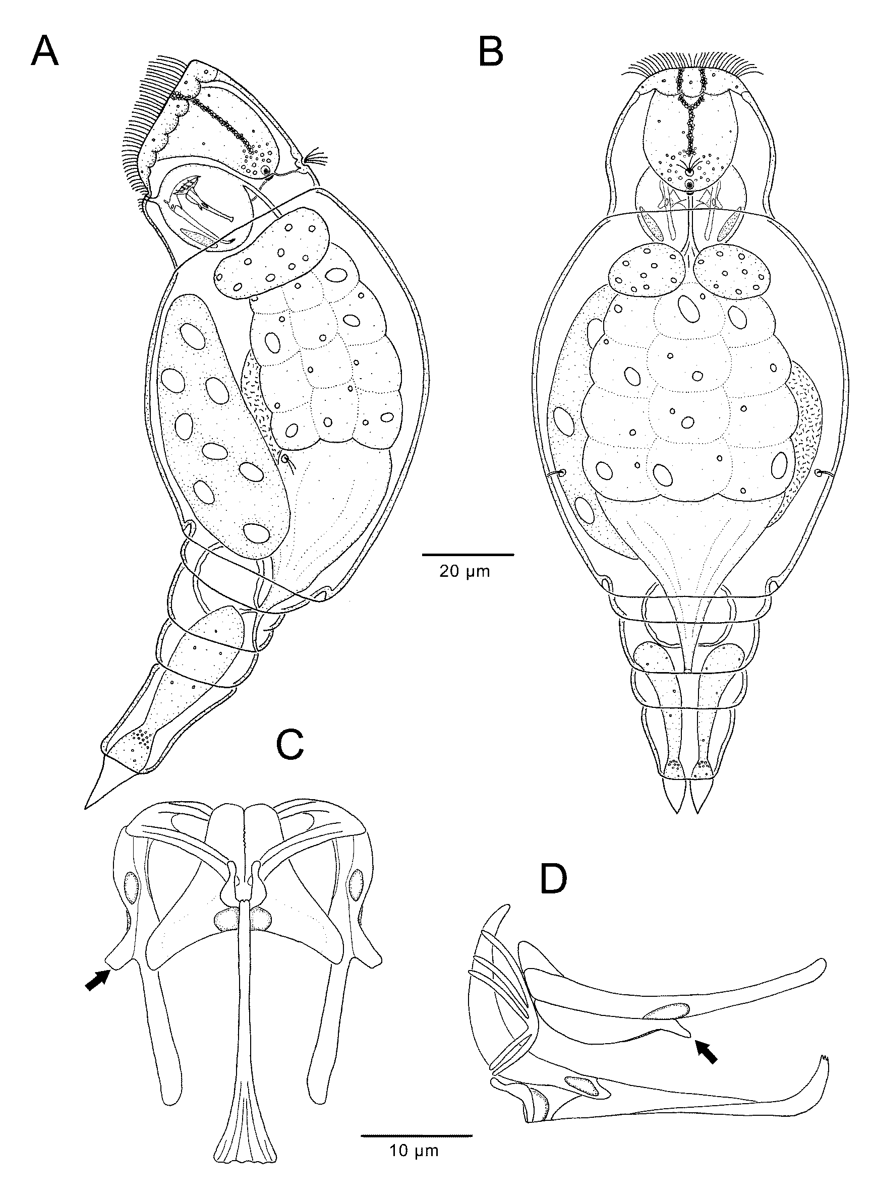
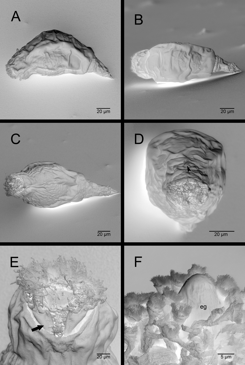
Habitus ( Figs 5 View FIGURE 5 A, B, 6A–C, 7A–E). Body stout, fusiform, hyaline, divided into 3 distinct regions: head, trunk and foot with toes. Epidermis hardly stiffened, with smooth surface. Head sloped, contractile and offset from trunk by weakly developed neck fold. Corona extending between apical and ventral head region, characterized by dense pseudotrochus of locomotory cilia. Pseudotrochus merges ventrally with cilia field running to mouth opening. This restricted posteriorly by paired lobe-like epidermal projection ( Fig. 7 View FIGURE 7 E). Locomotory cilia enclosing apical field on head; middle of apical field with 2 openings of retrocerebral organ. Trunk arched dorsally, much wider than head ( Figs 5 View FIGURE 5 B, 7B), and comprising 3 pseudosegments. First pseudosegment large, followed by narrower, shorter lumbar pseudosegment and preanal pseudosegment. Foot retractile, tapering conically, divided into 2 pseudosegments and 2 relatively short, pointed, conical toes ( Figs 5 View FIGURE 5 A, 7B), directed caudally during swimming process. Terminal foot pseudosegment twice length of first one.
Digestive system ( Figs 5 View FIGURE 5 A, B). Mouth opening in ventral part of corona leads to short, slender, ciliated buccal tube that discharges ventrally to spherical mastax. Oesophagus diverges from anterior third of mastax dorsally. Stomach multicellular, ciliated, with yellow-brownish, small and large egg-shaped droplets, occupying one-third of trunk ( Fig. 6 View FIGURE 6 A). Ciliated intestine offset from stomach. Anus discharges below preanal pseudosegment.
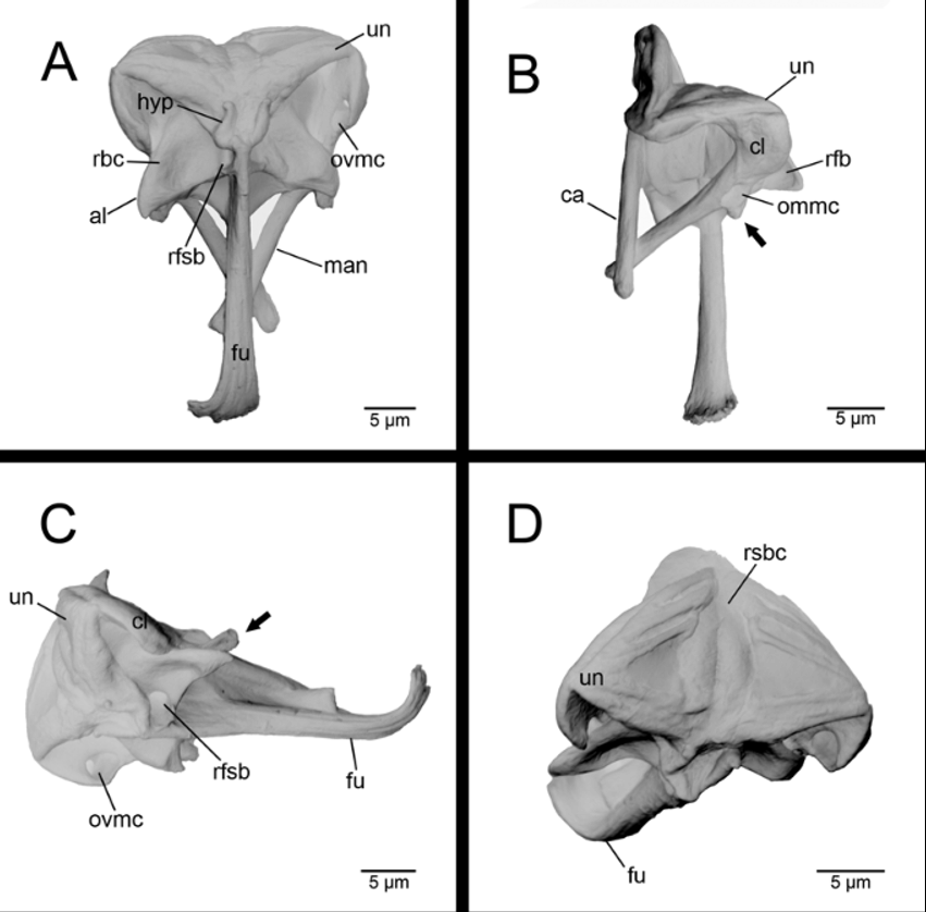
Trophi ( Figs 5 View FIGURE 5 C, D, 9A–D). Trophi virgate, bilaterally symmetrical with fulcrum residing almost in longitudinal axis of body.
Rami. Stout, in ventral view appearing triangular, arched upward anteriorly in sickle-shaped bend ( Fig. 10 View FIGURE 10 ), inner margins with fine denticles ( Fig. 5 View FIGURE 5 C). Ramus foramen subbasalis small, somewhat circular ( Figs 5 View FIGURE 5 C, 9C), directed ventrally. Ramus foramen basalis large, ovate, directed dorsally. Rami with broad, acuminate alulae distally ( Figs 5 View FIGURE 5 C, 9A).
Fulcrum. Long, rod-shaped, longitudinally striate; in ventral view slender, expanding distally; in lateral view flattened and curved upward distally.
Unci. Slightly curved plates, with 2 ventral and 3 more dorsal principal teeth; ventral and dorsal tooth group separated by triangular gap ( Fig. 5 View FIGURE 5 D, 9D).
Manubria. Slightly curved, tapering gradually from broad basal clava to distal end of long, rod-shaped cauda. Clava composed of 3 chambers that are not clearly distinguishable but show distinct openings. Ventral manubrial chamber with small, rounded, ventrally directed opening and features a small, knob-like protuberance ( Figs 5 View FIGURE 5 C, D, 9B) ventrolateral to median chamber of manubrium. Median manubrial chamber forms elongated cauda and shows small, oval, ventrolaterally directed opening ( Fig. 9 View FIGURE 9 B). Small dorsal manubrial chamber opens dorsolaterally, with ovate opening.
Hypopharynx. a fork-like structure at the proximal end of the fulcrum ( Fig. 9 View FIGURE 9 A).
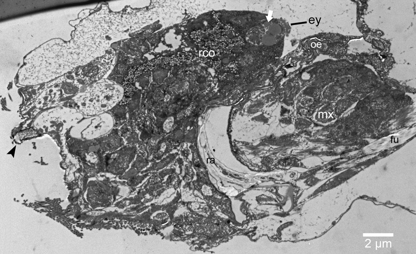
Nervous system and sensory organs. Cerebral ganglion rounded, extending to centre of head, followed by large, reasonably hyaline retrocerebral organ that cannot be discriminated from brain by light microscopy. A dark-red kidney-shaped eye present at posterior end of retrocerebral organ ( Fig. 10 View FIGURE 10 ). A fine connection inserts close to eye and courses down to mastax ( Figs 5 View FIGURE 5 A, 6A). Dorsal antenna consists of several cilia encircled by flat, rounded collar located in front of head fold ( Fig. 7 View FIGURE 7 D). Lateral antennae comprising a few cilia, with a flat, rounded collar, located just beyond midpoint of trunk.
Glandular system ( Figs 5 View FIGURE 5 A–B, 6B). Two lateral salivary glands visible, integrated into mastax complex. Both gastric glands large, ball-shaped, unstalked, with 9 nuclei each, diverging anteriorly from stomach. Retrocerebral organ lies behind cerebral ganglion and opens with 2 anterior ducts on apical field of corona ( Fig. 10 View FIGURE 10 ). In posteriormost region of retrocerebral organ, and immediately in front of the eye, is a vesicle-filled sphere. Pedal glands extend into preanal pseudosegment, with terminally constricted mucus reservoir above toes.
Protonephridial system ( Figs 5 View FIGURE 5 A, B). Protonephridia paired, with distinct terminal organs (exact number and position not determined). Collecting tubules open into contractile bladder that in turn empties into terminal part of intestine (cloaca).
Reproductive system ( Figs 5 View FIGURE 5 A, B, 6A, B). Germovitellarium unpaired, with 8 nuclei, located in the ventral region of trunk, often associated with a large, dark egg.
Measurements. Total length 150–255 µm, maximum dorsoventral extent 70 µm, maximum width 60–80 µm, foot length 28 µm, toe length 12–26 µm, trophi length 35 µm, ramus length 16 µm, manubrium length 21 µm, manubrium width 7 µm and fulcrum length 24 µm.
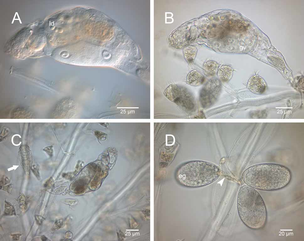
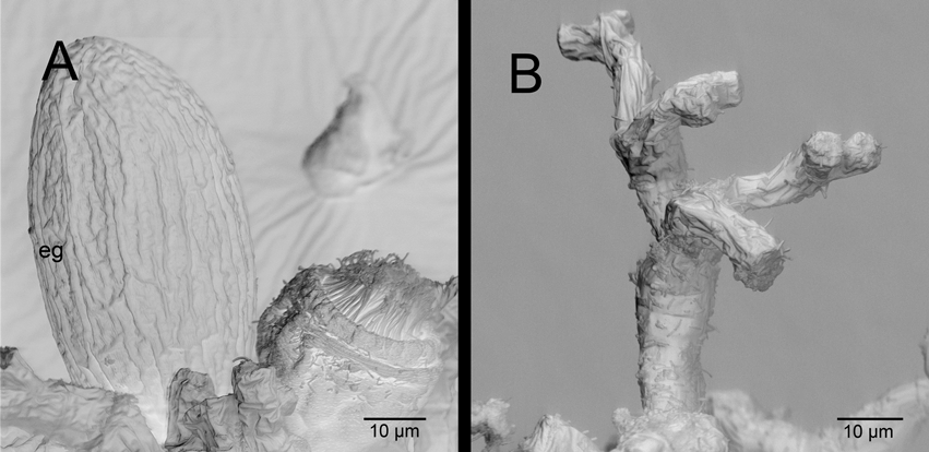
Ecology. The species is limnosaprobic and free-living, occurring in running, acidic, brackish water and is present in both colder and warmer seasons. Distribution is cosmopolitan ( Nogrady et al. 1995). The species is necrophagic and includes smaller sessile peritrich ciliates in its diet, especially Carchesium . Several generations, including juveniles and eggs, often live on a single ciliate colony ( Figs 6 View FIGURE 6 C, D). In one instance, six specimens were observed living on a single Carchesium colony. Specimens are well protected in the colony, which encloses the rotifers following disturbances. Feeding specimens attach with their pedal glands, stretch and approach the body of an individual ciliate. The ciliate is eaten completely, leaving only its stalk ( Figs 6 View FIGURE 6 A, 8B). Usually 2– 3 eggs are deposited and fixed to the substratum, with a stalk formed by an adhesive secretion from the pedal glands ( Fig. 6 View FIGURE 6 D). Smooth ( Fig. 7 View FIGURE 7 F) as well as sculptured eggs ( Fig. 8 View FIGURE 8 A) were found. Specimens were also found in the middle of stalked ciliates growing on Macrocyclops . Ehrenberg (1838) reported the species on Epistylis digitalis , Carchesium polipinum , Zoothamnium and piercing Volvox globator .
Budde (1925) also found specimens in Volvo x and in the shells of dying and dead crustaceans ( Daphnia , Chydorus, Entomostraca ), where they always feed towards the head, deposit 2– 3 eggs and then leave the individual. The species was also found on trichopteran eggs, on Hydra ( De Beauchamp 1905) and on snail eggs ( Wulfert 1935).
No known copyright restrictions apply. See Agosti, D., Egloff, W., 2009. Taxonomic information exchange and copyright: the Plazi approach. BMC Research Notes 2009, 2:53 for further explanation.
|
Kingdom |
|
|
Phylum |
|
|
Class |
|
|
Order |
|
|
Family |
|
|
Genus |
Pleurotrocha petromyzon ( Ehrenberg, 1830 ) (Notommatidae)
| Wilts, Eike F., Bininda-Emonds, Olaf R. P. & Ahlrichs, Wilko H. 2009 |
Pleurotrocha laurentina:
| Harring 1913 |
Proales laurentius
| Jennings 1896 |
Notops laurentinus
| Jennings 1894 |
Proales petromyzon:
| Hudson & Gosse 1886 |
Notommata petromyzon:
| Ehrenberg 1838 |
Notommata gibba
| Ehrenberg 1832 |
Notommata petromyzon
| Ehrenberg 1830 |