
ICHTHYOPHIIDAE, Taylor, 1968
|
publication ID |
https://doi.org/10.1111/j.1096-3642.2012.00838.x |
|
persistent identifier |
https://treatment.plazi.org/id/03DB87B7-FFFE-FFB2-FF00-910FFD896759 |
|
treatment provided by |
Marcus |
|
scientific name |
ICHTHYOPHIIDAE |
| status |
|
ICHTHYOPHIIDAE View in CoL ( FIGS 5 View Figure 5 , S 9–S View Figure 9 11 View Figure 11 )
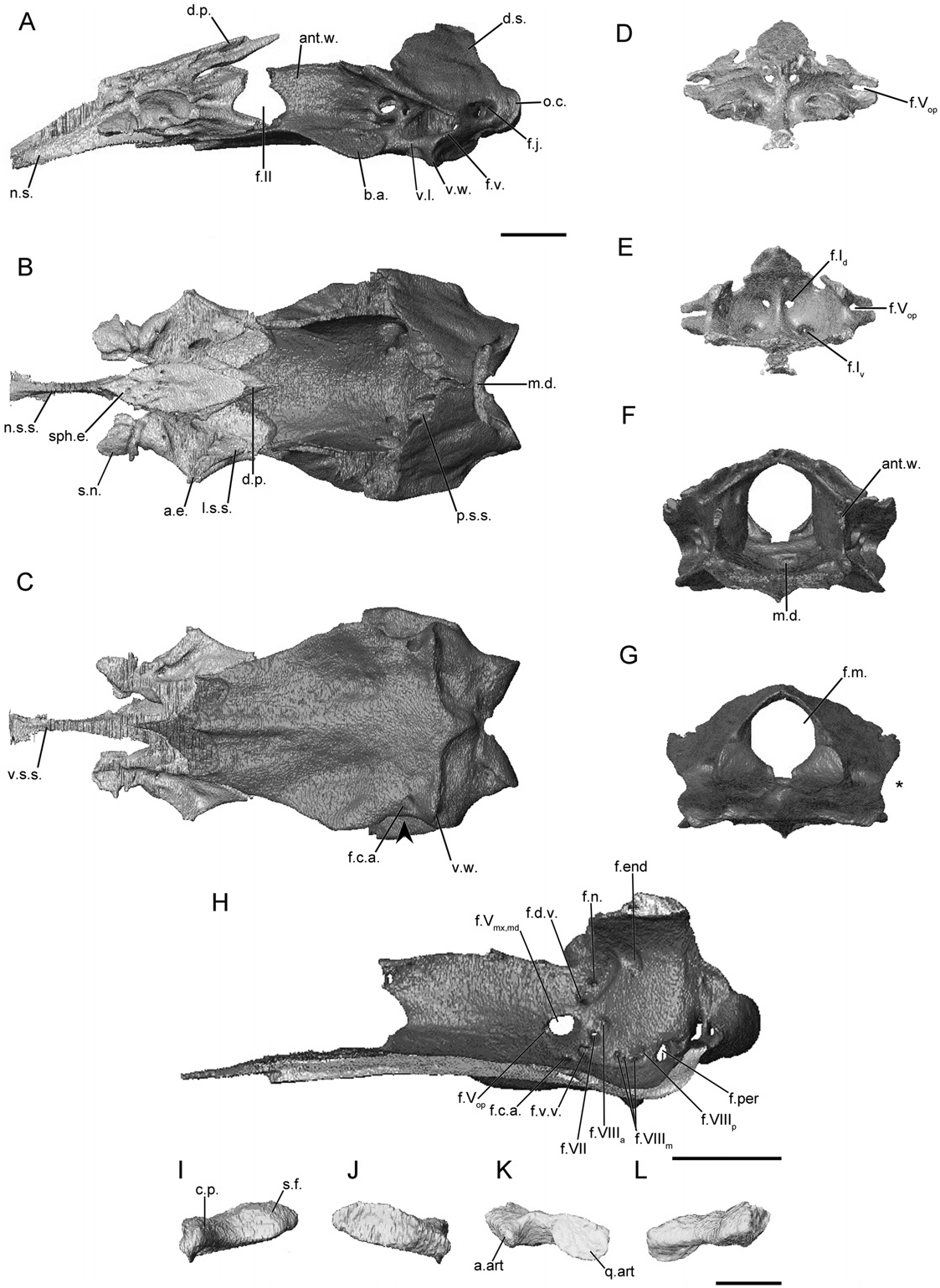
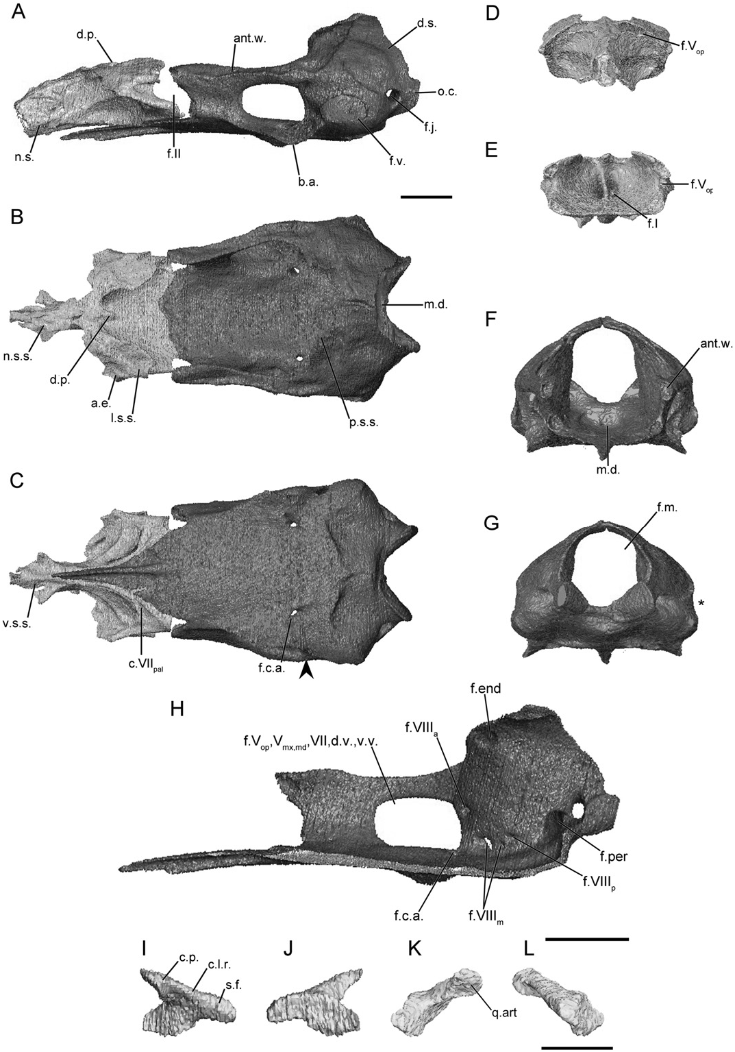
The main body of the sphenethmoid is short in ichthyophiids, accounting for less than half to roughly 30% of the total sphenethmoid length ( Fig. 5A View Figure 5 ). The lateral wall of the main body is capped by a variably broad sutural surface that receives the frontal only in Cau. asplenia , or both the frontal and parietal in the other species. The anterolateral corner bears a very broad ossified process in all ichthyophiids ( Fig. 5B View Figure 5 ) except for Cau. asplenia in which there is only a slight expansion ( Fig. S9B View Figure 9 ). A thin dorsomedial process is present that is not exposed dorsally. The process does not extend beyond the level of the lateral wall of the sphenethmoid ( Fig. 5B View Figure 5 ). The posterior margin of the lateral wall is slightly incised by the optic foramen in all species ( Fig. 5A View Figure 5 ), except for Uraeotyphlus narayani in which it is more deeply so ( Fig. S11A View Figure 11 ). The posterior margin of the floor of the sphenethmoid is deeply incised, exposing a great area of the os basale ventral to it ( Fig. 5B View Figure 5 ). In Cau. asplenia and the species of Ichthyophis there is a median extension of the margin of the floor (not visible here).
The nasal septum is tall and blade-like ( Fig. 5A View Figure 5 ). The dorsal sutural surface is uniformly narrow, except for U. narayani in which it is slightly broader at the base ( Fig. S11B View Figure 11 ). The nasal septum tapers in height distally, and terminates posterior to the external naris. Sola nasi are present, except for U. narayani . Additionally in U. narayani a pair of anterolaterally projecting flanges extends from the ventral surface of the nasal septum ( Fig. S11C View Figure 11 ).
The dorsal pair of anterior foramina is not confined within the wall of the sphenethmoid in Cau. asplenia and the species of Ichthyophis ( Fig. 5E View Figure 5 ) and form troughs instead. The ventral foramina are located somewhat laterally from the midline ( Fig. 5E View Figure 5 ), except for U. narayani in which they are much closer to the midline ( Fig. S11E View Figure 11 ). A single anterolateral foramen is present in the anterolateral corner of the main body ( Fig. 5E View Figure 5 ).
In dorsal view the antotic wall is angled towards the midline anteriorly and is vertical in anterior view. The dorsal surface is capped by a narrow sutural surface. In the species of Ichthyophis and U. narayani the surface expands slightly at the level of the antotic foramina before becoming continuous with the surface along the anterior margin of the otic-occipital ( Fig. 5B View Figure 5 ). The anterior margin of the antotic wall is weakly to deeply incised by the posterior margin of the optic foramen ( Fig. 5A View Figure 5 ). The antotic region is perforated by three foramina, constituting Pattern 4. Both trunks of the trigeminal nerve plus the abducens nerve exit through a large subcircular foramen ( Maddin, 2011). A dorsal vein exits through a foramen posterior and dorsal to the large foramen (except for U. narayani ), and the facial nerve and a ventral vein exit through a common foramen just posterior and slightly ventral to the large foramen ( Fig. 5H View Figure 5 ).
The dorsal surface of the otic-occipital complex of the os basale is tilted slightly posteroventrally. It tapers towards the midline and bears an anterior sutural surface for the parietal (except for Cau. asplenia ). A laterally extending shelf, located dorsal to the fenestra vestibuli, is present ( Fig. 5A View Figure 5 ). The fenestra vestibuli is elongate anteroposteriorly, and in posterior view it incises the lateral margin of the otic-occipital complex to varying degrees amongst ichthyophiid species ( Fig. 5G View Figure 5 ). A large articular facet receives the footplate of the stapes. The occipital condyle projects only slightly posterior to the otic capsule, and its margin is continuous with that of the dorsal surface of the complex when viewed laterally. The jugular foramen is present in the base of the condyle, but it is only partially visible in lateral view ( Fig. 5A View Figure 5 ). The foramen magnum is circular in outline ( Fig. 5G View Figure 5 ).
The medial wall of the otic capsule is perforated by seven or eight foramina ( Fig. 5H View Figure 5 ). Four foramina common to all species examined here (endo- and perilymphatic foramina, and anterior and posterior vestibulocochlear nerve foramina) are present in similar locations. Variably within the family three or four small foramina transmit the medial branch of the vestibulocochlear nerve ( Maddin, 2011).
The anterior margin of the floor of the os basale comes to a rounded point in Cau. asplenia and the species of Ichthyopis ( Fig. 5C View Figure 5 ), and is pointed in U. narayani ( Fig. S11C View Figure 11 ). Anteriorly it extends only to the level of the main body of the sphenethmoid and does not pass beneath the nasal septum in Cau. asplenia and the species of Ichthyophis ; however, it reaches to within the anterior half of the nasal septum in U. narayani . The posterior median depression is weakly developed except for U. narayani in which it is well defined. An expansion of the floor of the os basale corresponds to the position of the basicranial articulation ( Fig. 5A, B View Figure 5 ), but the latter are not well developed. There is little to no constriction of the lateral margin of the floor when viewed ventrally, and there is no wing-like projection ventral to the otic capsules ( Fig. 5A, C View Figure 5 ), except for U. narayani ( Fig. S11A,C View Figure 11 ). The posterior margin of the floor terminates in a rounded point ( Cau. asplenia and U. narayani ) or arched ridge ( Ichthyophis ) that extends almost to the ventral margin of the foramen magnum.
The carotid artery enters the floor through a foramen just anterior to the otic capsule ( Fig. 5C View Figure 5 ). It passes only a short distance anteriorly before dividing into lateral and medial canals, which terminate at foramina just anterior to the otic capsule on either side of the antotic walls.
The footplate of the stapes is elongate anteroposteriorly and its long axis is orientated roughly horizontally ( Fig. 5I, J View Figure 5 ). Its anterior margin is thickened and closely applied to the anterior margin of the fenestra vestibuli. The remaining margins of the footplate closely approach the fenestra margins, except in U. narayani in which the stapes is extensively inset. In Ic. beddomei there is a dorsal process on the footplate not seen in other species ( Fig. S10I View Figure 10 ). The columellar process is robust and long in ichthyophiids. It is pierced by a foramen, except for U. narayani in which the stapes is imperforate.

No known copyright restrictions apply. See Agosti, D., Egloff, W., 2009. Taxonomic information exchange and copyright: the Plazi approach. BMC Research Notes 2009, 2:53 for further explanation.