
Myronides tomohonense, Hennemann, 2021
|
publication ID |
https://doi.org/ 10.11646/zootaxa.5073.1.1 |
|
publication LSID |
lsid:zoobank.org:pub:AA3269D1-CA2F-4528-BC9D-3A4C75D05BD9 |
|
persistent identifier |
https://treatment.plazi.org/id/03DB87EE-FFC6-9D16-FF40-5EFDFB0CF7D9 |
|
treatment provided by |
Plazi |
|
scientific name |
Myronides tomohonense |
| status |
sp. nov. |
Myronides tomohonense n. sp.
( Fig. 22 View FIGURE 22 )
Myronides pfeifferae, Günther, 1938: 74 .
Hennemann, 1998: 119.
HT, ♀: Tomohon , Celebes, X.94, Sar.; Myronides pfeifferae Westw., K. Günther det. [ NHMB] .
PT, ♀: Tomohon , Celebes, VI .94, Sar.; Myronides pfeifferae Westw., K. Günther det. [NHMB].
PT, ♀: Lokon, Mittelregion , Celebes, V.94, Sar.; Myronides pfeifferae Westw., K. Günther det. [ NHMB] .
PT, ♂: Tomohon , Celebes, V.94, Sar.; Myronides pfeifferae Westw., K. Günther det. [ NHMB] .
PT, ♂: Tomohon , Celebes, 2.V.94, Sar.; Myronides pfeifferae Westw., K. Günther det. [ NHMB] .
PT, ♂: Sarasin, IX.1894, Masarang-Kette, Nord-Celebes; Myronides pfeifferae Westw., K. Günther det. [NHMB].
Etymology: Named after the type-locality, Tomohon in the Minahasa Regency of the Province Sulawesi Utara in northeastern Sulawesi.
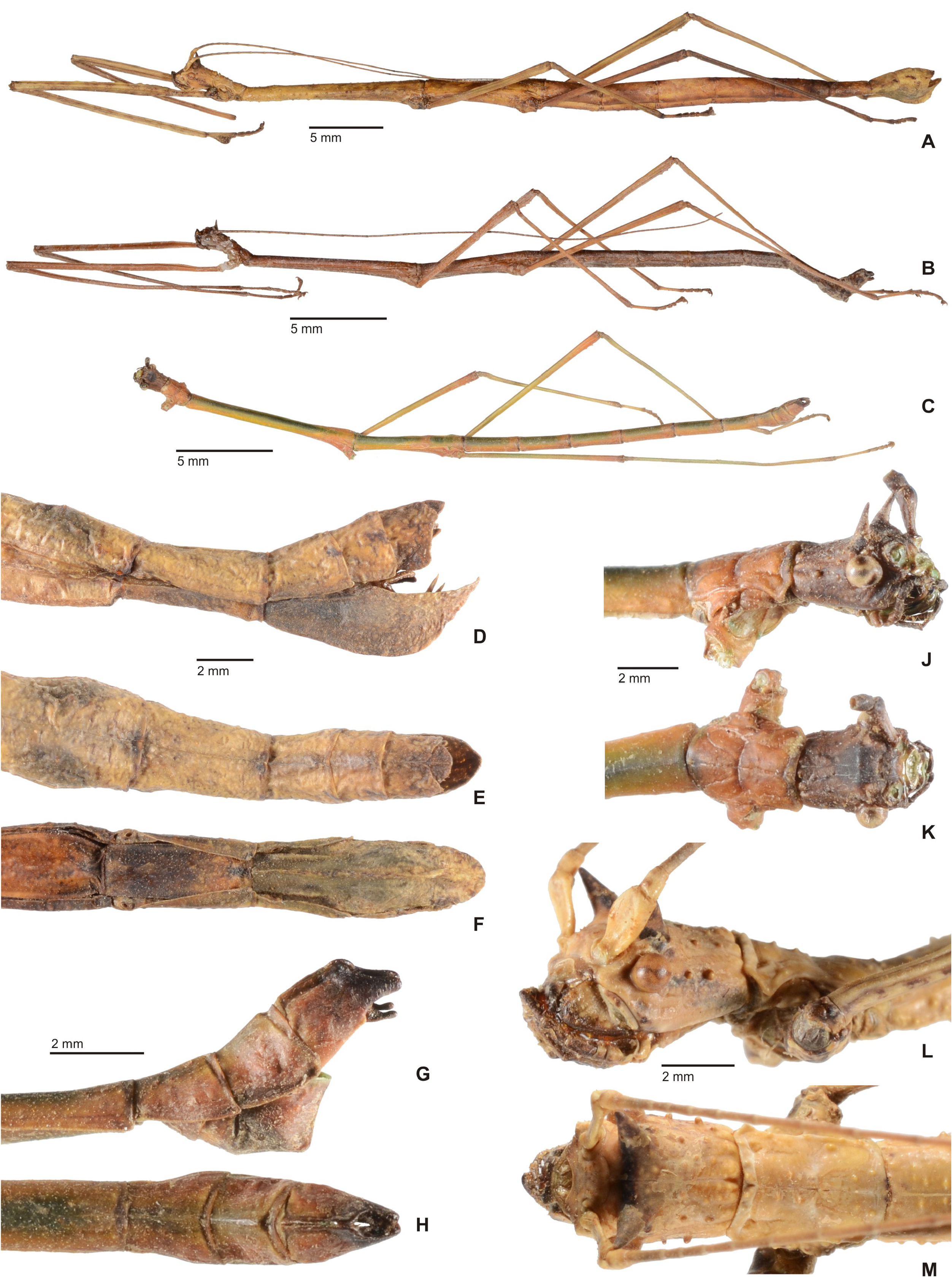
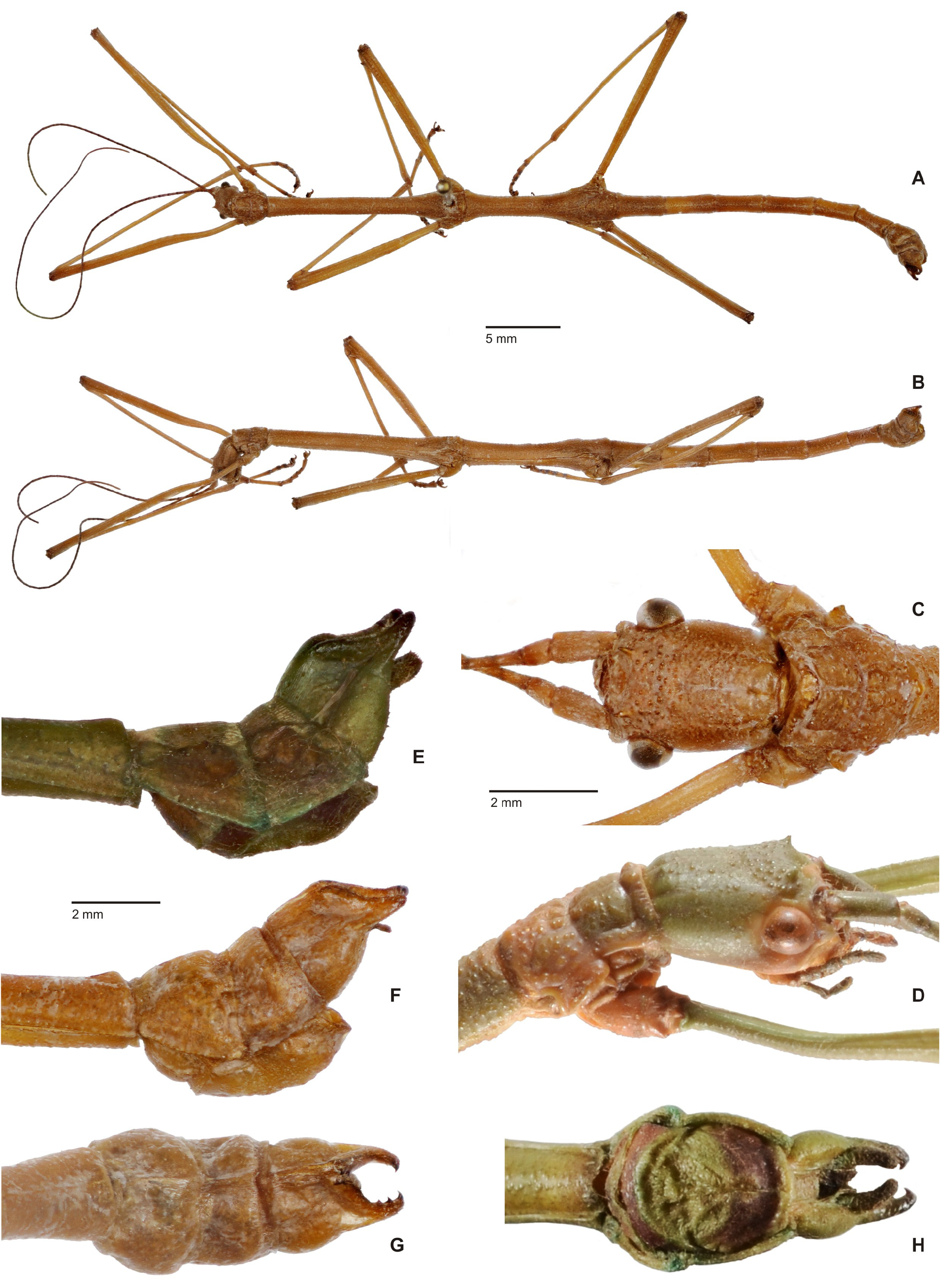
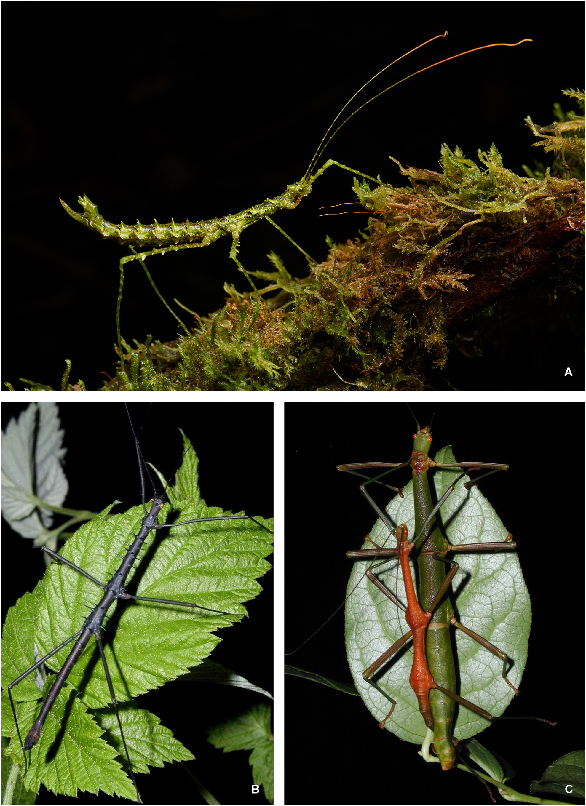
Differential diagnosis: This new species is the only known representative of Myronides Stål, 1875 on the island of Sulawesi and the so far only known species in the genus in which ♀♀ have a prominent dorsal lobe on the probasitarsus ( Fig. 22A View FIGURE 22 ) and in which ♂♂ have a complex colouration with tones of brown, orange, green and yellow ( Fig. 22C View FIGURE 22 ). Females are similar to those of the type-species M. pfeifferae ( Westwood, 1859) but may be separated by the much more prominent, longer and acutely pointed pair of cephalic horns, which extend by more than half the height of head capsule ( Fig. 22L View FIGURE 22 ), notably smaller sub-apical teeth on the two outer ventral carinae of the femora, larger subgenital plate, which extends considerably beyond the apex of the abdomen ( Fig. 22D View FIGURE 22 ) and large dorsal lobe of the probasitarsi ( Fig. 22A View FIGURE 22 ). Males are well characterized within the genus and readily distinguished from those of M. pfeifferae by their complex colouration, having a bold dark green longitudinal streak along the dorsal body surface that is interrupted at the borders of each body segment (♂♂ of pfeifferae are uniformly buff to brown). Moreover, the pair of cephalic horns are much larger in this species ( Fig. 22J View FIGURE 22 ) and the sub-apical ventral teeth of the femora are less pronounced.
Description: All type specimens have provisionally been conserved in spirits, so the colouration described may not be true for the live insects. One of the ♀ paratypes is very pale straw to yellow and here omitted from the colour descriptions, because this colouration is obviously artificial. The colouration of the ♂♂ is described based on only one specimen that appears to exhibit close to the natural colour patterns when alive.
♀ ( Fig. 22A View FIGURE 22 ). Medium-sized for the genus (body length 102.0–111.0 mm), form fairly stocky with a short median segment and a prominent, acutely pointed pair of cephalic spines. Body surface sparsely tuberculose and granulose. Colour ochraceous with irregular brown markings and sometimes with a slight greenish wash, the head darker brown in one of the paratypes. Cephalic spines and the intervening dark brown, the genae with a weakly defined longitudinal postocular streak. Larger tubercles of the head and thorax dull yellow. Apex of basitarsi and all following tarsomeres dark brown. Antennae with a slightly dull orange hue, the two basal segments coloured like body, III – VI and the 15 or so apical antennomeres with a greyish brown wash. Eyes dull ochre with a faint longitudinal dark ocular stripe .
Head: Longer than wide, sub-cylindrical, broadest at the eyes and gently narrowing toward the posterior ( Fig. 22M View FIGURE 22 ). Frons with two small C-shaped impressions between bases of antennae. Between the eyes with a very prominent pair of acutely pointed and strong, slightly anteriad directed spines, which extend by more than half the height of head capsule ( Fig. 22L View FIGURE 22 ). Vertex flat with a fine, impressed coronal line and with a few scattered nodes, genae with two obtuse nodes. Eyes circular in outline, strongly projecting and their diametre contained about 2.2 in length of genae. Antennae reaching to anterior of abdominal segment II. Scapus compressed dorsoventrally, somewhat deflexed laterally with both lateral margins weakly rounded; almost 2x longer than wide. Pedicellus oval in crosssection and about 1/3 the length of scapus.
Thorax: Pronotum slightly shorter but about equal in length to head, rectangular in outline and 1.5x longer than wide. Transverse median sulcus shallow, gently curved and not reaching lateral margins of segment; anterior and lateral margins with a few low tubercles and some paired tubercles on dorsal surface ( Fig. 22M View FIGURE 22 ). Mesothorax elongate, slender and about 5.5x longer than prothorax. Mesonotum with a fine longitudinal median carina and unevenly set with tubercles and nodes of variable sizes. Mesopleurae sparsely nodose. Mesosternum obtusely keeled longitudinally with with some small, scattered nodules along the medio-longitudinal keel. Metanotum a little less than half the length of mesonotum, about 3.2x longer than wide; sculpturing like mesonotum. Metapleurae and sternum sparsely nodulose.
Abdomen: Median segment 1.7x longer than wide and a little less than half the length of metanotum; sculpturing alike. Abdomen excluding median segment equal in length to complete thorax; entire dorsal surface with a fine longitudinal median carina and irregularly tuberculose and nodulose. Segment II almost 1.8x longer than median segment, II–V roughly equal in length, VI and VII slightly shortening; All roughly uniform in width, rectangular and on average 2x longer than wide. Tergum VI occasionally somewhat widened medially and with a pair of obtuse, dorso-lateral swellings (e.g. in the HT). VII shorter and narrower than all preceding segments. Sterna sparsely granulose, Preopercular organ on VII formed by a pair of low, wart-like swellings near posterior margin ( Fig. 22F View FIGURE 22 ). Tergum VIII somewhat less than half the length of VII, IX shorter and slightly wider than long. Anal segment longer than IX, strongly convex longitudinally and with a very large, triangular posteromedian excavation ( Fig. 22E View FIGURE 22 ); the lateral margins roundly deflexed in the basal portion ( Fig. 22D View FIGURE 22 ). Epiproct distinct, scale-shaped and slightly projecting over posterolateral angles of anal segment ( Fig. 22E View FIGURE 22 ). Cerci very small, conical and somewhat compressed laterally. Gonapophysis VIII strongly up-curving and almost reaching apex of anal segment, gonoplacs distinct and digitiform ( Fig. 22D View FIGURE 22 ). Subgenital plate strongly keeled medio-longitudinally, strongly convex and bulgy in the median portion ( Fig. 22D View FIGURE 22 ), the posterior margin obtusely rounded and projecting slightly beyond apex of abdomen ( Figs. 22E–F View FIGURE 22 ).
Legs: All moderately long and slender, the profemora about as long as mesothorax, metafemora almost reaching to posterior margin of abdominal segment IV and metatibiae just not reaching apex of abdomen. Anterodorsal carina of profemora moderately deflexed and weakly undulate and wavy in the basal two thirds. All femora unarmed except for three small sub-apical teeth which decrease in size towards the apex of femur. Medioventral carina moderate. Probasitarsus almost as long as remaining tarsomeres combined and with a prominent, rounded dorsal lobe in median portion. Meso- and metabasitarsus slender and a little longer than following three tarsomeres combined.
♂ ( Figs. 22B–C View FIGURE 22 ). Fairly small for the genus (body length 73.0– 74.2 mm), apterous, shape slender with a prominent pair of acutely pointed cephalic spines. Body surface very minutely and sparsely granulose. Colourful ( Fig. 22C View FIGURE 22 ); head dark brown, pronotum, posterior portions of all body segments, the lateral surfaces of the abdomen and all coxae reddish mid brown, the dorsal surface of the body dull green. Lateral portions of meso- and metanotum yellowish green. Femora yellowish green with apical quarter red, the tibiae greyish green and somewhat darker at the apex. Anterior margin of anal segment blackish. Antennae dull reddish brown. Eyes ochre with a dark longitudinal ocular stripe.
Head: Generally as in ♀♀ but posterior portion more strongly narrowed and the nodes less numerous ( Fig. 22K View FIGURE 22 ); portion between the eyes mor strongly swollen and rounded and the cephalic pair of horns more acutely pointed ( Fig. 22J View FIGURE 22 ). Eyes large, very prominently projecting and their diametre contained about 1.6x in length of genae. Antennae reaching to posterior margin of abdominal segment V. Scapus less broadened than in ♀♀.
Thorax: Pronotum roughly of same dimensions as head, the transverse median sulcus very prominent, gently curved and almost expanding over entire width of segment, a distinct impressed longitudinal median line present; tubercles just very weakly defined to obsolete ( Fig. 22K View FIGURE 22 ). Mesothorax very elongate and slender and just slightly widened and swollen posteriorly; about 5.6x longer than prothorax. Mesonotum with a very weak and fine longitudinal median carina and mesosternum very weakly tectinate longitudinally. Metanotum a little more than 1/3 the length of metanotum, surface alike.
Abdomen: Median segment about half the length of metanotum, roughly 3x longer than wide and gently narrowed medially. Segment II 1.3x longer than median segment and very slightly longer than III–V, VI–VII decreasing in length with VII only 3/5 the length of II. II–V on average about 3.5x longer than wide, VII only about 2.5x longer than wide; II–VII uniform in width and all terga with a very fine and indistinct longitudinal median line. Tergum VIII roughly half the length of VII and widened in the posterior portion, IX notably shorter with lateral margins roundly deflexed; medio-longitudinal carina more distinct than on preceding terga. Anal segment strongly tectiform and split to form two movable hemi-terga ( Fig. 22H View FIGURE 22 ); the apical portion strongly narrowed in lateral aspect to form an obtuse, gently in-curving digitiform process ( Fig. 22G View FIGURE 22 ). The interior surface of these two processes set with several minute black denticles ( Fig. 22H View FIGURE 22 ). Cerci small, strongly constricted medially with the apex slightly club-shaped. Poculum strongly convex, cup-shaped, obtusely rectangular in lateral aspect with the posterior half carinate medio-longitudinally and the posterior margin rounded and slightly projecting over posterior margin of tergum IX ( Fig. 22G View FIGURE 22 ).
Legs: All long, slender and unarmed except for 2–3 small sub-apical denticles on the two outer ventral carinae of the femora. Profemora about equal to combined length of pro- and mesothorax, metafemora reaching about half way along abdominal segment V and metatibiae projecting considerably beyond apex of abdomen. Basitarsi slender, very elongate and at least equal in length to remaining tarsomeres combined.
Comments: Eggs unknown.
Distribution: So far only known from the very northeastern portion of Sulawesi, the Minahasa Regency.
Genus Neomyronides n. gen.
( Figs. 23–24 View FIGURE 23 View FIGURE 24 )
Type-species: Pseudostheneboea aberrans emiri Hennemann, 1998: 104 , by present designation.
Pseudostheneboea, Hennemann, 1998: 102 , figs. 7–10, pl. 1: 7 –8, pl. 2: 5,6 & 8 (in part).
Zompro, 2005: 260.
Otte & Brock, 2005: 293 (in part).
Staelonchodes, Günther, 1938: 77 , fig. 15.
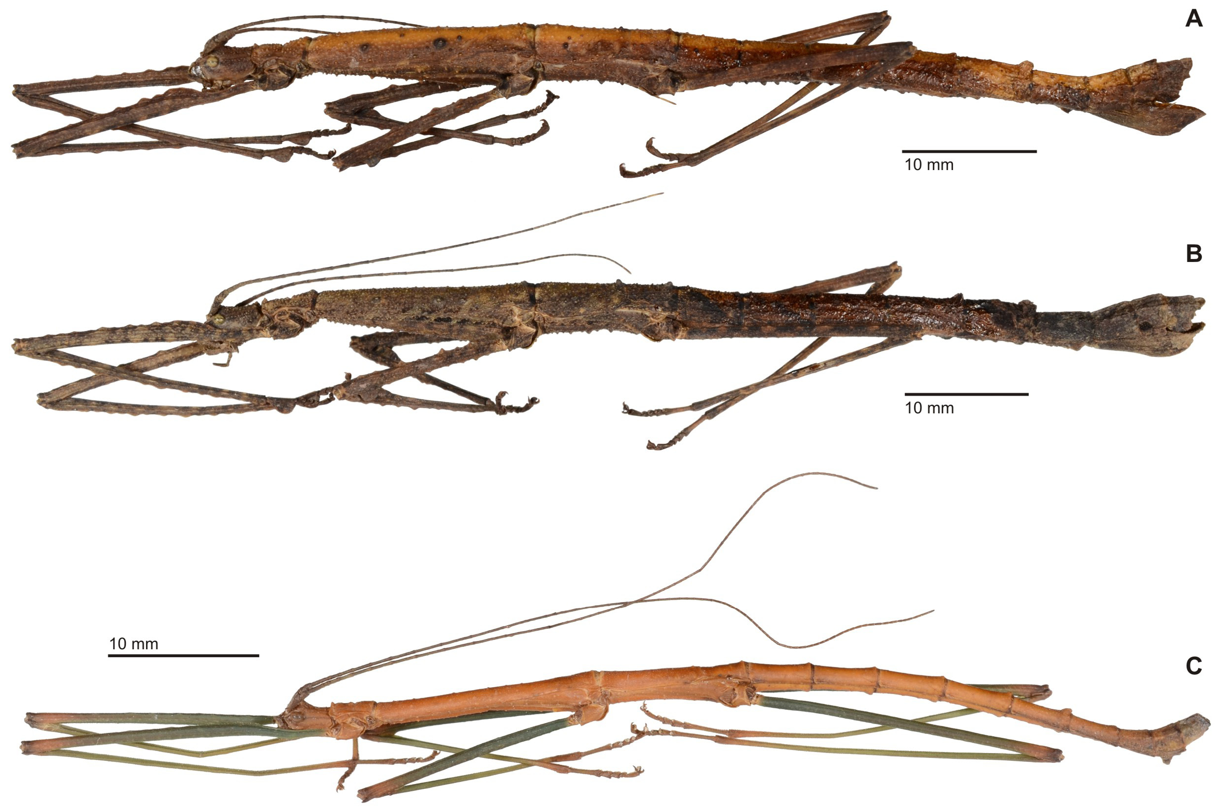
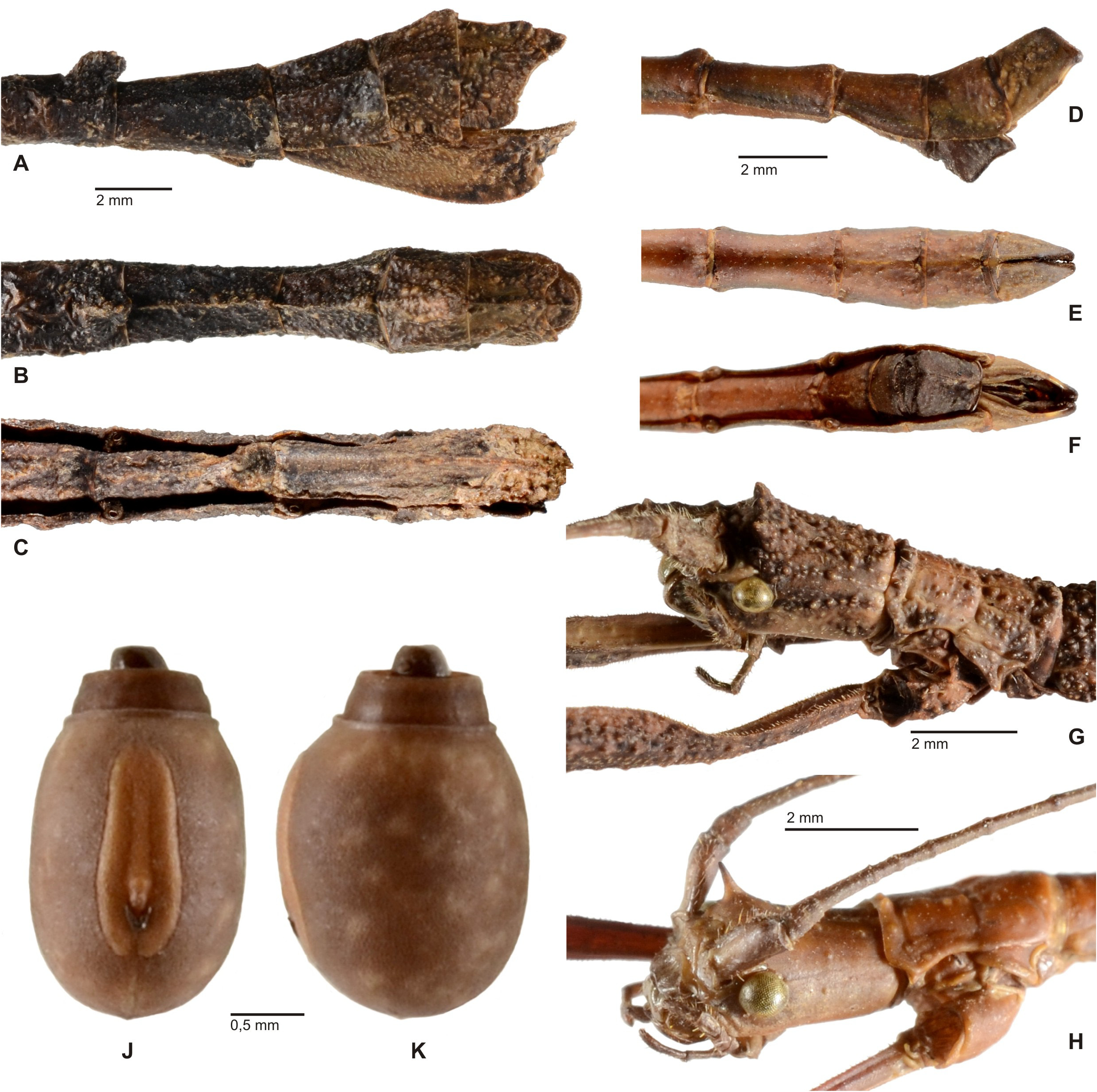
Diagnosis: ♀, ♂. Fairly small (body length ♂♂ 51.0– 62.5 mm, ♀♀ 73.5–88.0 mm) and stocky Lonchodinae ; very similar in appearance to Myronides Stål, 1875 and Mithrenes Stål, 1877 ( Fig. 23 View FIGURE 23 ). Distinct sexual dimorphism with ♂♂ much smaller, more slender and less sculptured than ♀♀. Colouration various shades of brown and ochre; intra-specifically variable in ♀♀ and ♂♂ might have green legs. Body surface of ♀♀ densely granulose and tuberculose, in ♂♂ smooth with only a few scattered granules on thorax. Head considerably longer than wide and narrowed towards the posterior; vertex flattened and between eyes with a pair of ± distinct spines ( Figs. 24G–H View FIGURE 24 ). Antennae elongate, consisting of about 60 antennomeres and reaching to median segment (♀♀) or abdominal segment IV–V (♂♂); IV–XV gradually increasing in length the remaining much shortened and XIV with a small, knob-like subbasal dorsal swelling. Prothorax about equal in length to head, rectangular and with a moderate median sulcus. Mesothorax 4.5–5.0x longer than prothorax; very slender in ♂♂ and very gently widening towards the posterior in ♀♀. Mesonotum of ♀♀ usually with a variable number of enlarged, blunt tubercles. Metanotum a little more than 1/3 the length of mesonotum and about 1.8x (♀♀) to 2.2x (♂♂) longer than median segment; with a distinct tubercle posteromedially. Meso- and metasternum in ♀♀ granulose and irregularly set with conical tubercles of variable sizes. Abdomen slightly longer than head and thorax combined. Median segment slightly longer than wide and with a somewhat enlarged tubercle posteromedially. Segments II–VI in ♀♀ very indistinctly increasing in length, all longer than wide and ± parallel-sided, VI about 1.5x longer than wide. VII gently narrowed towards the posterior. Terga III–VI with a posteromedian tubercle, which increases in size towards VI where it forms a laterally compressed, rounded lobe of variable size ( Fig. 24A View FIGURE 24 ). Sternum VII with a distinct Preopercular organ formed by a laterally compressed, wart-like swelling some distance before posterior margin ( Fig. 24C View FIGURE 24 ). Terminalia of ♀♀: Terga VIII–X much shorter than previous and decreasing in length, IX ± swollen and raised posteromedially. Anal segment tectinate with anterolateral angles rounded and ± deflexed; posterior margin roundly excavated. Epiproct rounded, scale-shaped and slightly projecting over apex of anal segment ( Fig. 24B View FIGURE 24 ). Cerci very small and hidden under anal segment. Subgenital plate strongly convex, bulgy and with a very prominent ledge-like longitudinal keel in median portion; the posterior portion irregularly tuberculose and the posterior margin slightly projecting beyond anal segment ( Fig. 24A View FIGURE 24 ). Terminalia of ♂♂: Segments II–VI considerably longer than VII and roughly uniform in length; terga V and VI with a rounded posteromedian swelling. VII–X much shorter than previous. Anal segment tectiform and split over entire length to form two hemi-terga ( Fig. 24E View FIGURE 24 ) that are broad and angular in lateral aspect ( Fig. 24D View FIGURE 24 ); the lower apical portion minutely dentate interiorly. Cerci small, cylindrical and concealed under anal segment. Poculum of moderate size, convex, cup-shaped with posterior margin rounded and ± reaching to posterior of tergum IX. Legs of moderate length, profemora longer than mesothorax, mesofemora shorter (♀♀) or longer (♂♂) than mesothorax and hind legs slightly (♀♀) to distinctly (♂♂) projecting beyond apex of abdomen. In ♂♂ unarmed, except for minute sub-apical spines on two outer ventral carinae of femora and occasionally with a single enlarged lobes on posterodorsal and posteroventral carina of mesofemora. In ♀♀ all carinae except ventral carinae of tibiae multi-lobate (indistinct in metafemora although). Mesofemora in particular with an enlarged sub-apical lobe on posteroventral carina. Tarsi slender in ♂♂, probasitarsus with a prominently raised and rounded dorsal carina in ♀♀.
Egg ( Figs. 24J–K View FIGURE 24 ): Small (overall length 2.6 mm), ovoid with dorsal surface of capsule much more convex than ventral surface and almost round in cross-section; about 1.4x longer than wide. Anterior portion somewhat narrowed. Capsule surface very minutely and evenly granulose. Micropylar plate elongate, about ¾ the length of capsule with posterior half slightly widened and posterior end indented; open interiorly. Micropylar cup distinct. Operculum almost circular and strongly raised and with a small, blackish, knob-like capitulum in centre.
Differential diagnosis: Very close to the Wallacean Myronides Stål, 1875 and Sulawesian endemic Paramanduria n. gen. and morphologically similar to the Philippine Mithrenes Stål, 1877 . From Myronides it differs by the averaging smaller size and more stocky shape with relatively stouter legs and shorter tarsi as well as the lack of medioventral keel on the mesosternum. Females are readily distinguished by the lobate front and mid legs, distinct rounded dorsal lobe of the probasitarsus and tuberculate posterior portion of the subgenital plate. Also, ♀♀ oc-casionally bear a struma or lobe-like posteromedian swelling on abdominal tergum VI, which is never present in Myronides . Males differ by the broader hemi-terga of the anal segment, which lack the finger-like postero-apical protrusion typically seen in Myronides , as well as the much smaller cerci. Eggs at once differ from those of Myronides by the strongly raised operculum ( Figs. 24J–K View FIGURE 24 ). From Paramanduria n. gen. ♀♀ readily differ by the lack of a beak-like ovipositor and the dorsally lobed probasitarsus. The Philippine endemic Mithrenes was revised by Hennemann & Conle (2007), who provided a new generic description. From this genus both sexes differ by the lack of a median keel on the mesosternum and averaging more stocky shape. ♀♀ can furthermore be distinguished by the much more prominent Preopercular organ on abdominal sternum VII as well as the dorsally lobed probasitarsus and ♂♂ differ by the lack of a downward-angled finger-like posteroventral appendage of the hemi-terga of the anal segment. The eggs significantly differ from those of Mithrenes by the simple ovoid shape of the capsule ( Figs. 24 View FIGURE 24 J-K).
Comments: The three Sulawesian species here comprised in this new genus have previously been attributed to not particularly closely related genera of the subfamily Lonchodinae , that either are endemic in Sri Lanka ( Prisomera Gray, 1835 ; see Hennemann, 2002: 39), Assam in north-east India ( Pseudostheneboea Carl, 1913 ) or distributed throughout Sundaland ( Staelonchodes Kirby, 1904 ).
Etymology: The prefix “ Neo -” means “New Myronides ” to indicate close relation to that genus. Neuter.
Distribution: Sulawesi (endemic).
Species included:
1. Neomyronides aberrans ( Günther, 1938: 77, fig. 15 (♂) [ Staelonchodes ]. HT, ♂: Assumpatu-Thal, Patuka, P. F. Sarasin 17.Sept.02; Typus; Staelonchodes aberrans n. sp. K. Günther det.[NHMB, VI.D.134]. n. comb.
Distribution: Central Sulawesi, Prov. Sulawesi Tengah, Valley of Asumbatu, Patuka [NHMB].
2. Neomyronides emiri ( Hennemann, 1998: 104, figs. 7–10, pl. 1: 7 –8, pl. 2: 5, 6 & 8) [ Pseudostheneboea ? aberrans emiri ]. HT, ♂: S-Sulawesi, Tana Toraja, Strasse von Rantepao nach Palopo 13km, ca. 1100 m, leg. F. Hennemann 13.–17.VIII.1995 [ MNHU, No. 7911]; AT, ♀: S-Sulawesi, Tana Toraja, Strasse von Rantepao nach Palopo 13km, ca. 1100 m, leg. F. Hennemann 13.–17.VIII.1995 [ MNHU, No. 7912]; PT, 3 ♂♂: S-Su-lawesi, Tana Toraja, Strasse von Rantepao nach Palopo 13km, ca. 1100 m, leg. F. Hennemann 13.–17.VIII.1995 [ MNHU, No’s 7913–7915]; PT, 3 ♀♀, 9 ♂♂: S-Sulawesi, Tana Toraja, Strasse von Rantepao nach Palopo 13km, ca. 1100 m, leg. F. Hennemann 13.–17.VIII.1995 [coll. FH, No’s 0300-1 to 12]; PT, ♀: S-Sulawesi, Tana Toraja, leg. Tajuddin X.1995 – II.1996 [coll. FH, No. 0300-13]; PT, ♂: ex Zucht F. Hennemann (F1) VI.1996 [coll. FH, No. 0300-14]; PT, ♂: Celebes, Loka am Lompa Batang-Geb. , leg. Sarasin [ NHMB]. n. stat., n. comb .
Comments: This species has originally been described as a subspecies of N. aberrans ( Günther, 1953) but re-examination has shown the ♂♂ of emiri to exhibit more than prolific features for regarding it as a separate species. Hence, it is here raised to species status (n. stat.). It is here selected as the type-species of the newly described genus Neomyronides n. gen. because it is the only species of that genus, which is known from both sexes and the eggs. Illustrations are provided below to complement the generic description.
Distribution: S-Sulawesi, Prov. Sulawesi Selatan, Rantepao Palopo km39, ca. 900 m [coll. FH]; S-Sulawesi, Prov. Sulawesi Selatan, Tana Toraja, 700 m [MNHU, coll. FH]; S-Sulawesi, Prov. Sulawesi Selatan, Gunung Lompobattang, Loka [NHMB].
3. Neomyronides obsolefactum (Günther, 1935: 4, pl. 1: 2 (♂ )) [ Prisomera ]. HT, ♂: Tanke Salokko 1500 m; S-O. Celebes, Mengkoka-Geb., 1.1932, G. Heinrich; Typus [ MNHU]. n. comb .
Distribution: SE-Sulawesi, Mengkoka Mountains, Gunung Tanke Salokko [MNHU].
Genus Paramanduria n. gen.
( Figs. 25–26 View FIGURE 25 View FIGURE 26 )
Type-species: Neopromachus celebensis Günther, 1935: 14 , pl. 2: 10, by present designation.
Neopromachus, Günther, 1935: 14 , pl. 2: 10 (♀).
Günther, 1938: 58 (in part).
Hennemann, 1998: 120.
Zompro, 2005: 258.
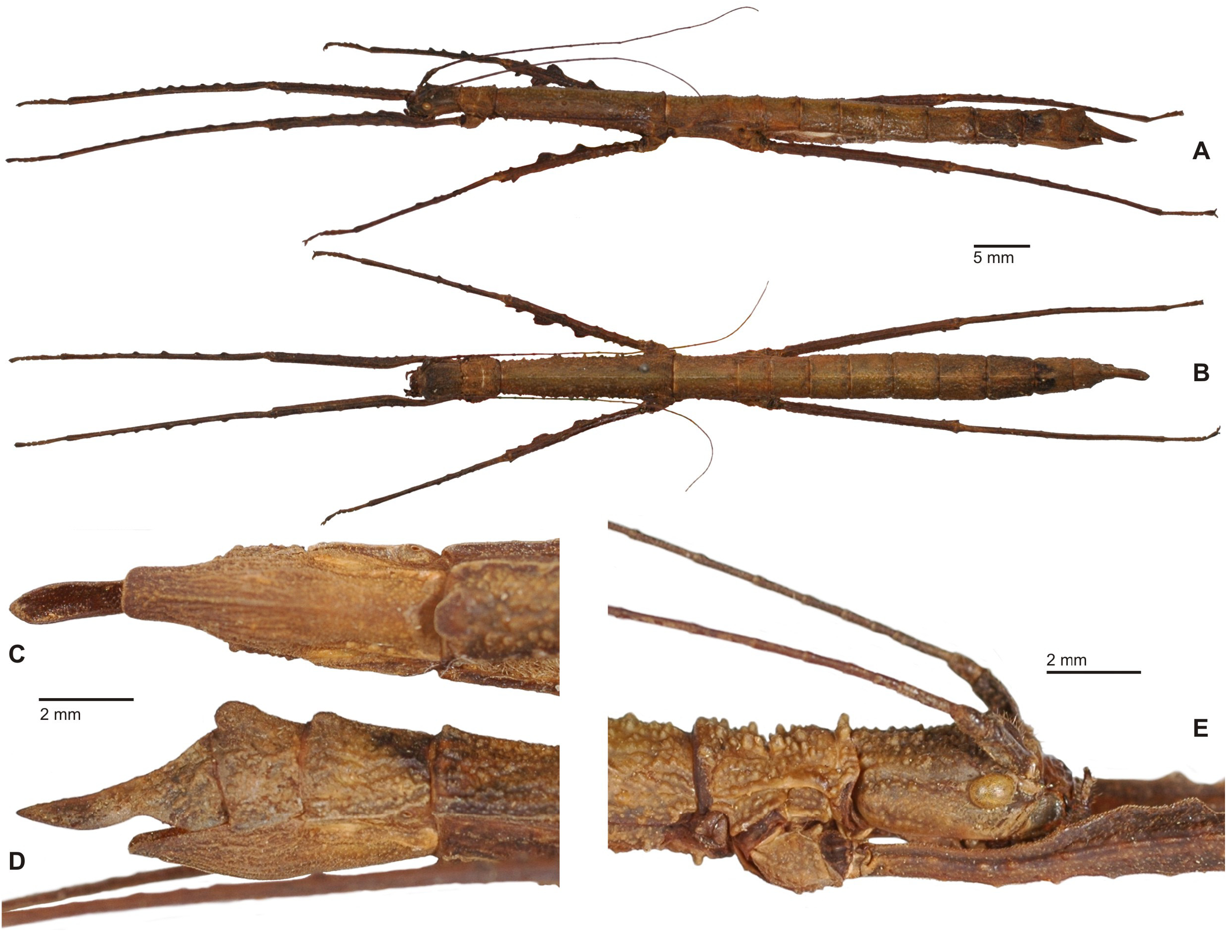
Diagnosis: ♀ ( Fig. 25 View FIGURE 25 ). Fairly small (body length 70.0 mm) and stocky Lonchodinae ( Figs. 25A–B View FIGURE 25 ) with a birdbeak like ovipositor (figs. 25C–D). General colour brown. Entire body granulose and tuberculose to a variable degree and with a blunt longitudinal median keel along most of dorsal body surface. Head distinctly longer than wide, roundly rectangular in dorsal aspect, the vertex flattened ( Fig. 25E View FIGURE 25 ) and with an impressed coronal line; between eyes with a pair of blunt swellings. Antennae slender, reaching to abdominal segment II and conisting of about 40 antennomeres; scapus compressed dorsoventrally and somewhat deflexed laterally, pedicellus cylindrical, IV–XXX increasing in length, the apical ten antennomeres much shorter. Antennomere XIV with a shiny, knob-like sub-basal dorsal swelling. Pronotum with a prominent, impressed transverse sulcus and at least two enlarged median tubercles at anterior margin. Mesothorax about 4.5x longer than prothorax and almost uniform in width; mesonotum with a variable number of enlarged blunt spiniform tubercles near lateral margins. Metanotum roughly 1/3 the length of mesonotum and almost 2x longer than median segment. Meso- and metapleurae with several enlarged spiniform tubercles; sterna less densely granulose or tuberculose than terga. Abdomen longer than head and thorax combined. Median segment roughly quadrate. Segments II–VI very slightly increasing in length and parallel-sided, II quadrate and VI about 1.3x longer than wide. VII slightly longer than all previous and gently gradually narrowing towards the posterior. Sternum VII with a prominent Preopercular organ that is formed by a scale-like median projection at posterior margin ( Fig. 25C View FIGURE 25 ). Terga VI–IX with a posteromedian hump, which is indistinct on VI but forming a prominent, rounded bi-dentate projection on IX. Anal segment strongly narrowed and declining in lateral aspect, the apex strongly elongated into a lanceolate projection that is somewhat narrowed medially and pointed apically ( Fig. 25D View FIGURE 25 ). Cerci very small and hidden under anal segment. Epiproct very small and also hidden under apical projection of anal segment. Subgenital plate moderately convex, keeled longitudinally with apex elongated, sub-angular and reaching about half way along apical projection of anal segment ( Fig. 25C View FIGURE 25 ). Legs of moderate length, profemora a little longer than mesothorax, mesofemora about equal in length to mesothorax and hind legs projecting beyond apex of abdomen. All carinae to a variable degree lobulate or bluntly dentate. Profemora curved and compressed basally. Protibiae with distinct blunt teeth dorsally. Mesofemora with large lobes on anteroventral carina ( Fig. 25B View FIGURE 25 ) that increase in size towards the apex of femur and somewhat smaller but enlarged lobes on anterodorsal carina. Basitarsi about equal in length to following three tarsomeres combined, probasitarsus with dorsal carina gently raised apically.
Differential diagnosis: Apparently close to the Philippine endemic Manduria Stål, 1877 and Neomyronides n. gen. but also morphologically similar to the principally New Guinean Neopromachus Giglio-Tos, 1912 . Females, the only sex known, share most features with the Sulawesian endemic Neomyronides n. gen. but readily differ by the presence of a beak-like ovipositor that is shaped by the apically elongated and lanceolate anal segment and elongat-ed subgenital plate ( Figs. 25C–D View FIGURE 25 ), as well as the just very slightly apically raised dorsal carina of the probasitarsus. The conspicuous dorsal sub-basal swelling on antennomere XIV is much more developed than in Neomyronides n. gen.. The ovipositor and prominent uni-lobate Preopercular organ on abdominal sternum VII ( Fig. 25C View FIGURE 25 ) are shared with Manduria but the type-species of this new genus differs by the more stocky appearance, not longitudinally tectinate mesosternum, broadened and sub-angular apex of the subgenital plate, distinctly multi-lobate legs (only single dorsal lobes are present exclusively on the mesofemora and mesotibiae in Manduria ) and much longer tarsi. In addition to the geographic separation the prominent Preopercular organ on abdominal sternum VII in this new genus represents the main feature that distinguishes ♀♀ from Neopromachus .
Comments: Already Günther (1935) was in doubt about the generic position of this Sulawesian species. The author commented that it takes on a very isolated position within Neopromachus Giglio-Tos, 1912 and that he believed it has in fact no particularly close phylogenetic relationship to the New Guinean species of that genus and rather represents a peculiar faunistic element of the island of Sulawesi “ Wenngleich generische Unterschiede zwischen dieser merkwürdigen neuen Art und all den übrigen Species des Genus Neopromachus Giglio-Tos nicht zu finden sind, so macht sie doch unter ihnen allen einen sehr aberranten und isolierten Eindruck. [...] Aber ich glaube doch, dass die vorliegende neue Art, die ich generisch von Neopromachus nicht zu trennen vermag, phylogenetisch mit den neuguineeischen Arten nicht zusammenhängt und eine selbständige Fauneneigentümlichkeit der Insel Celebes darstellt. (Günther (1935: 15)”. Indeed, morphology of the ♀♀ rather supports close relation to the Sulawesian endemic Neomyronides n. gen. and the Philippine Manduria Stål, 1877 than to the New Guinean Neopromachus . Günther (1938: 79) described Prisomera nodosum based on a ♂ and nymph from the valley of Bone, South Sulawe-si ( Fig. 26 View FIGURE 26 ). He estimated this species might represent the previously unknown ♂ of P. celebensis (Günther, 1935) and if that was the case a new genus would have to be established for incorporating these two species: “ Diese Art stellt vielleicht die ♂♂ von Neopromachus celebensis K. Gthr. 1936 dar; das würde, wenn es sich so verhält die Er-richtung einer neuen Gattung bedingen. ( Günther, 1938, 79, footnote 2)”. The presence of tooth-like lobes on both the posterodorsal and both outer ventral carinae of the mesofemora and the conspicuous sub-basal dorsal swelling on antennomere XIV suggest nodosum might be congeneric to celebensis . However, they obviously represent two distinct species and without having at hand for examination ♂♂ and ♀♀ of either species from the same locality, this hypothesis cannot be confirmed at the present state of our knowledge. Consequently, nodosum is here attributed to Paramanduria n. gen. with doubt and is not incorporated in the generic description presented above. Any broader discussion, differentiation and incorporation of the ♂♂ in the generic description must await availability of additional material and the still unknown eggs.
Etymology: The name refers to the strong visual resemblance and assumed close relation to the Philippine genus Manduria Stål, 1877 . Feminine.
Distribution: Sulawesi (endemic).
Species included:
1. Paramanduria celebensis (Günther, 1935: 14, pl. 2: 10 (♀ )) [ Neopromachus ]. HT, ♀: Celebes, Matinangeb., 600m, X.1930, G. Heinrich leg.; Typus [ MNHU]. n. comb .
Distribution: N-Sulawesi, Prov. Sulawesi Tengah, Matinan Mountains [MNHU].
2. Paramanduria (?) nodosum ( Günther, 1938: 79, figs. 16–17 (♂ )) [ Prisomera ]. HT, ♂: Sarasin 8.I.1894, BoneTal ca. 200M, Nord-Cel.; Typus; Prisomera nodosum n. sp. K. Günther det. [NHMB, VI.D.136]; PT, ♂ nymph: N-Celebes, Bone-Tal ca. 700m, leg. Sarasin 14.I.1894; det. Günther [SMTD]. n. comb.
Distribution: S-Sulawesi, Sulawesi Selatan, Bone valley [NHMB, SMTD].
Genus Periphetes Stål, 1877
( Fig. 27 View FIGURE 27 )
Type-species: Phasma graniferum Westwood, 1859: 35 , pl. 3: 4, by original designation.
Comments: A discussion of this genus and list of species included has been presented by Hennemann & Conle (2007: 53), who described a new species from the Philippines and established new synonymies of the most common species in Sulawesi, P. forcipatus ( Bates, 1865) . Precise type data of the latter taxa can be found in Hennemann & Conle (2007: 54) and are therefore omitted in the list of species below.
Distribution: Philippines, Sulawesi & Sangihe.
Species recorded from Sulawesi:
1. Periphetes borealis n. sp.
Distribution: NE-Sulawesi, Prov. Sulawesi Utara, Minahasa, Masarang Mountains [NHMB]; NE-Sulawesi, Prov. Sulawesi Utara, Minahasa, Mount Mahawu, summit ca. 1300 m [NHMB].
2. Periphetes forcipatus ( Bates, 1865: 338) [ Lonchodes ]. ( Fig. 67C View FIGURE 67 )
= Lonchodes duivenbodei Kaup, 1871: 30 ., pl. 2: 3 (♀). [Synonymised by Hennemann & Conle, 2007: 53]
= Dixippus furcatus Brunner View in CoL v. Wattenwyl, 1907: 279. [Synonymised by Hennemann & Conle, 2007: 53]
= Periphetes sangirensis Dohrn, 1910: 408 . [Synonymised by Hennemann & Conle, 2007: 53]
= Periphetes duivenbodei elongatus Günther, 1938: 58 , 75. [Synonymised by Hennemann & Conle, 2007: 53]
Distribution: S-Sulawesi, Prov. Sulawesi Selatan, Tana Toraja, Rantepao, 700 m [coll. FH]; S-Sulawesi, Prov. Sulawesi Selatan, Bungadidi [coll. FH]; S-Sulawesi, Prov. Sulawesi Selatan, Saddan-Toraya [coll. OC]; SSulawesi, Prov. Sulawesi Selatan, Bungadidi [coll. FH]; SE-Sulawesi, Prov. Sulawesi Tenggara, Buton Island [coll. FH, coll. OC]; Central Sulawesi, Prov. Sulawesi Tengah, Lake Poso [NHMB]; N-Sulawesi, Sulawesi Utara, Minhasa, Menado [OXUM, HLDH]; N-Sulawesi, Prov. Gorontalo, Kota Gorontalo [HLDH].
3. Periphetes parastatidon Günther, 1935: 11 , pl. 1: 7 (♀) & 8 (♂).
Distribution: S-Sulawesi, Prov. Sulawesi Selatan, Gunung Latimojong , 1500 m [MNHU].

| NHMB |
Natural History Museum Bucharest |
| VI |
Mykotektet, National Veterinary Institute |
No known copyright restrictions apply. See Agosti, D., Egloff, W., 2009. Taxonomic information exchange and copyright: the Plazi approach. BMC Research Notes 2009, 2:53 for further explanation.
|
Kingdom |
|
|
Phylum |
|
|
Class |
|
|
Order |
|
|
Family |
|
|
Genus |
Myronides tomohonense
| Hennemann, Frank H. 2021 |
Dixippus furcatus
| Hennemann, F. H. & Conle, O. V. 2007: 53 |
Pseudostheneboea
| Hennemann, F. H. 1998: 102 |
Myronides pfeifferae, Günther, 1938: 74
| Hennemann, F. H. 1998: 119 |
| Gunther, K. 1938: 74 |
Staelonchodes, Günther, 1938: 77
| Gunther, K. 1938: 77 |
Periphetes duivenbodei elongatus Günther, 1938: 58
| Hennemann, F. H. & Conle, O. V. 2007: 53 |
| Gunther, K. 1938: 58 |
Periphetes sangirensis
| Hennemann, F. H. & Conle, O. V. 2007: 53 |
| Dohrn, H. 1910: 408 |
Lonchodes duivenbodei
| Hennemann, F. H. & Conle, O. V. 2007: 53 |
| Kaup, J. J. 1871: 30 |