
Rhaphidosoma atkinsoni Bergroth, 1893
|
publication ID |
https://doi.org/ 10.11646/zootaxa.4303.2.5 |
|
publication LSID |
lsid:zoobank.org:pub:43229076-7D67-44E5-AEF5-6056E5B1F04B |
|
DOI |
https://doi.org/10.5281/zenodo.6040292 |
|
persistent identifier |
https://treatment.plazi.org/id/03DC87EA-FFA6-FF85-FF06-F9B0FAF6FBB2 |
|
treatment provided by |
Plazi |
|
scientific name |
Rhaphidosoma atkinsoni Bergroth, 1893 |
| status |
|
Rhaphidosoma atkinsoni Bergroth, 1893 View in CoL
( Figs. 4–40 View FIGURES 1 – 5 View FIGURES 6 – 12 View FIGURES 13 – 16 View FIGURES 17 – 26 View FIGURES 27 – 31 View FIGURES 32 – 40 )
Rhaphidosoma atkinsoni Bergroth, 1903: 63 View in CoL (original deScription)
Rhaphidosoma atkinsoni: DiStant (1904: 330) View in CoL (diagnoSiS, illuStrationS), HaridaSS (1985: 245) (feeding, ovipoSition), HaridaSS (1988: 49) (ultraStructure of egg), Maldonado CaprileS (1990: 271) (catalogue, diStribution), AmbroSe (2006: 2399) (liSted).
Raphidosoma [inadvertent error] atkinsoni View in CoL : BiSWaS & Mitra (2014: 15) (liSted).
Rhaphidosoma atkinsoni: Mukherjee & HaSSan (2016: 593) View in CoL (diagnoSiS, image).
Redescription. Apterous male and female. Body strongly elongate, narrow, in male almost 14 times longer than its maximum width at metacoxae, female slightly broader than male in thoracic region and almost twice as broad in midabdominal region. Body of male subparallel posteriad of prothorax, with only slight dilations at meso- and metathoracic regions.
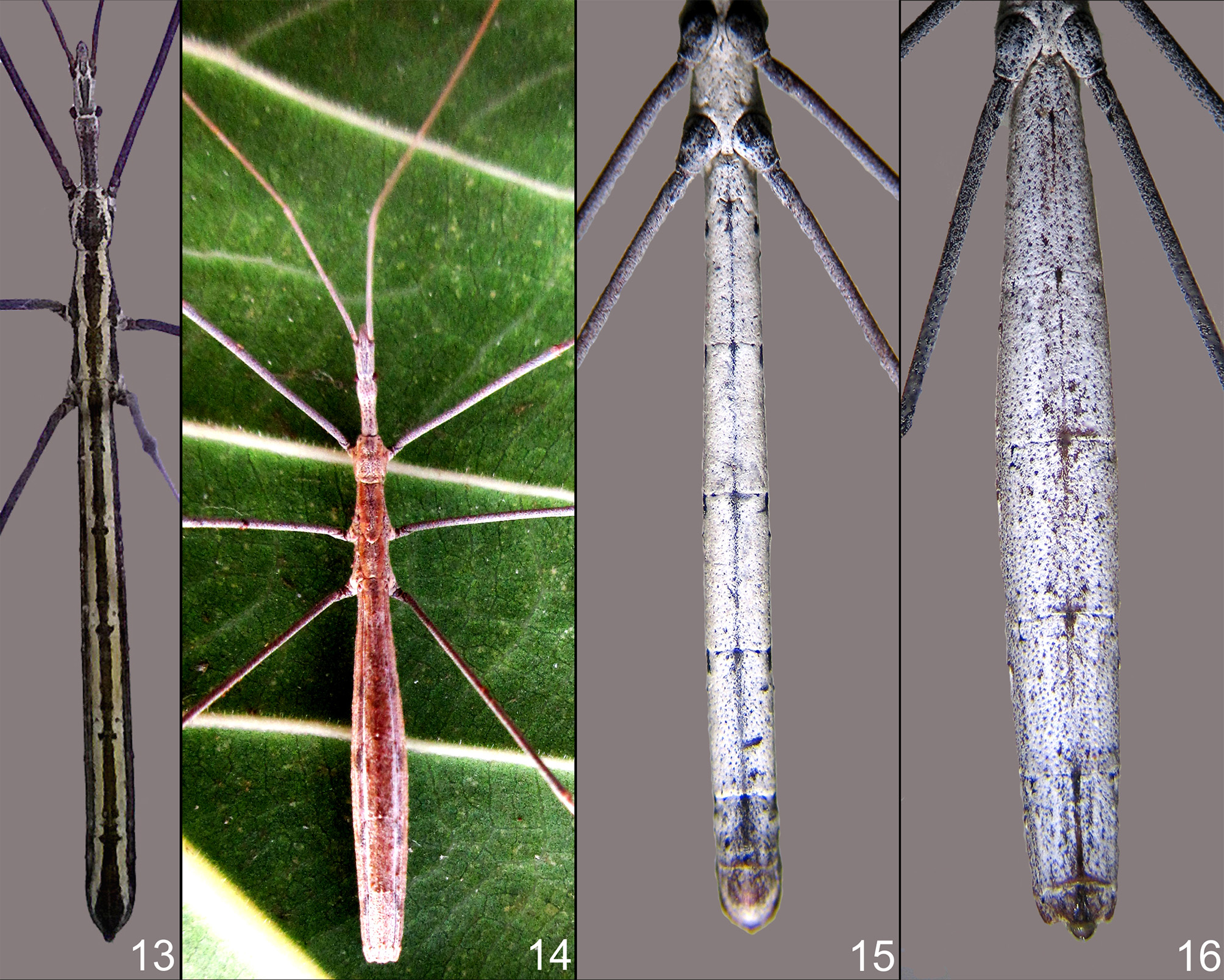
Colouration, integument and vestiture. Dark brown, covered with dense, white, scale-like setae that form complete bands laterally along entire length of body except head. Male with a median dark band covered with only sparse setae, exposing dark brown ground colour of integument; a lateral band of colourless setae and another dark band marginally ( Fig. 13 View FIGURES 13 – 16 ). Median dark band with transverse delicate wrinkles and fine tubercles at places; each abdominal tergite also with rounded depression on either side of median dark line which is devoid of white setae. Female more diffuse earth-brown, without distinct lateral bands ( Fig. 14 View FIGURES 13 – 16 ). Coloration of setae as well as relative density of setae is different in male and female. Segmental boundaries obscured by setae. Head also densely covered with white setae and many dark brown fine tubercles on its whole surface. A pair of dark lines (which are bare areas with few setae in male and relatively more setae in female) that converge posteriorly to form a ‘V’ is present in front of eyes and behind antenniferous tubercles. Antennae dark brown and sparsely covered with white setae, thus appearing spotted. Entire venter more densely covered with white setae, with minor difference in male and female; coxae partly dark brown, underside of head and thorax also with sparse, fine, brown granules. Granules on abdomen sparse, barely visible due to dense setae in male, more clear due to relatively sparse setae in female. A ventromedian, partly dark brown line present along all abdominal sternites. Pygophore with sparse setae hence dark brown, similarly female genital segments darker with very few setae ( Figs. 15, 16 View FIGURES 13 – 16 ). Abdominal segments III– VII each with a round dark brown patch at anterolateral angle, these patches prominent in male. Spiracles on first abdominal segment dorsal, dark brown, remaining spiracles situated on laterosternites but visible from dorsal side as minuscule protuberances on either side, except for second abdominal spiracle which is very minute. Rostrum brown with very sparse setae on first visible segment; second visible segment longest. All legs brown, relatively sparsely covered by scale-like setae; setae on legs also denser in female than in male.
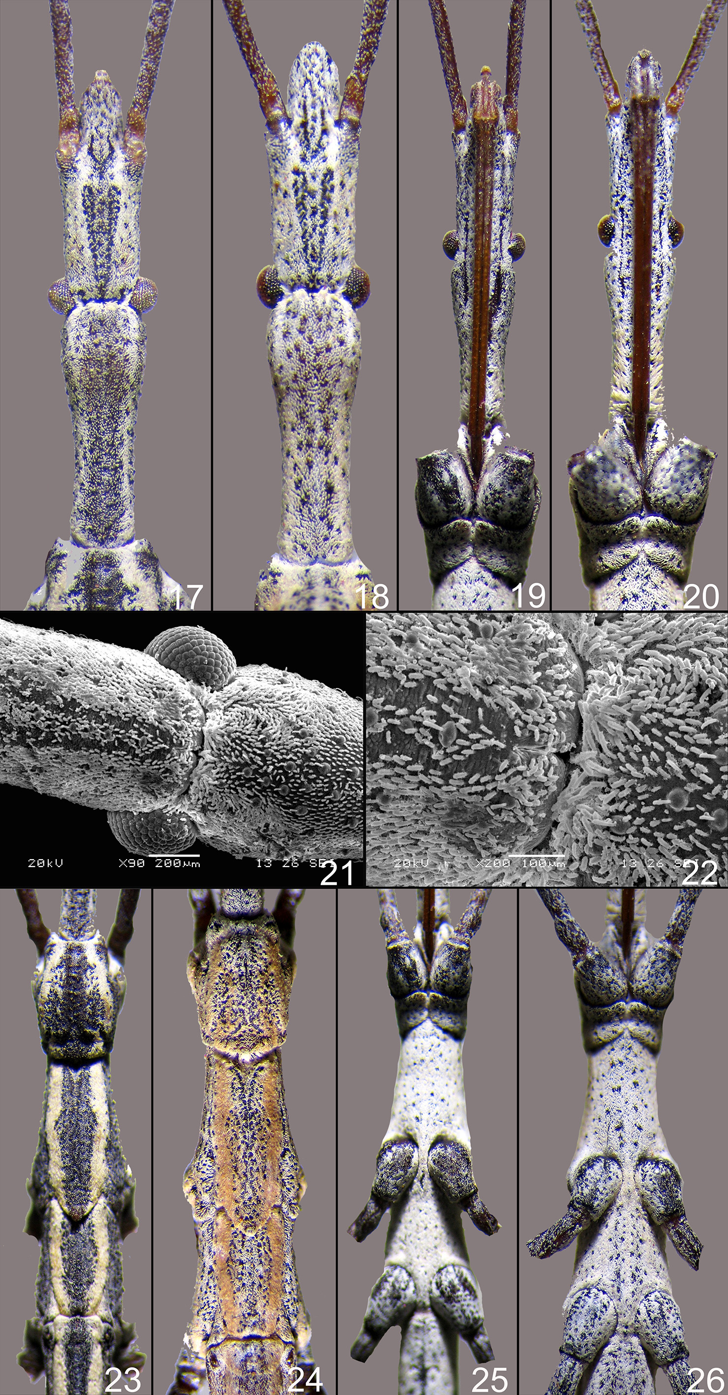
Structure. Head. Elongate, with a deep transverse interocular sulcus close to posterior margin of eyes. Area in front of sulcus rather flat, posteriad of sulcus slightly laterally dilated, in lateral view tumescent above anteriorly, narrowed behind and slightly concave posteriorly. Eyes moderate, semiglobular, situated laterally. Antenniferous tubercles prominent, slightly divergent, exposed from above. Clypeus and mandibular plates indistingushable from above due to setae, but clypeus is slightly raised and mandibular plates are slightly oblique. Apex of head without distinct spine ( Figs. 17, 18 View FIGURES 17 – 26 ). Antennae long, first segment longest, second slightly shorter than first, third and fourth much shorter than first. Labium long, reaching procoxae, its first visible segment almost as long as preantennal region of head, second visible segment longest ( Figs. 19, 20 View FIGURES 17 – 26 ). Ocelli completely lacking, untraceable even with SEM; few setae, especially on midline, are situated on tubercles, and some setae are slightly longer than others ( Figs. 21, 22 View FIGURES 17 – 26 ).
Thorax. Pronotum tumescent above, with anterior margin concave and posterior margin straight, and with short, median, longitudinal sulcus visible in part; subdivided into a subtrapezoid, anteriorly slightly narrowed anterior lobe and a very short, rim-like, dorsally flat posterior lobe, by a transverse sulcus very close to base. Mesonotum also with a median very shallow longitudinal sulcus, its surface partly granular. Scutellar area slightly raised above at tip; metanotum with median raised area or blunt carina along its length. Metanotum shorter than mesonotum, with posterior margin sinuate and posterolateral corners slightly produced. Granules on pro-, meso- and metanota more prominent laterally ( Figs. 23, 24 View FIGURES 17 – 26 ). Prosternum narrow, with deep median sulcus to receive labium. Ventral (pleural) parts of posterior lobe of prothorax encircling coxae posterolaterally and closely approaching but not meeting each other, with a noticeable gap in the midline ventrally. Meso- and metasterna densely clothed with setae, provided with few dark brown granules. Area between mesocoxae slightly elevated, boundary between meso- and metasterna unnoticeable. Mesosternum produced between metacoxae to meet abdominal sternite, their boundaries indistinct. Posterior part of metasternum slightly sulcate ( Figs. 25, 26 View FIGURES 17 – 26 ).
Legs. All legs long, slender, uniformly coloured. All coxae swollen. Hind legs with longest femora and tibiae. Tarsi with 3 segments and sharp claws.
Abdomen. First abdominal tergite short, medially raised, with small anteriorly directed tubercle and with lateral spiracle. Remaining segments long but segmental boundaries indistinct due to setae. Connexivum narrow, spiracles small but distinct. Median part of each tergite shining in male; third and fourth tergites with small mid-dorsal tubercle.
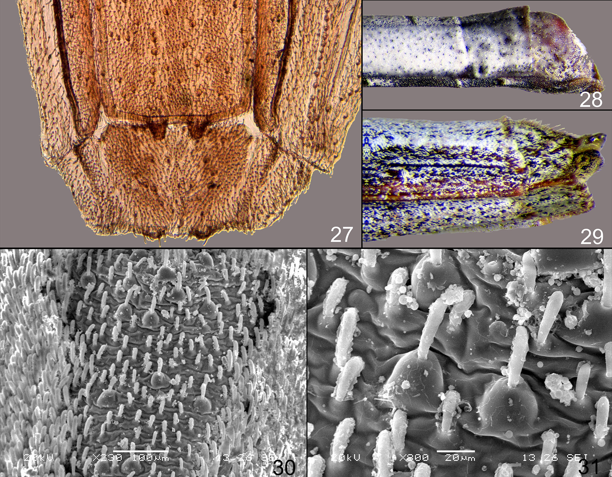
Pygophore with sparse setae, eighth segment completely telescoped into seventh segment and not visible ventrally. In male posterior border of tergites III–VI medially slightly elevated as small shining tubercle. In female tergite V with a small medio-dorsal tubercle at posterior border, tergite VI with a pair of small tubercles and tergite VII with a pair of posteriorly directed blunt tubercular projections on either side of midline ( Fig. 27 View FIGURES 27 – 31 ); tergite VIII broader than long; tergite IX slightly sloping downward, not fully visible from above, while only extreme apex of tergite X exposed ( Figs. 28, 29 View FIGURES 27 – 31 ). Male abdominal tergites with median part slightly wrinkled and covered with broad setae, some of which have a globular base; and with lateral area covered with dense setae ( Figs. 30, 31 View FIGURES 27 – 31 ).
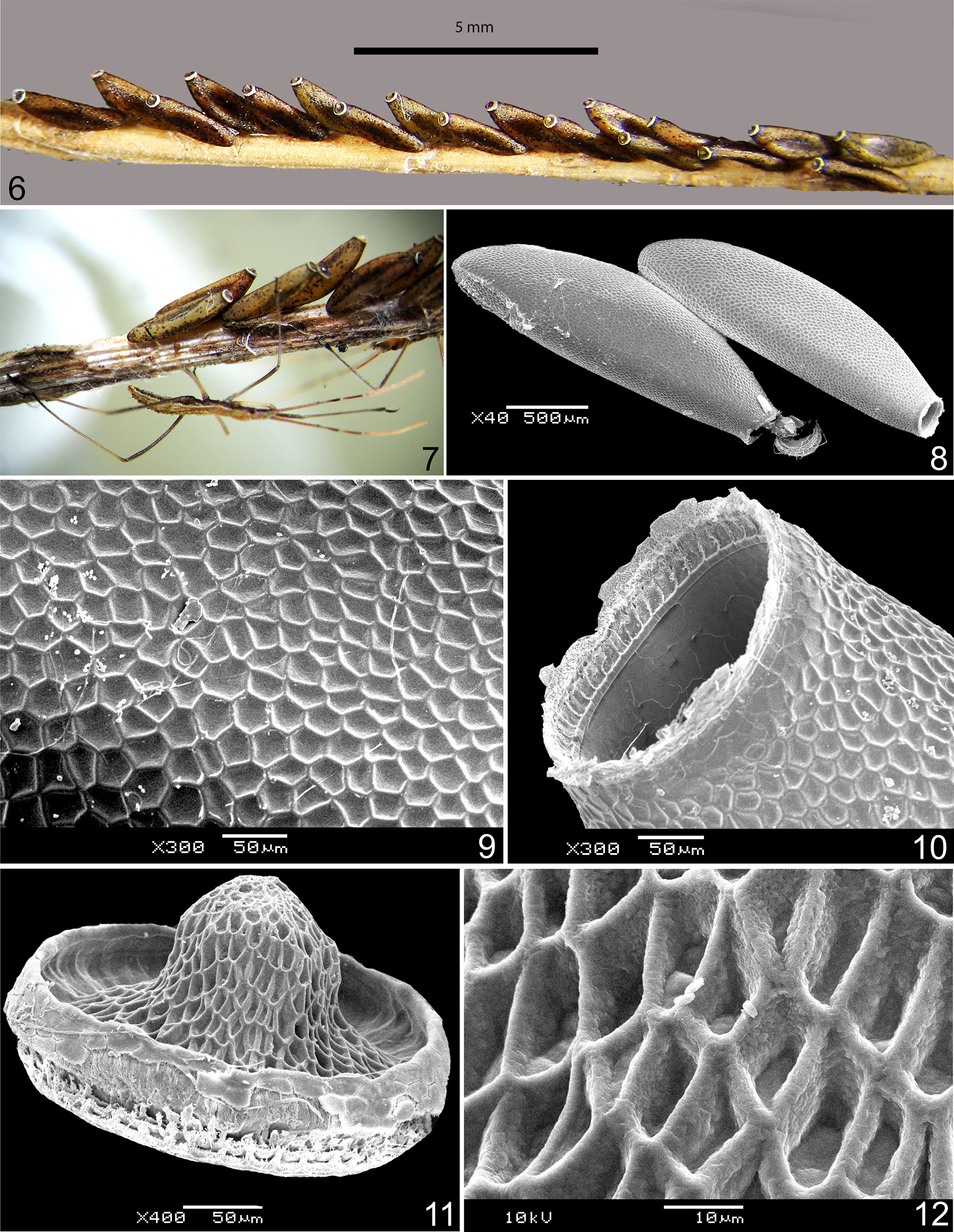
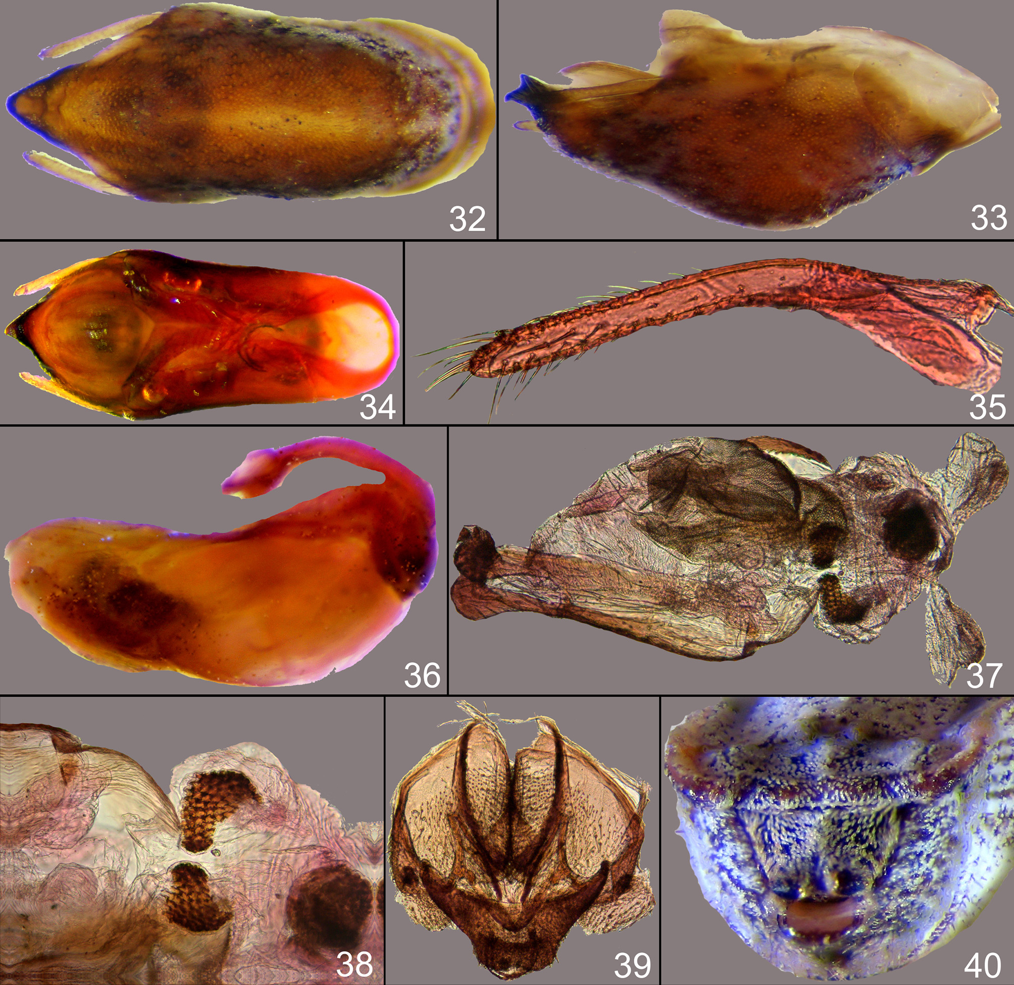
Male genitalia. Pygophore ventrally convex and sclerotized, its superior tip produced into a dark brown tubercle visible in ventral view ( Fig. 32 View FIGURES 32 – 40 ). In lateral view pygophore appears boat-shaped, the tubercle at superior tip appears bidentate ( Fig. 33 View FIGURES 32 – 40 ). Pygophore dorsally membranous and its shape resembles a ‘slipper’ in dorsal view ( Fig. 34 View FIGURES 32 – 40 ). Parameres small, symmetrical, sparsely covered with long setae, almost straight distally ( Fig. 35 View FIGURES 32 – 40 ) and projecting out slightly behind in intact pygophore. Phallus oblong oval in lateral view ( Fig. 36 View FIGURES 32 – 40 ), endosoma with three sclerotised, spiny areas ( Figs. 37, 38 View FIGURES 32 – 40 ).
Female terminalia. Boundaries between valvulae and valvifers obscure due to setae. Ninth tergite downwardly sloping, with blunt, black and shining tubercle at each corner ( Fig. 39, 40 View FIGURES 32 – 40 ).
Measurements in millimetres (average of 2 ♂♂ / average of 2 ♀♀): total length 25.1 / 23.5, length of head anteriad of transverse sulcus 2.2 / 1.9, length of head posteriad of transverse sulcus 2.3 / 2.0, total length of head 4.5 / 3.9, width across eyes 1.0 / 1.0, length of prothorax at midline 1.0 / 1.1, width of prothorax 1.1 / 1.1, length of mesothorax 1.5 / 1.5, width of mesothorax 1.5 / 1.6, length of metathorax 0.8 / 0.8, width of metathorax 1.8 / 1.8, width of abdomen at level of first spiracle seen from above 1.1 / 1.2, length of pro coxa 0.7 / 0.7, length of pro femur 9.7 / 8.5, length of pro tibia 10.7 / 8.9, length of pro tarsus with claw 0.7 / 0.5, length of meso coxa 1.0 / 1.0, length of meso femur 9.25 / 7.8, length of meso tibia 9.1 / 7.85, length of meso tarsus with claw 0.7 / 0.5, length of meta coxa 0.8 / 1.0, length of meta femur 12.7 / 11.2, length of meta tibia 14.85 / 14.1, length of meta tarsus with claw 0.8 / 0.6, length of first segment of antenna 7.7 / 6.7, length of second segment of antenna 3.5 / 2.6, length of third segment of antenna 3.2 / 2.6, length of fourth segment of antenna 3.0 / 2.8, total length of labium 4.3 / 4.3, length of second visible segment of labium 3.3 / 3.2, length of third visible segment of labium 0.44 / 0.44
No known copyright restrictions apply. See Agosti, D., Egloff, W., 2009. Taxonomic information exchange and copyright: the Plazi approach. BMC Research Notes 2009, 2:53 for further explanation.
|
Kingdom |
|
|
Phylum |
|
|
Class |
|
|
Order |
|
|
Family |
|
|
Genus |
Rhaphidosoma atkinsoni Bergroth, 1893
| Pansare, Pratik P., Joshi, Nikhil U., Boyane, Swapnil S. & Ghate, Hemant V. 2017 |
Rhaphidosoma atkinsoni:
| Mukherjee 2016: 593 |
Rhaphidosoma atkinsoni: DiStant (1904: 330)
| AmbroSe 2006: 2399 |
| Maldonado 1990: 271 |
| DiStant 1904: 330 |