
Notodromas trulla, Smith, Robin J. & Kamiya, Takahiro, 2014
|
publication ID |
https://doi.org/10.11646/zootaxa.3841.2.4 |
|
publication LSID |
lsid:zoobank.org:pub:FF9AB04E-BEA7-4782-9D69-190C53827EA7 |
|
DOI |
https://doi.org/10.5281/zenodo.5629707 |
|
persistent identifier |
https://treatment.plazi.org/id/03DD2F79-EF1A-FFB2-06C5-FB73FD16FA34 |
|
treatment provided by |
Plazi |
|
scientific name |
Notodromas trulla |
| status |
sp. nov. |
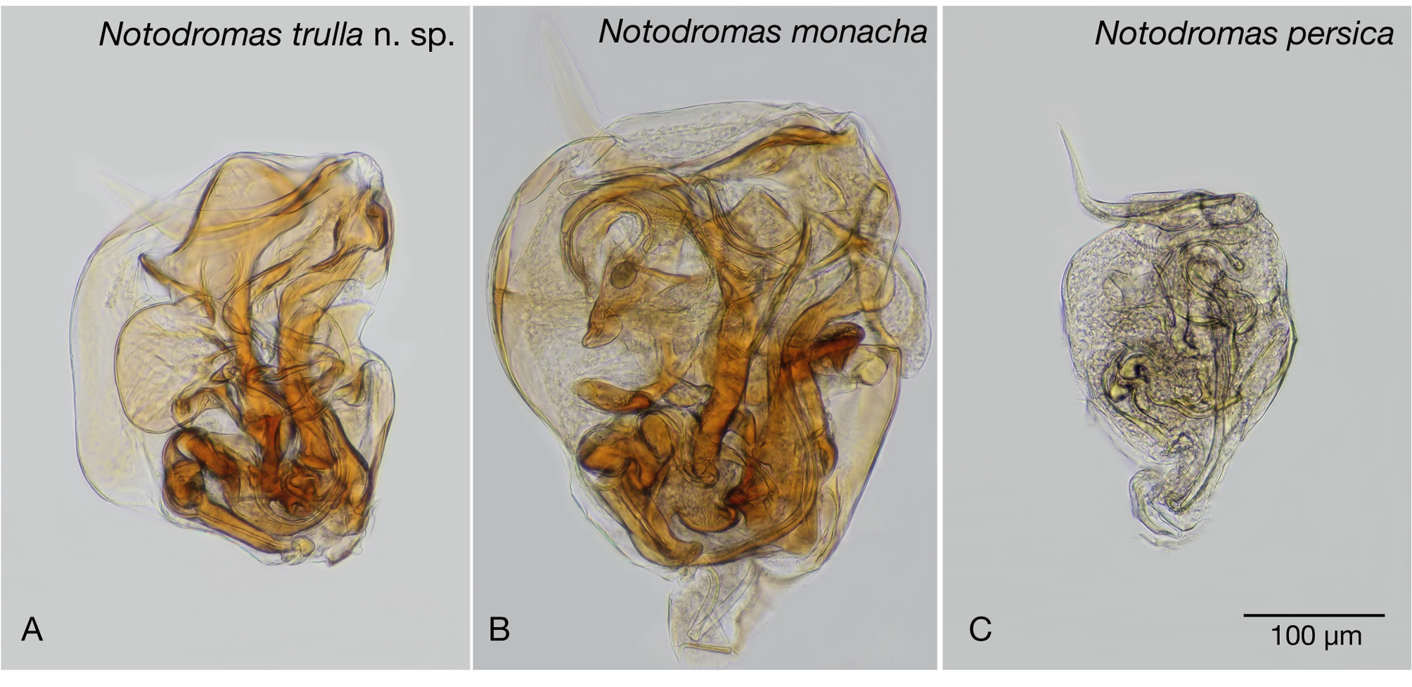
Notodromas trulla n. sp. ( Figures 2–9 View FIGURE 2. A & B View FIGURE 3. A – F View FIGURE 4 View FIGURE 5 )
1927 Notodromas monacha O. F. Müller—Komai : 1193, fig. 2302.
?1933 Notodromas monacha O. F. Muelle—Brehm : 297.
?1937 Notodromas monacha— Onoda & Murakoshi: 174–175, fig. 3. 1947 Notodromas monacha O. F. Müller—Ueno : 870, fig. 2494.
?1948 Notodromas monacha Müller—Uchida : 248, fig. 1397.
?1957 Notodromas monacha O. F. Muller—Ueno : 142, plate 71, fig. 15.?1959 Notodromas monacha (O. F. Müller) 1776 —Hanai: 433, 434. 1965 Notodromas monacha O. F. Müller—Ueno , in Ueno & Hanai: 456, fig. 425. 1977 Notodromas monacha O. F. Müller, 1776 —Hanai et al.: 21.
1979 Notodromas monacha O. F. Müller—Ueno , in Ueno & Hanai: 386, fig 1360. 1989 Notodromas monacha (O. F. Müller) —Okubo & Ida: 105, pl. 1A–D. 2000 Notodromas monacha ( O. F. Müller, 1776) —Okubo: 106, figs 9a–e. 2003 Notodromas monacha ( O. F. Müller, 1776) —Okubo: 43–48, figs 1 & 2. 2004 Notodromas monacha ( O. F. Müller, 1776) —Okubo: 16, figs 4g –l. 2014 Notodromas sp.—Smith: 167, figs 29.1g –j.
Type locality. a pond covering approximately 15 m x 15 m, with a maximum depth of 0.3 m, in Yuhidera Kenmin Nature Park, Yuhidera-cho, Kanazawa City, Ishikawa Prefecture, Japan (36º 34' 26.2” N, 136º 42' 19.1” E).
Type material. Holotype—dissected male ( LBM 1430005748). Allotype—dissected female ( LBM 1430005749). Paratypes—dissected males ( LBM 1430005750, LBM 1430005751), dissected female ( LBM 1430005752), whole dried females ( LBM 1430005753, LBM 1430005754, LBM 1430005755, LBM 1430005756), whole dried males ( LBM 1430005757, LBM 1430005758).
Material examined. (material collected by the authors, unless stated otherwise) 12 males and 18 females from small, shallow pools with duckweed in an abandoned rice field with bulrushes in one corner in Hiranai-machi, Aomori Prefecture (40º 56' 44.03” N, 140º 51' 54.9”), 26 September 2004. 162 males and females from a pool at Tsubakizaki, Ishikawa Prefecture (37º 12' 21.1” N, 136º 57' 57.9” E) collected by Eriko Tanaka between July 2004 and September 2005. One individual from a rice field in Kaimochi, Anamizu Town, Ishikawa Prefecture (37º 12' 21.0” N, 136º 55' 30” E), collected by Eriko Tanaka, September 2004. Three individuals from a rice field in Maenami, Anamizu Town, Ishikawa Prefecture (37º 11' 28.5” N, 136º 57' 57.9” E) collected by Eriko Tanaka, September and November 2004. Seventy-four males and 162 females from the type locality, 23 July 2004. Fourhundred and eighteen males and 431females from the type locality, 24 July 2010.
Derivation of name. From the Latin trulla , the diminutive form of trua, meaning stirring spoon or skimmer, referring to the small, but distinctive, spoon-shaped seta on the male's fifth limb right palp ( Fig. 7A & B).
Diagnosis. Male and female carapaces sexually dimorphic. Female carapace with posterior flanges on both valves, flange on right valve partially overlapping left. Aesthetasc Y of antenna very long, extending beyond end of first segment of endopodite. Fifth limbs with two a-setae, and setae b and d present. Male fifth limb palps asymmetrical, with right palp much longer than left. Male fifth limb right palp with sub-apical, stiff seta with rounded, spoon-shaped end. Female fifth limb palps symmetrical and elongate, each with one short distal seta. Claws Gp and Ga of caudal rami approximately similar in length. Male hemipenis with indentation on inner edge, and internally with one rounded lobe towards outer edge and one rounded lobe towards distal edge. Distal-inner edge with large, shallow-curved, hook-like projection, orientated lengthwise towards distal-outer edge.
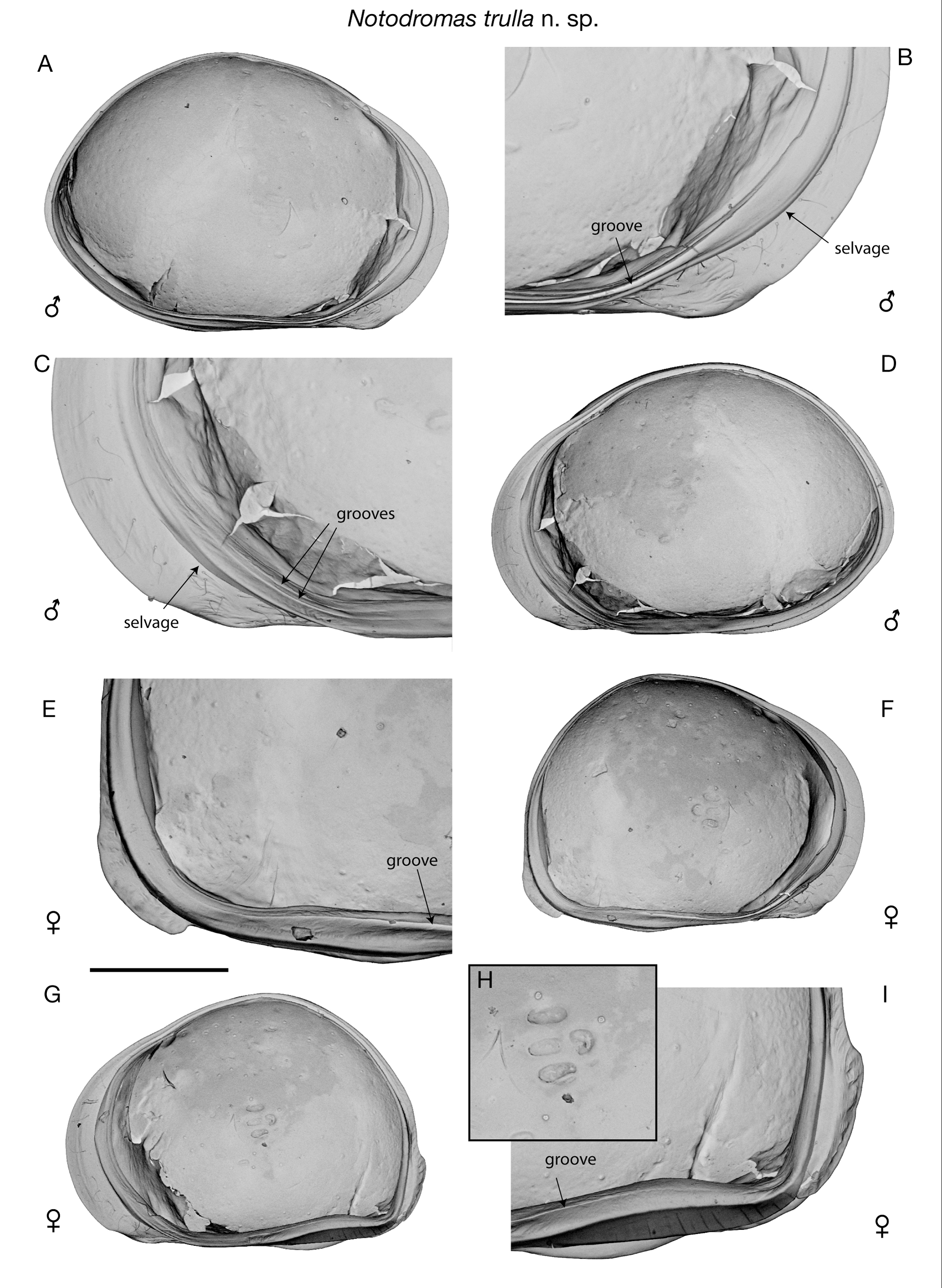
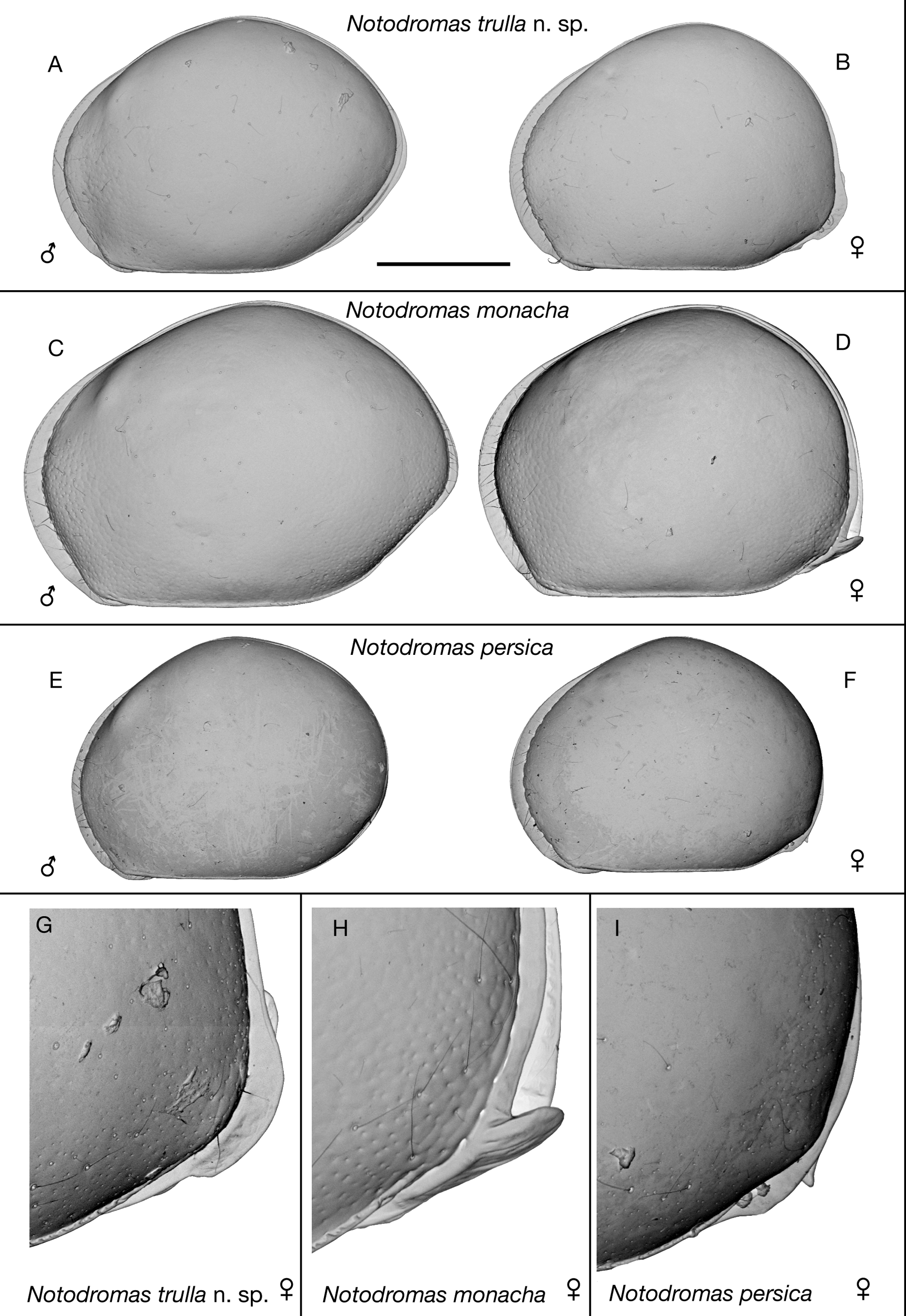
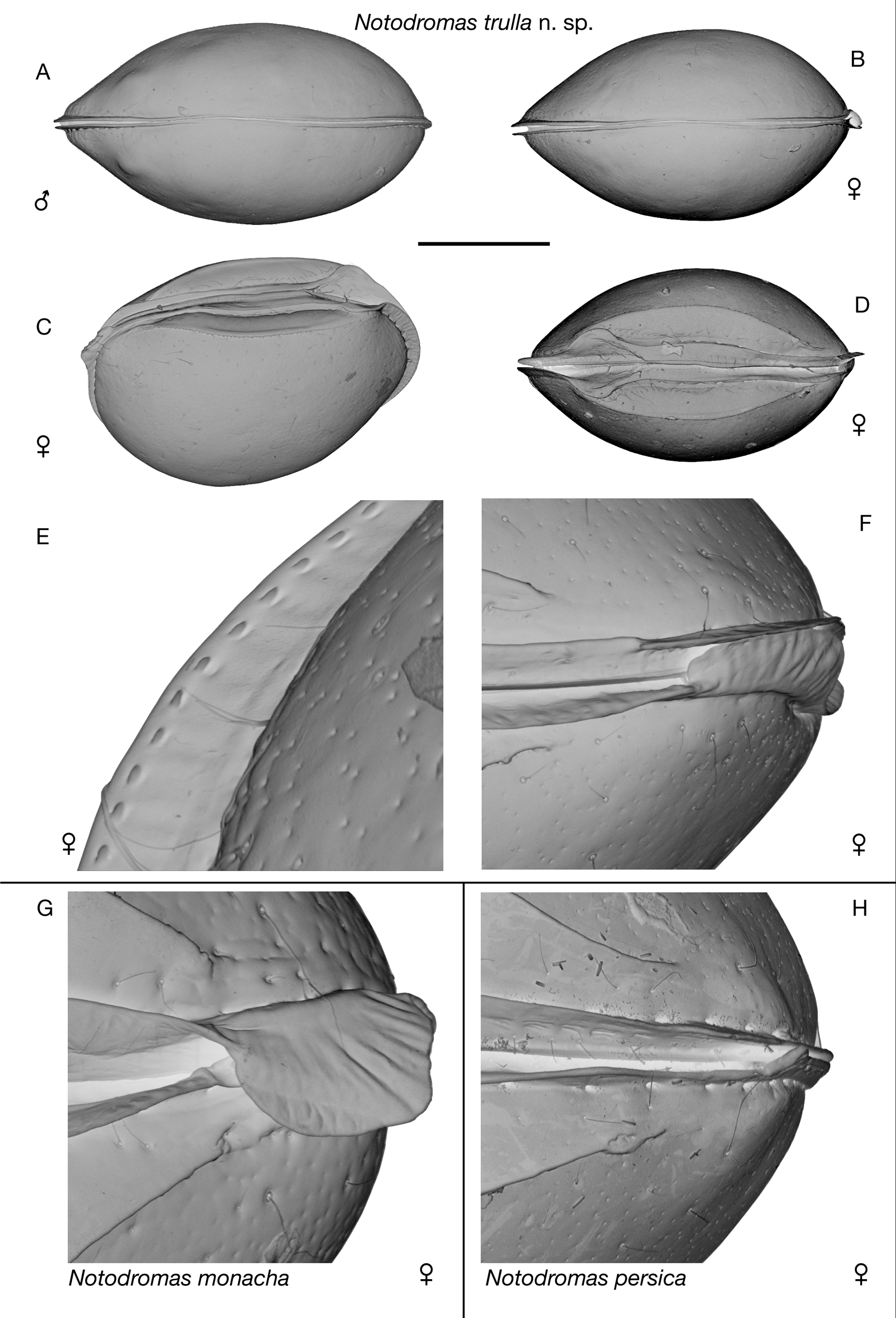
Description. Male carapace length 847–913 µm, height 610–663 µm (height/length=0.69–0.75). Female carapace length 786–851 µm, height 585–637 µm (height/length=0.73–0.75). Male carapace sub-pentagonal in lateral view ( Fig. 2A View FIGURE 2. A & B ). Dorsal margin unevenly curved with slight hump at mid-length. Hinge adont. Anterior and posterior margins both unevenly curved with apex at about mid-height; posterior margin more angular at apex than anterior. Ventral margin straight with distinctive angle at contact point with anterior margin. Flanges running along both anterior and posterior margins, anterior one wider than posterior one. Eye-spots visible anterior of hinge. Dorsal view with rounded posterior, and more pointed, slightly beak-shaped anterior ( Fig. 3A View FIGURE 3. A – F ). Maximum width approximately mid-length. Surface generally smooth, but with slight pitting towards anterior margin.
Female carapace lateral view with rounded, dome-like dorsal margin ( Fig. 2B View FIGURE 2. A & B ). Anterior margin similar to male, but apex of posterior margin much lower than in male. Posteriorly, with widened flange on both valves, right flange partially overlapping left ( Figs 2 View FIGURE 2. A & B G, 3F, 4E–G & I). Dorsal view of female slightly less elongate and eye spots more subdued compared to male ( Fig. 3B View FIGURE 3. A – F ).
Both sexes with sharply defined oval concavity on ventral surface ( Figs 3C & D View FIGURE 3. A – F ). Outer lists running either side of venter margins, diverging outwards towards anterior margin around mouth area. Outer side of anterior flange of both sexes with series of regularly spaced, tiny, elongate tubercles running parallel and adjacent to outer margin, each tubercle with slight concavity along both long sides (resembling stitching) ( Fig. 3E View FIGURE 3. A – F ).
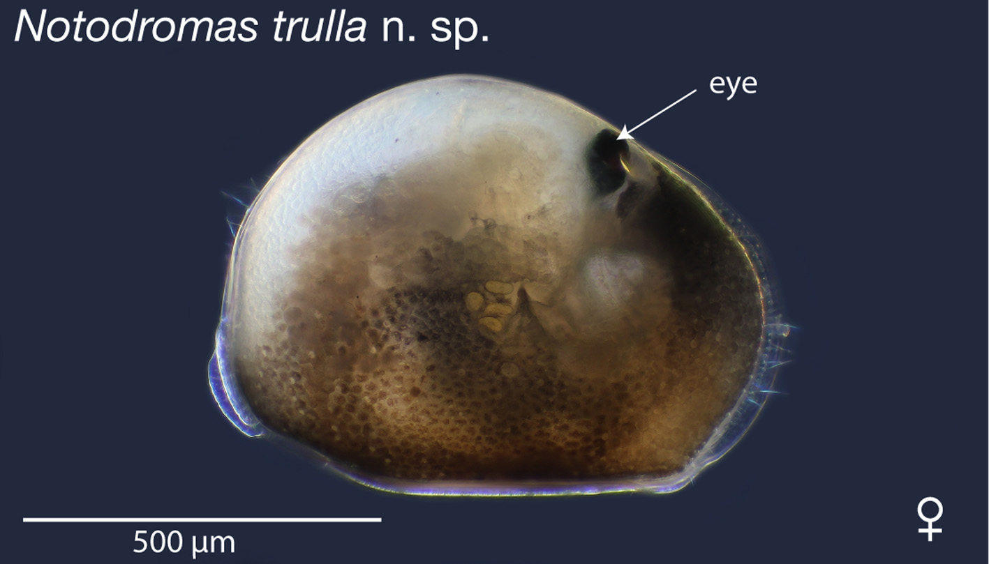
Internally, valves with inwardly displaced selvages along anterior margins ( Figs 4 View FIGURE 4 A–D, F & G). Calcified inner lamella narrow, with deep groove along ventral margin of right valve ( Fig. 4 View FIGURE 4 B). Left valve with two very shallow grooves running along anterior-ventral margin (observed with SEM) ( Fig. 4 View FIGURE 4 C). Adductor muscle scars with three small scars arranged in line sub-vertically, and one additional scar offset to posterior side ( Fig. 4 View FIGURE 4 H). Two, small mandibular scars positioned towards ventral-anterior side of adductor scars. Valves with two or three indistinct scars near anterior part of dorsal margin. Colour yellowish white with dark brown pigmentation on lower half towards ventral margin, and dark brown/blackish patch near anterior margin just below eye ( Fig. 5 View FIGURE 5 ).
Antennule with seven articulated segments ( Fig. 6 A). First segment with one robust dorsal seta and two long ventral setae. Second segment small, wider than long, with one robust dorsal seta. Third segment elongate, with one medium-length dorsal-apical seta and one very short ventral-apical seta. Fourth and fifth segments both quadrate, each with two long dorsal-apical setae and one very short ventral-apical seta. Sixth segment elongate, with four long and one tiny apical setae. Seventh segment elongate and very narrow, with two long, and one short setae and aesthetasc ya.
Antennal first segment (protopodite) small and rounded with one proximal and two distal setae on ventral edge ( Fig. 6 B). Second segment (protopodite) elongate, with very long ventral-apical seta. Exopodite with small rounded base supporting one long, one medium-length and one short setae. First segment of endopodite with very long and robust Y aesthetasc on ventral edge, extending beyond end of segment, one long seta on apical-ventral edge, and five long and one short natatory setae on apical inner edge. Second segment of endopodite with two short apical seta on outer edge and one long and two short apical setae on inner edge. Third segment of endopodite with three short, two medium-length (one with shaped end, Fig. 6 C) and one longer apical setae on outer edge. Final segment of endopodite very elongate and thin, apically with one claw, one very thin seta and aesthetasc y3.
Mandibular palp four segmented ( Fig. 6 D). First segment (protopodite) with branchial plate on outer edge, and with one long setulous seta, one long and one shorter smooth setae, and shorter, thick alpha seta on inner edge. Second segment (endopodite) with 4 + 1 + beta setae on inner edge and two setae on outer edge. Beta seta robust and setulous, shorter than other five inner setulous setae. Third segment with rounded inner edge covered in frill of setules, four short, stout apical setae, one long setulous apical seta towards inner edge, and three slightly shorter sub-apical setae on outer edge. Fourth segment with one long, three medium-length and three short apical setae. Mandibular coxa slender dorsally, with robust teeth ventrally.
Maxillula with two-segmented palp, three endites ( Fig. 6 E) and branchial plate. First segment of palp with subapical robust, brush-like seta, and four apical setae. Second segment with six apical setae. Third endite with six robust, stubby setae apically, each with stiff setules, and three brush-like setae in sub-apical position.
Fifth limb with two short a-setae on proximal, dorsal edge of basis ( Fig. 6 F). Endite of basis with approximately 10 short, stubby, hirsute setae, and one long, hirsute seta (seta d) on ventral edge. Long, hirsute seta (seta b) on basis adjacent to anterior side of palp. Female fifth limb palp symmetrical, elongate, tapering distally to one small apical seta. Male fifth limb palps asymmetrical ( Figs 7A & E). Right palp considerably longer than left, tapering slightly distally to large clasper. Distal end of palp with stiff seta protruding from sub-apical hole; seta with flattened, spoon-shaped end ( Fig. 7B). Clasper somewhat variable in shape and length, and flexible towards distal end, typically curled into hook-shape ( Figs 7A, C & D). Left palp tapering proximally, ventral edge with small bump, dorsal edge with concavity towards distal end, apically with small projection on ventral corner, and large, sickle-shaped clasper.
Sixth limb very small, five segmented ( Fig. 6 G). First segment without setae. Second segment with robust eseta, distally hirsute. Third and fourth segments with relatively short f- and g-setae respectively. Final segment with relatively long h1 and h3 setae; h1 about 80% and h3 approximately 40% length of claw h2.
Seventh limb with four segments ( Fig. 6 H). First two segments sub-equal in size and shape, both elongate and straight. First segment with setae d1, d2 and dp present. Second segment with robust e-seta. Third segment elongate and slightly curved with robust f-seta at mid-length. Final segment tiny and rounded, with long h2, slightly shorter h1, and long, reflexed h3 setae.
Caudal ramus unevenly curved, with maximum curvature at approximately mid-length ( Fig. 7F). Distally with long and slender Ga and Gp claws, approximately similar in length, and seta sp, approximately half length of Gp. Seta sa absent. Caudal ramus attachment branched towards dorsal end ( Fig. 6 I).
Male sexual organ sub-quadrate ( Figs 7G, 8A View FIGURE 8. A ). Inner edge with indentation approximately mid-length. Outer margin straight in middle, rounded towards each end. Distal margin sloping with hump at approximately midlength. Distal-inner edge with large, shallow-curved, hook-like projection, orientated lengthwise towards distalouter edge. Internally, with two large rounded lobes, one in distal position (labeled 'a' in Fig. 7G) and one positioned at mid-length towards outer margin (labeled 'b' in Fig. 7G).
Zenker organ ( Fig. 9) with numerous spines projecting from central tube. End plates funnel shaped, supported by chitinous rods radiating from central tube.
| LBM |
Laboratorio de Biologia Molecula Depto de Biologia Celular |
No known copyright restrictions apply. See Agosti, D., Egloff, W., 2009. Taxonomic information exchange and copyright: the Plazi approach. BMC Research Notes 2009, 2:53 for further explanation.
|
Kingdom |
|
|
Phylum |
|
|
Class |
|
|
Order |
|
|
SubOrder |
Cypridocopina |
|
Family |
|
|
Genus |
Notodromas trulla
| Smith, Robin J. & Kamiya, Takahiro 2014 |
Notodromas monacha ( O. F. Müller, 1776 )
| O. F. Muller 1776 |
Notodromas monacha ( O. F. Müller, 1776 )
| O. F. Muller 1776 |
Notodromas monacha ( O. F. Müller, 1776 )
| O. F. Muller 1776 |