
Typton fapespae, Almeida, Alexandre O., Anker, Arthur & Mantelatto, Fernando L., 2014
|
publication ID |
https://doi.org/ 10.11646/zootaxa.3835.1.6 |
|
publication LSID |
lsid:zoobank.org:pub:98E76FDF-C0D5-4FF4-BDB0-A3FECD5D2B63 |
|
DOI |
https://doi.org/10.5281/zenodo.6140847 |
|
persistent identifier |
https://treatment.plazi.org/id/274B3BF8-944B-4589-8C41-ABE9287E4369 |
|
taxon LSID |
lsid:zoobank.org:act:274B3BF8-944B-4589-8C41-ABE9287E4369 |
|
treatment provided by |
Plazi |
|
scientific name |
Typton fapespae |
| status |
sp. nov. |
Typton fapespae View in CoL sp. nov.
( Figs. 1–5 View FIGURE 1 View FIGURE 2 View FIGURE 3 View FIGURE 4 View FIGURE 5 )
Typton gnathophylloides View in CoL — Nalesso et al. 1995: 96; Duarte & Nalesso 1996: 143; Amaral et al. 2010: 249 [not T. gnathophylloides Holthuis, 1951 View in CoL ]
Type material. Brazil, coast of São Paulo. Holotype: male (pocl/cl 3.0/ 3.7 mm), São Sebastião, off Praia do Cabelo Gordo, 23°49’38.91”S 45°25’17.73”W, free diving, depth: less than 5 m, in sponge growing on large rock, coll. M. Tavares, J.B. Mendonça, A. Anker and P.P.G. Pachelle, 30 Oct. 2013 [ MZUSP 31127]. Paratypes: 2 males (pocl/cl 2.6/ 3.3 mm, 2.8/ 3.4 mm), 2 ovigerous females (pocl/cl 2.3/ 2.7 mm, 3.4/4.0 mm), same collection data as for the holotype [ MZUSP 31128–31131]; 1 male (pocl/cl 2.4/ 2.9 mm), Ubatuba, Praia do Cedro, 23º27’33.76”S 45º02’3.50”W, scuba diving, depth: 7–8 m, in Mycale (Zygomycale) angulosa , coll. F. Zara, 31 Aug. 2012 [ CCDB 4486]; 1 female (pocl/cl 1.9/ 2.3 mm), Ubatuba, Praia de Itaguá, 23º27’05”S 45º02’48”W, scuba diving, depth: less than 3 m, in Schizoporella errata ( Waters, 1878) (Gymnolaemata: Schizoporellidae ), coll. I. Leone et al., 5 Apr. 2011 [ CCDB 3413].
Additional material. 2 males (pocl/cl 4.2/ 4.9 mm), 2 females (pocl/cl 4.0/ 4.5 mm), Ubatuba, Praia Lamberto, 23°29’56.91”S 45°7’2.50”W, 5 Sep. 1975 [ ZUEC CRU 14]; 2 males (pocl/cl 4.1/ 4.6 mm, 3.6/ 4.1 mm), 3 specimens in poor condition, São Sebastião, Praia do Araçá, 23°48’78.1”S 45°24’46.9”W, 26 Jan. 1975 [ ZUEC CRU 20]; 9 males (pocl/cl 1.5–2.9 mm / 2–3.4 mm), 7 females (pocl/cl 2.8–4.2/3.6–4.7) São Sebastião, Praia do Araçá, 23°48’78.1”S 45°24’46.9”W, 14 Sep. 1974 [ ZUEC CRU 21]; 11 males (pocl/cl 2.8–4.8 mm / 3.4–5.1 mm), 4 females (pocl/cl 4.1–4.9 mm / 4.6–5.4 mm), São Sebastião, Praia do Araçá, 23°48’78.1”S 45°24’46.9”W, 6 Jul. 1974 [ ZUEC CRU 22]; 1 male (pocl/cl 4.2/ 4.6 mm), 1 female (pocl/cl 3.8/ 4.4 mm), Ubatuba, Praia Lamberto, 23°29’56.91”S 45°7’2.50”W, 10 Jun. 1974 [ ZUEC CRU 31].
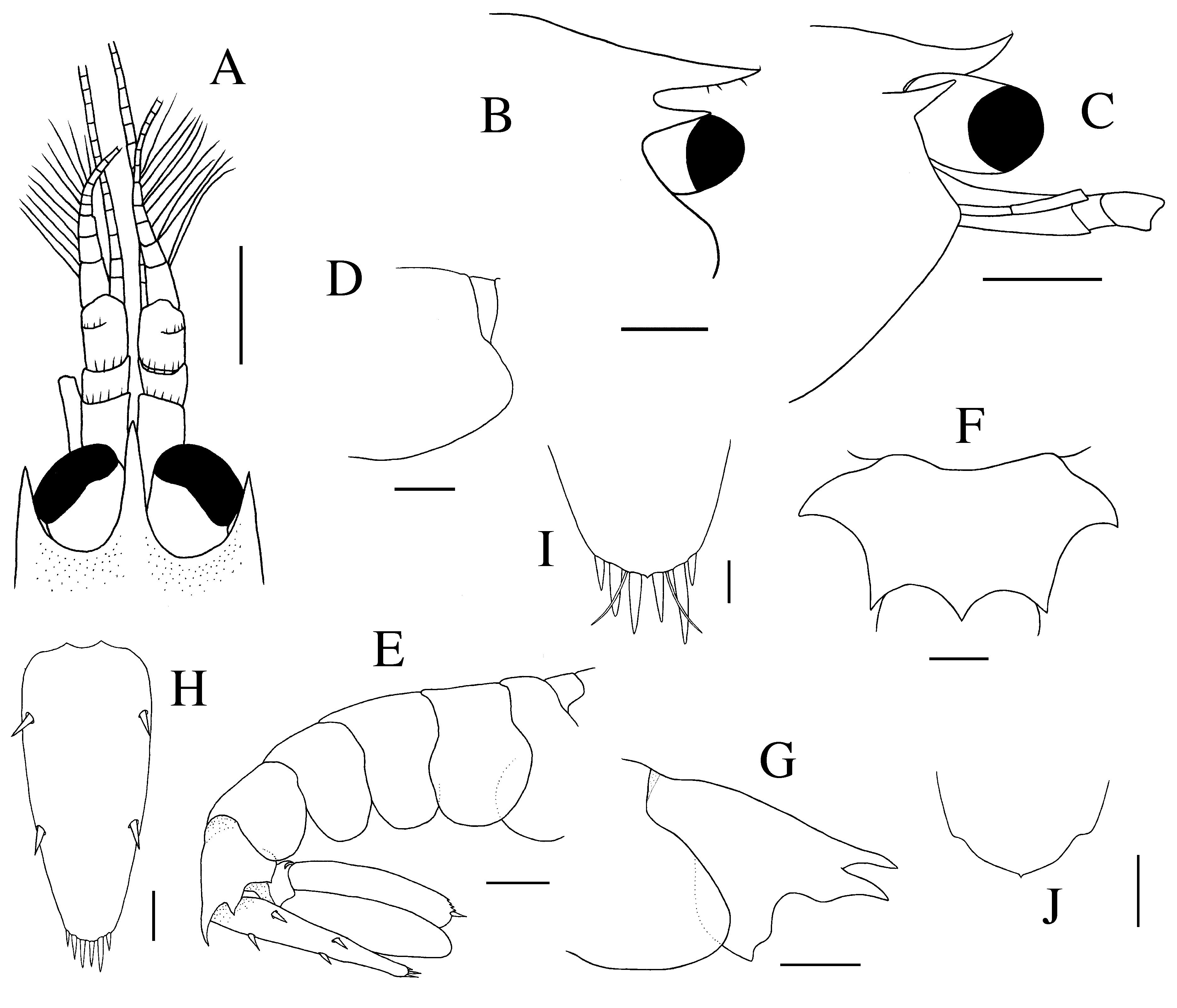
Description. Carapace smooth, glabrous, slightly depressed, longer than deep. Rostrum arrow-shaped in dorsal view ( Fig. 1 View FIGURE 1 A), usually reaching base of cornea and sometimes extending beyond distal margin of eyes and half-length of dorsally visible part of first article of antennular peduncle, straight to slightly curving upwards, tip with single small seta ( Fig. 1 View FIGURE 1 A–C). Paraorbital processes ( Fig. 1 View FIGURE 1 A–C) well developed, usually exceeding halflength of rostrum and half-length of cornea in lateral view, reaching level of most-anterior pterygostomial margin, non-divergent in dorsal view, pointing straight-forward or slightly upward in lateral view ( Fig. 1 View FIGURE 1 A–C). Orbit ( Fig. 1 View FIGURE 1 B, C) feebly demarcated, without distinct infero-orbital angle or antennal spine; pterygostomial margin ( Fig. 1 View FIGURE 1 B, C) slightly produced anteriorly, rounded to somewhat angular. Posteroventral margin of carapace rounded, with slight cardiac concavity ( Fig. 1 View FIGURE 1 D).
Abdomen ( Fig. 1 View FIGURE 1 E) elongate, smooth, glabrous; first five somites ( Fig. 1 View FIGURE 1 E) with rounded pleura; posterodorsal margin of sixth somite ( Fig. 1 View FIGURE 1 F) with strong, sharp, subtriangular, median tooth; posterolateral angle of sixth somite ( Fig. 1 View FIGURE 1 E–G) produced into strong acute tooth, posteroventral angle blunt, not posteriorly produced; first to third and sixth sternites unarmed, fourth and fifth each with low, rounded, median process.
Telson ( Fig. 1 View FIGURE 1 H) moderately slender, about 2.2 times as long as anterior width, posterior margin width about 0.35 times width of anterior margin; lateral margins slightly convex, tapering distally; dorsal surface with two pairs of well-developed spines, each about 0.1 of telson length, inserted at about 0.25 (anterior pair) and 0.6 (posterior pair) telson length; posterior margin ( Fig. 1 View FIGURE 1 I, J) broadly rounded with minute median point; three pairs of spines present as following: small lateral spines and longer intermediate and submedian spines, intermediate being 2.0–2.8 times as long as lateral and 1.3–1.6 times as long as submedian ( Fig. 1 View FIGURE 1 I).
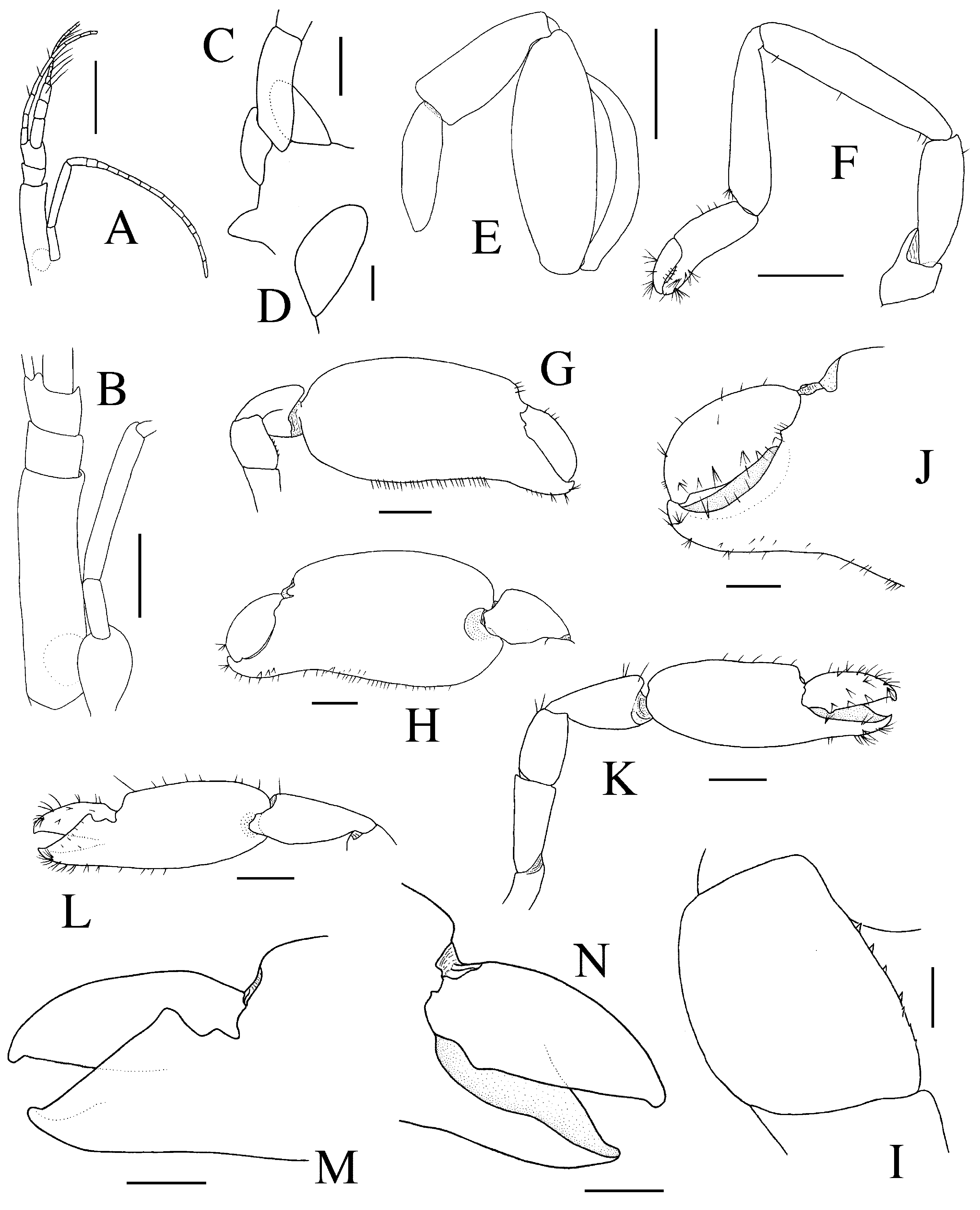
Antennular peduncle ( Figs. 1 View FIGURE 1 A, 2A) slender, with proximal article subcylindrical, 3.8 times as long as distal width; statocyst well-developed, with statolith; stylocerite short, narrow; intermediate article shorter than distal article, their combined length equal to about 0.4 of proximal article length; lateral (= upper) flagellum ( Figs. 1 View FIGURE 1 A, 2A) short, first two articles of rami fused, stout, with 7–8 groups of aesthetascs, short ramus distally obsolete, longer ramus with 5–6 segments; mesial (= lower) flagellum long, with 9–10 segments. Antenna with antennal gland tubercle mesially; basicerite stout, unarmed laterally; scaphocerite ( Fig. 2 View FIGURE 2 C, D) vestigial, about 1.8 times as long as greatest width, terminally obliquely rounded, margins non-setose; carpocerite ( Fig. 2 View FIGURE 2 B) slender, not reaching end of antennular peduncle; flagellum ( Fig. 2 View FIGURE 2 A) short, about 2.6 times as long as carpocerite.
Mouthparts not dissected. Third maxilliped ( Fig. 2 View FIGURE 2 E) with broad antepenultimate article, with both dorsal and ventral margins strongly convex; penultimate article about half as long as antepenultimate article, much more slender, with straight margins, ultimate article most slender, tapering towards apex; exopod well developed.
First pereiopod ( Fig. 2 View FIGURE 2 F) slender; ischium about three times as long as wide, unarmed; merus about five times as long as broad, slightly longer than carpus; carpus subcylindrical, broadening distally, about four times as long as broad and 1.5 times as long as chela; carpo-propodal brush lacking; chela with palm subcylindrical, slightly compressed; fingers robust, subspatulate, about 0.4 of chela length, with feebly bidentate tips.
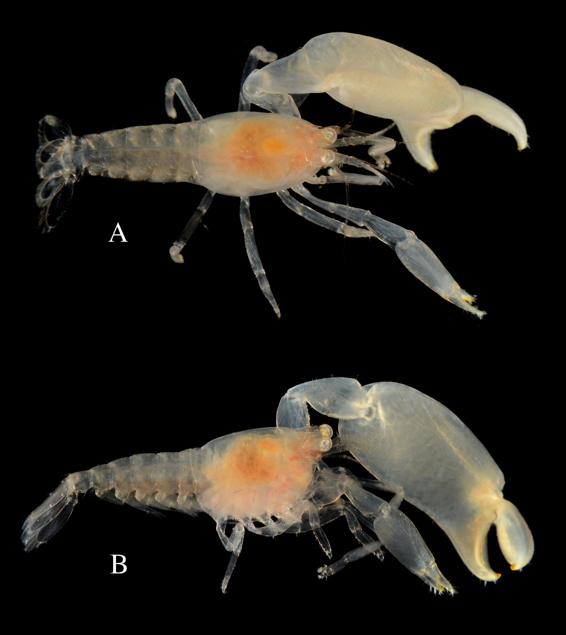
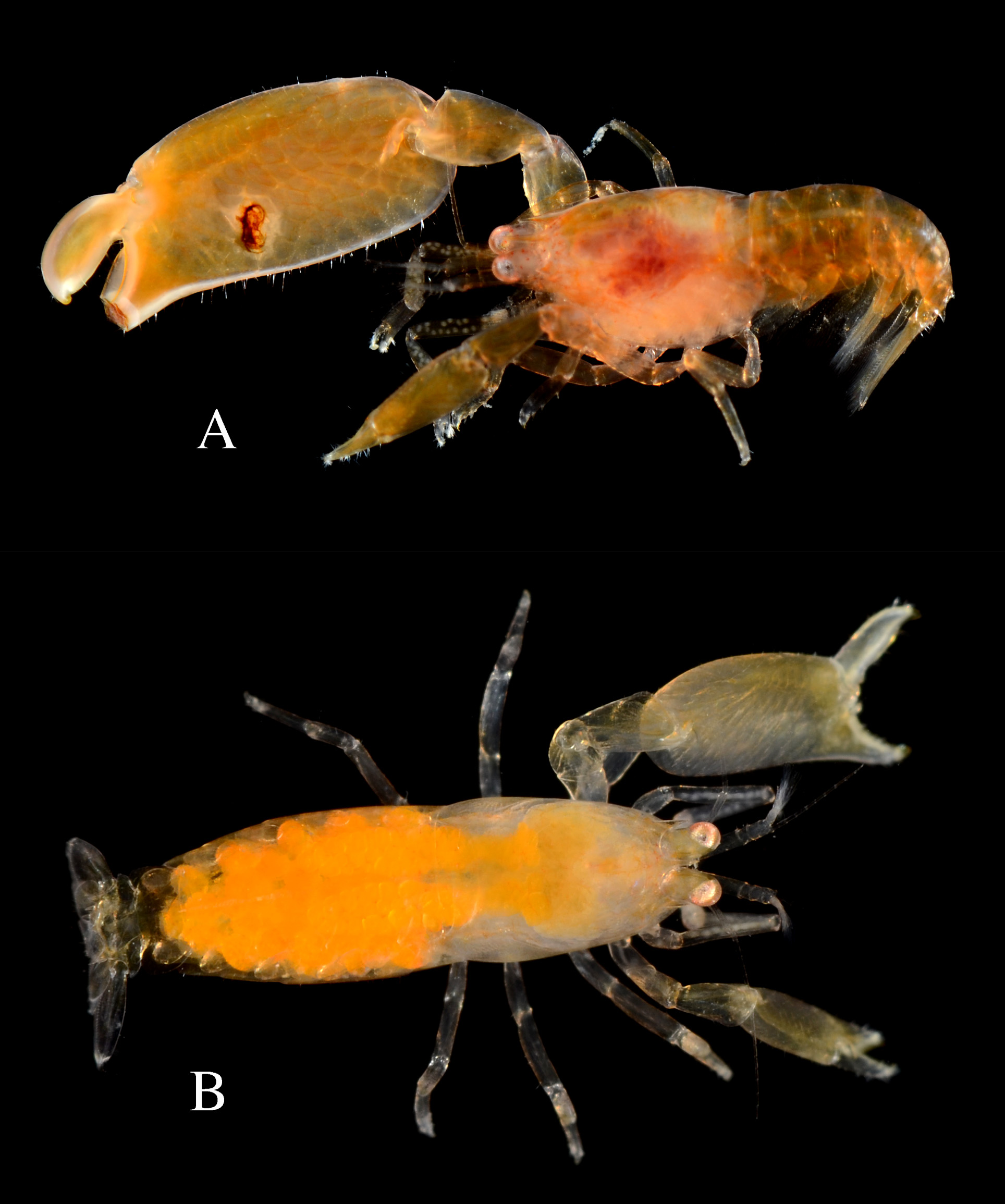
Major second pereiopod ( Figs. 2 View FIGURE 2 G–J, 4, 5) greatly enlarged, much longer and more robust than minor second pereiopod; largest adult males with massive chela equaling combined length of cephalothorax and abdomen ( Figs. 4 View FIGURE 4 , 5 View FIGURE 5 A), females with much smaller chela, equaling or slightly exceeding length of cephalothorax ( Fig. 5 View FIGURE 5 B); ischium subequal to merus in length, unarmed; merus ( Fig. 2 View FIGURE 2 I) about 1.3 times as long as broad, about 0.6 length of carpus and 0.2 length of palm, ventral surface with small, conical, distally more or less sharp teeth; carpus ( Fig. 2 View FIGURE 2 G, H) 0.3–0.4 length of palm, smooth, 1.6 times as long as maximal width, narrow proximally, distally widening, distoventral surface somewhat excavated; chela ( Fig. 2 View FIGURE 2 G, H, J) very large, with palm smooth, slightly compressed laterally, suboval in cross-section, flattened medially, slightly tapering distally, about twice as long as maximal height; dactylus ( Fig. 2 View FIGURE 2 J) laterally compressed, about twice as long as maximal height, about 0.4 times length of palm; tip strongly curved, corneous; cutting edge conspicuously thickened, forming large bulge; pollex about as long as dactylus; tip curved, corneous; surface opposed to dactylus with relatively large and deep groove accommodating dactylar bulge, mesial and lateral margins entire, concave.
Minor second pereiopod variously but always significantly smaller than major second pereiopod ( Figs. 2 View FIGURE 2 K–N, 4, 5); ischium ( Fig. 2 View FIGURE 2 K) about 1.4 times length of merus, unarmed; merus ( Fig. 2 View FIGURE 2 K) 1.5 times as long as broad, about 0.7 of carpal length and 0.4 of palm length, with minute conical teeth on ventral surface; carpus ( Fig. 2 View FIGURE 2 K, L) about 0.7 length of palm, smooth, about twice as long as maximal height, much wider distally; palm ( Fig. 2 View FIGURE 2 K, L) smooth, non-setose, suboval in cross-section, about 1.8 times as long as maximal height; dactylus ( Fig. 2 View FIGURE 2 K–N) about 0.6 length of palm, compressed, with curved tip; cutting edge sharp, with blunt proximal tooth laterally; pollex excavated on surface opposed to dactylus; lateral edge sharp, nearly entire; mesial edge projecting proximally as strong subtriangular tooth; finger tips simple.
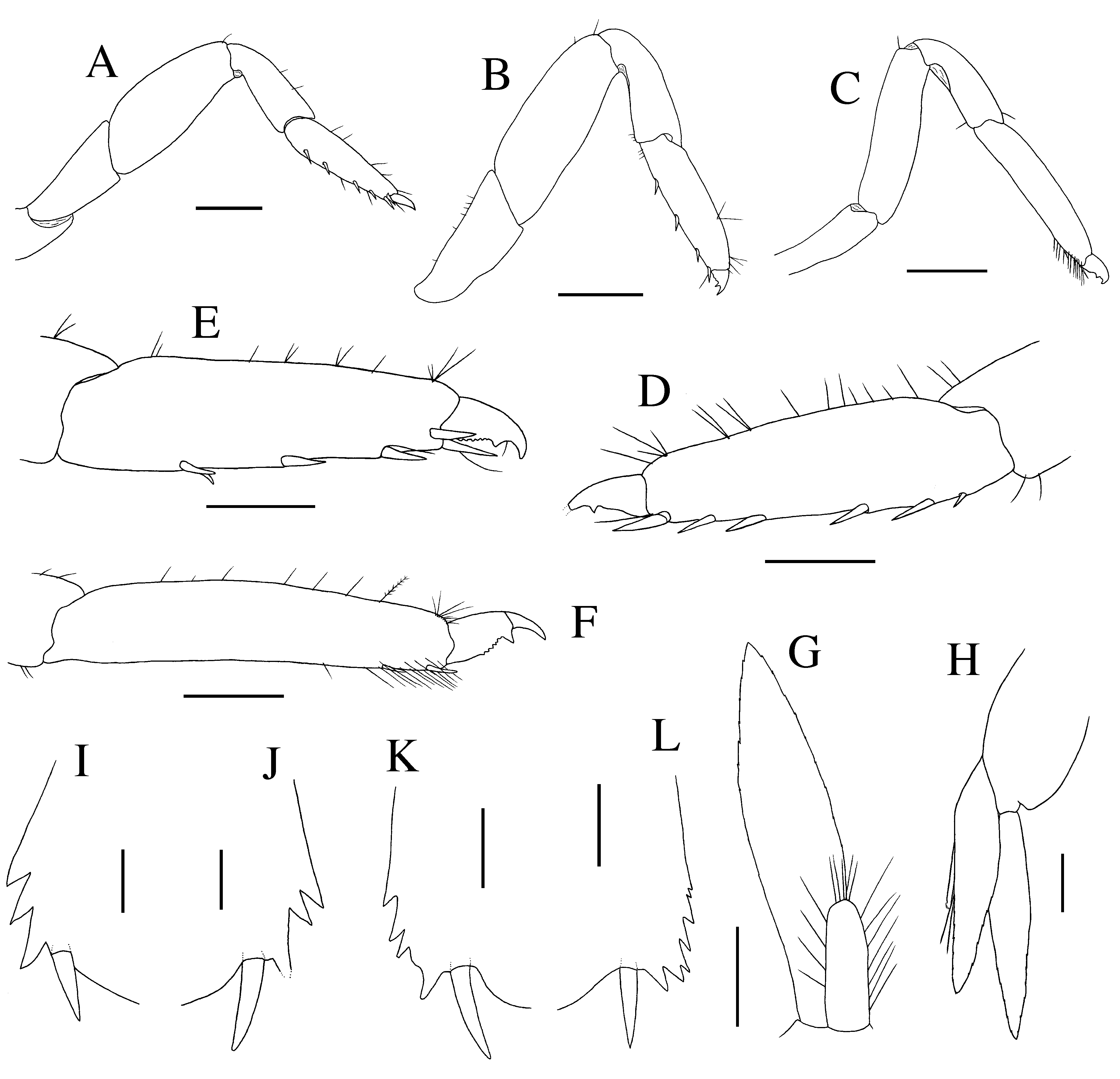
Third pereiopod ( Fig. 3 View FIGURE 3 A, D) stout, with ischium about 0.8 times length of merus, unarmed; merus ( Fig. 3 View FIGURE 3 A) about twice as long as broad and 1.2 times length of carpus, unarmed; carpus ( Fig. 3 View FIGURE 3 A) about 0.9 length of propodus, nearly three times as long as distal width, unarmed; propodus ( Fig. 3 View FIGURE 3 A, D) about 2.8 times as long as wide, ventral margin with four or five irregularly spaced spines, distal pair of stouter spines fringing base of dactylus; dactylus ( Fig. 3 View FIGURE 3 A, D) biunguiculate, compressed, about 0.25 of propodus length and 2.2 times longer than basal width, ventral margin of corpus slightly concave, crenulated, with acute, triangular, ventrally pointing secondary unguis; terminal unguis distinctly demarcated, about half as long as corpus, strongly curved, not crenulated proximally. Fourth pereiopod ( Fig. 3 View FIGURE 3 B, E) generally similar to third pereiopod; merus ( Fig. 3 View FIGURE 3 B) about 2.4 times as long as wide; carpus ( Fig. 3 View FIGURE 3 B) slightly longer than propodus, about three times as long as distal width; propodus ( Fig. 3 View FIGURE 3 B, E) about three times as long as wide; ventral margin with three or four spines and distal pair of stouter spines; dactylus ( Fig. 3 View FIGURE 3 B, E) biunguiculate, nearly twice as long as basal width; ventral margin of corpus conspicuously crenulated, with triangular, ventrally pointing secondary unguis; terminal unguis equal or slightly shorter than half-length of corpus.
Fifth pereiopod ( Fig. 2 View FIGURE 2 C, F) noticeably more slender than third and fourth, with ischium about 0.6 length of merus, unarmed; merus ( Fig. 2 View FIGURE 2 C) about 3.5 times as long as wide and 1.4 length of carpus, unarmed; carpus ( Fig. 2 View FIGURE 2 C) 0.8 times length to propodus, about 3.3 times as long as distal width, unarmed; propodus ( Fig. 2 View FIGURE 2 C, F) about 4.5 times as long as broad; ventral margin with one subdistal and one distal spines; distolateral surface with brush of grooming setae; dactylus ( Fig. 2 View FIGURE 2 C, F) biunguiculate, compressed, about 0.2 of propodus length, 2.3 times as long as basal width; ventral margin of corpus slightly convex, distally conspicuously crenulated, with strong, subacute, ventrally pointing secondary unguis; terminal unguis well demarcated, more slender than that of third and fourth pereiopods, about half-length of corpus, curved, not crenulated proximally.
First male pleopod ( Fig. 3 View FIGURE 3 G) with endopod not reaching half-length of exopod, about three times as long as proximal width, elongate, with rounded tip, lateral margin with four longer plumose setae, apex and mesial margin with several shorter setae. Second male pleopod ( Fig. 3 View FIGURE 3 H) with endopod about 0.8 length of exopod, with appendix interna at about 0.4 length of mesial margin; appendix masculina with corpus obsolete, being essentially represented by two stiff setulose setae protruding beyond appendix interna.
Uropods ( Figs. 1 View FIGURE 1 E, 3I –L) with protopod unarmed; exopod slightly longer than telson, with lateral margin slightly convex, non-setose; distolateral margin ( Fig. 3 View FIGURE 3 I–L) serrated, with 2-6 acute or subacute teeth; distolateral angle with acute tooth, reaching approximately half-length of stout spine; diaeresis inconspicuous; endopod slightly longer than exopod; proximal parts of lateral and mesial margins non-setose.
Variation. The most obvious variable character is the length of the rostrum, which can extend beyond the distal margin of the cornea ( Fig. 1 View FIGURE 1 A, B) or reach only to the middle of the cornea ( Fig. 1 View FIGURE 1 C, 5). The paraorbital processes can be about as long as half-length of cornea ( Fig. 1 View FIGURE 1 A, B) or significantly shorter ( Fig. 1 View FIGURE 1 C, 5A). In addition, both the rostrum and the paraorbital processes are variable in their direction, pointing either straightforward or slightly upward (cf. Fig. 1 View FIGURE 1 B and 1C). The pterygostomial region varies from broadly rounded ( Fig. 1 View FIGURE 1 B) to more angularly protruding ( Fig. 1 View FIGURE 1 C). The number of spines on the ventral margin of the P3 and P4 propodi varies from four to five and three to four, respectively. There is also a significant variation in the number of teeth on the distolateral margin of the uropodal exopod, ranging from 2–6 (not including terminal tooth adjacent to strong spine) ( Fig. 3 View FIGURE 3 I–L).
Colour pattern. Uniform translucent yellowish to pale orange, with some scattered reddish and yellowish chromatophores ( Figs. 4 View FIGURE 4 , 5 View FIGURE 5 ); cornea silvery reddish; ovaries and freshly laid eggs yellow-orange ( Fig. 5 View FIGURE 5 B).
Type locality. Brazil, São Paulo, São Sebastião (Praia do Cabelo Gordo).
Etymology. The new species is named after Fundação de Amparo à Pesquisa do Estado de São Paulo (FAPESP) for supporting studies on the origins, diversity and distribution of the flora and fauna of the State of São Paulo, especially through the Biota-FAPESP Program.
Distribution. Presently known only from São Sebastião and Ubatuba, northern coast of the state of São Paulo, southeastern Brazil.
Biology. Like most if not all species of the genus, T. fapespae sp. nov. appears to be an obligate associate of demosponges. All São Sebastião specimens collected by us were extracted from an unidentified sponge, whereas the male paratype from Ubatuba was found inside Mycale (Zygomycale) angulosa . The female paratype from Ubatuba was extracted from a colony of the bryozoan Schizoporella errata , most likely having escaped from a cryptic sponge growing inside or attached to the bryozoan colony. The specimens collected from the tangled matrix of tubes of the chaetopterid polychaete Phyllochaetopterus socialis and reported as T. gnathophylloides by Nalesso et al. (1995) were likely associated to cryptic sponges. Additional material reported by Duarte & Nalesso (1996) as T. gnathophylloides was also found inside Mycale (Zygomycale) angulosa , suggesting that this sponge may be the main host of T. fapespae sp. nov.
While photographing the São Sebastião specimens of T. fapespae sp. nov., the second author (AA) noticed that most shrimps in an irritated state produced a fairly strong snapping sound, presumably by rapidly closing the major second pereiopod, in a way similar to that of Periclimenaeus Borradaile, 1915 . Typton spongicola , a close eastern Atlantic relative of T. fapespae sp. nov. (see below), is also known to produce snapping sounds ( Johnston 1946). In both species, the pollex of the major chela of P2 is deeply depressed accommodating a large flat bulge of the dactylus ( Fig. 2 View FIGURE 2 J; see also Bruce 2009: fig. 5A, B), a configuration that may be responsible for a mechanically induced snapping sound. Thorough studies determining the exact nature and physical properties of the snapping sound are needed for the snapping species of Typton , but also for Periclimenaeus and several other pontoniine genera.
Remarks. Only three other species of Typton have a strong median tooth on the posterodorsal margin of the sixth abdominal somite, viz. T. hephaestus (eastern Pacific), T. holthuisi (central Atlantic), and T. spongicola (eastern Atlantic). Typton fapespae sp. nov. can be distinguished from the poorly described and illustrated T. hephaestus by the presence of several (2–6) teeth on the distolateral margin of the uropodal exopod, above the terminal tooth adjacent to a stout spine [ Fig. 3 View FIGURE 3 I–L; this margin has no teeth in T. hephaestus (see Holthuis 1951)]. The hammer-shaped dactylus of the major second pereiopod in T. hephaestus provides another character separating the two species. However, both features (uropodal exopod and major P2) were indirectly inferred from the description of T. vulcanus , which was described by Holthuis (1951) as "extremely close", with "the only distinct difference between the two forms [being] the shape of the posterior margin of the sixth abdominal segment". A redescription of T. hephaestus is indeed highly desirable.
The serrated distolateral margin of the uropodal exopod characteristic of T. fapespae sp. nov. is also present in T. spongicola and T. holthuisi . However, T. fapespae sp. nov. differs from these two species by the subquadrate merus of the major second pereiopod bearing a field of small but conspicuous, sharp denticles on the ventral surface ( Fig. 2 View FIGURE 2 E, H); the merus is more rectangular and ventrally unarmed in T. spongicola and T. holthuisi (cf. Bruce 2009; De Grave 2010). Additional distinguishing features of T. fapespae sp. nov. are the more angular pterygostomial region (cf. Fig. 1 View FIGURE 1 C and Bruce 2009: figs. 1, 2A and De Grave 2010: fig. 1A); the stouter dactylus of the major second cheliped (about 2.1 times as long as maximal depth in T. fapespae sp. nov. vs. 2.6 times in T. spongicola and 2.4 times in T. holthuisi ); and the more robust third and fourth pereiopods (e.g., with P3 merus about twice as long as wide in T. fapespae sp. nov. vs. four times in T. spongicola and almost three times in T. holthuisi ). The new species differs specifically from T. spongicola by the lower positioned paraorbital processes (cf. Fig. 1 View FIGURE 1 B, C and Bruce 2009: fig. 2A); the shape of the antennal scaphocerite (cf. Fig. 2 View FIGURE 2 D and Bruce, 2009: fig. 3D); and the stronger median tooth on the posterodorsal margin of the sixth abdominal somite, reaching beyond lateral teeth in T. fapespae sp. nov. vs. not reaching beyond them in T. spongicola (cf. Fig. 1 View FIGURE 1 F, G and Bruce 2009: figs. 1, 3J). Typton fapespae sp. nov. differs specifically from T. holthuisi by the pollex of the minor second pereiopod ending in a simple tip (vs. with bifid tip in T. holthuisi , cf. Fig. 2 View FIGURE 2 J, L and De Grave 2010: fig. 2J); the dactyli of the ambulatory pereiopods (P3–5) furnished with much smaller denticles on the corpus and with an unarmed unguis (vs. with much larger denticles on the corpus and some denticles also on the unguis in T. holthuisi , cf. Fig. 3 View FIGURE 3 D, E and De Grave 2010: fig. 3D, F); the shorter dorsolateral spines of the telson (about 0.10 telson length in T. fapespae sp. nov. vs. 0.14 in T. holthuisi , cf. Fig. 1 View FIGURE 1 H and De Grave 2010: fig. 1I); and the presence (in most specimens) of a minute median tooth on the posterior margin of the telson (absent in T. holthuisi , cf. Fig. 1 View FIGURE 1 J and De Grave 2010: fig. 1J). Regarding its size, T. fapespae sp. nov. (pocl 1.9–3.4 mm) is intermediate between the somewhat smaller T. holthuisi (pocl 1.4–1.5 mm, De Grave 2010) and much larger T. spongicola (pocl of ovigerous females 6.5–7.5 mm, Bruce 2009).
Typton fapespae sp. nov. can be easily distinguished from all other western Atlantic species of Typton , including all species previously reported from Brazilian waters ( T. carneus , T. distinctus , T. gnathophylloides , T. prionurus , T. tortugae ), by the presence of a strong median tooth on the posterodorsal margin of the sixth abdominal somite.
No known copyright restrictions apply. See Agosti, D., Egloff, W., 2009. Taxonomic information exchange and copyright: the Plazi approach. BMC Research Notes 2009, 2:53 for further explanation.
|
Kingdom |
|
|
Phylum |
|
|
Class |
|
|
Order |
|
|
Family |
|
|
Genus |
Typton fapespae
| Almeida, Alexandre O., Anker, Arthur & Mantelatto, Fernando L. 2014 |
Typton gnathophylloides
| Amaral 2010: 249 |
| Duarte 1996: 143 |
| Nalesso 1995: 96 |