
Euplanoida cf. pacificola (Plehn, 1896)
|
publication ID |
https://doi.org/ 10.11646/zootaxa.4700.1.2 |
|
publication LSID |
lsid:zoobank.org:pub:CB832B12-CD89-42A1-90CB-142B7819D912 |
|
persistent identifier |
https://treatment.plazi.org/id/03DD87FF-FFA9-FFE2-A79D-FB96CB33F896 |
|
treatment provided by |
Plazi |
|
scientific name |
Euplanoida cf. pacificola |
| status |
|
Euplanoida cf. pacificola View in CoL
( Figures 2 View FIGURE 2 A–K, 3A–E)
Type locality of nominal species. Valparaiso, Chile ( Plehn 1896: 153–155, pl. 10, Figs 7 View FIGURE 7 , 9 View FIGURE 9 , pl. 13, Fig. 10 View FIGURE 10 ; on bottom of a boat) .
Material examined. Fourteen specimens. Seven specimens as whole mounts: UMAR-PLAT 001, 1 spec. (Panteón Beach, under rocks, 3 m, Oct 28, 2012, coll. MRS); UMAR-PLAT 002, 002A, 2 spec. (Puerto Ángel Beach, under rocks, 3 m, Oct 28, 2012, coll. AR); UMAR-PLAT 003, 1 spec. juvenile (Agua Blanca Beach, under rocks in tidal pools, 0.5 m, Nov, 2013, col. MRS) ; UMAR-PLAT 004, 1 spec. (Camarón Beach, under rocks, 3 m, Dec 6, 2014, coll. ECL) ; UMAR-PLAT 005, 1 spec. (Estacahuite Beach, under rocks, 2 m, Oct 6, 2013, coll. MRS) ; UMAR-PLAT 006, 1 spec. (Yerbabuena Beach, under rocks, 12 m, Oct 9, 2013, coll. MRS) . Four specimens in histology sections: UMAR-PLAT 001A–F, 1 spec. (sagittal sections of reproductive structures in six slides; Panteón Beach, under rocks, 3 m, Oct 28, 2012, coll. AR); UMAR-PLAT 004A–F, 2 spec. (sagittal sections of reproductive structures in six slides); UMAR-PLAT 004G–J, 1 spec. (frontal sections of reproductive structures in four slides; Camarón Beach, under rocks, 0.5 m, Feb 25, 2017, coll. MRS) . Three specimens preserved in 70% ethanol: UMAR- PLAT 002 B (Puerto Ángel Beach, under rocks, 3 m, Oct 28, 2012, coll. AR & MRS) .
Description of external features
Color. In vivo the specimens are translucent, variable color from grayish or beige, to pink, light brown, orange ( Figs 2 View FIGURE 2 A–E); pharynx and intestinal branches are visible and according from the gut content could be varied to white, brown, pink or dark green ( Fig. 2A View FIGURE 2 ). In mature specimens, testes and ovaries are also visible scattered in the parenchyma, both light brown. In ethanol, specimens become white or beige, opaque in appearance.
Body. Body shape elongate or cuneiform ( Figs 2 View FIGURE 2 A–E), with 6–30 mm (n= 14, µ= 15.16 mm, SD= 6.28) long, and 2.2–10 mm (n= 13, µ= 5.86 mm, SD= 2.2) wide; the anterior margin is rounded and the posterior margin is slightly pointed ( Figs 2 View FIGURE 2 C–E).
Tentacles. Without nuchal tentacles.
Eyes. Without marginal eyes; the cerebral eyes are located at 1.3–7.3 mm (n= 5, µ= 3.82 mm, SD= 4.88) from the anterior margin of the body, these are distributed in the periphery of the brain and are arranged in two lines, which surpass the anterior and posterior region of the brain; tentacular eyes clustered in a circle, scarce ( Fig. 2F View FIGURE 2 ) the tentacular eyes become confused with the cerebral eyes as they join together, these are named cerebral-tentacular eyes.
Brain. Bilobed, 0.35 mm (n= 5) of long and wide, presents globuli cell masses of circular–oval shape, that are lodged in the anterior region of the brain ( Fig. 2G View FIGURE 2 ).
Digestive system. Pharynx ( Fig. 2H View FIGURE 2 ) elongated and highly branched, located at the central region of the body, at 1.3–7.3 mm (n= 5, µ= 3.96 mm, SD= 2.22) of distance from the anterior margin; pharynx measures 3–4.6 mm (n= 5, µ= 3.8 mm, SD= 0.66) long, and 0.6–1.6 mm (n= 5, µ= 0.92 mm, SD= 0.41) wide; the mouth in the last third of the pharynx, at a distance from the anterior margin of 6 mm, with 12–14 (n= 5, µ= 13, SD= 0.70) pharyngeal lobes.
Gonopores. Separated, located in the last third of the body. Male gonopore ( Figs 3 View FIGURE 3 A–C, E) at 6–12.7 mm (n= 5, µ= 5.14 mm, SD= 5.34) from the anterior margin and 0.8–3.6 mm (n= 5, µ= 1.15 mm, SD= 1.47) from the pharynx. The female gonopore ( Figs 3 View FIGURE 3 A–B) is positioned posterior to the male gonopore, at 0.05 mm of distance.
Description of internal features
Male reproductive system. Without prostatic vesicle; tripartite oval seminal vesicle with 0.25–0.5 mm (n= 5, µ= 0.22 mm, SD= 0.21) of diameter ( Figs 2K View FIGURE 2 , 3A, D View FIGURE 3 ); thickened spermiducal ducts that connect ventrally with the seminal vesicle ( Figs 2K View FIGURE 2 , 3A View FIGURE 3 ); all of these positioned anterior to the male gonopore; the spermiducal ducts internally present spermiducal oval-shaped vesicles ( Figs 2K View FIGURE 2 , 3 View FIGURE 3 A–D); these ducts and vesicles ascend to the anterior region of the pharynx, then bifurcate in the second third of the pharynx and reach the second third of the Lang’s vesicle.
The seminal vesicle connects to the penis papilla through an ejaculatory duct ( Fig. 3D View FIGURE 3 ); penis papilla conical, slightly pointed and unarmed ( Fig. 3C View FIGURE 3 ), which is 0.1 mm long and 0.05 mm wide; male atrium ( Fig. 3C View FIGURE 3 ) 1 mm long and 0.25 mm wide, and its walls are delimited by a glandular epithelium.
Female reproductive system. With cement glands ( Fig. 3A View FIGURE 3 ) around the female gonopore; a short female atrium directly connected to the vagina ( Fig. 3B View FIGURE 3 ), oriented towards the anterior region of the body, slightly scalloped and delimited by a ciliary epithelium; the vagina is connected by means of a Lang’s duct ( Figs 3B, E View FIGURE 3 ) with the very elongated Lang’s vesicle ( Figs 2I View FIGURE 2 , 3 View FIGURE 3 A–B, E), reaching 1–2.2 mm (n= 7, µ= 1.17 mm, SD= 0.76) of length and 0.2–0.85 mm (n= 7, µ= 0.26 mm, SD= 0.33) in width; it is located posteriorly to the female gonopore and oviduct; the latter is located around the pharynx.
Habitat. Littoral to sublittoral (12 m); under rocks, associated with green seaweed, bryozoans and chitons. Can be found in groups of 2– 4 specimens per rock (MRS personal observation).
Distribution. Euplanoida cf. pacificola was found at Camarón, Puerto Ángel, Estacahuite and Yerbabuena beaches, Oaxaca. Is the first record of the genus for the southern Mexican Pacific ( Fig. 12 View FIGURE 12 ).
Taxonomic remarks. This family includes nine genera: Euplana Girard, 1893 ; Diplopharyngeata Plehn, 1896 ; Semonia Plehn, 1896 ; Aprostatum Bock, 1913 ; Taenioplana Hyman, 1944 ; Euplanina Sopott-Ehlers & Schmidt, 1975 ; Euplanoida Faubel, 1983 , Paraprostatum Faubel & Sluys, 2007 and Namyhplana Brusa & Damborenea, 2013 . The specimens of this study were determined as belonging to the genus Euplanoida by the presence of a seminal vesicle, an unarmed penis papilla, a Lang’s vesicle and the absence of nuchal tentacles, marginal and frontal eyes ( Faubel 1983).
Euplanoida has six species: the type species, E. pacificola ( Plehn, 1896) , described from Valparaiso, Chile, has been recorded on the Peruvian coasts and in the Gulf of California ( Hyman, 1953a); E. elioti ( Laidlaw, 1903a) , described from Kenya and/or Zanzibar, Eastern Africa; E. malayana ( Laidlaw, 1903b) , described from Strait of Malacca, Indonesia; E. panangensis ( Laidlaw, 1903b) , described from Palau Island, Malaysia; E. concolor ( Meixner, 1907) , described from Musha Island, Djibouti and E. tropicalis ( Hyman, 1954) , described from Kapoho, Hawaii.
The specimens of the present study were determined as E. cf. pacificola , because of the presence of a prominent tripartite seminal vesicle of 0.25–0.5 mm (n= 5, µ= 0.22 mm, SD= 0.21) of diameter; symmetrical duct of spermiducal vesicles and spermiducals vesicles, both positioned ventrally to the oviduct and anteriorly to the male gonopores; a short penis papilla (L= 0.1 mm, W= 0.05 mm), unarmed; very elongated Lang’s vesicle (L= 1.16 mm); cerebral and tentacular eyes form join as cerebral-tentacular eyes; absence of tentacles, marginal and frontal eyes ( Figs 2 View FIGURE 2 A–K, 3A–E).
The separation between Euplanoida cf. pacificola and the nominal species is difficult, mainly by the ambiguous information of the original description, the possible artifices in the process of fixation and/or stages of life of the specimens, and the color variations between the specimens reviewed by Plehn (1896), Hyman (1953a) and the present study (see Table I View TABLE I ).
Type specimens of Euplanoida pacificola were collected on the keel of the boat, “am Kiel des Schiffes haftend”, from Valparaiso, Chile, and also recorded in the Peruvian coasts ( Plehn 1896). The specimens from Perú were determined as a variation of E. pacificola , according to Plehn (1896: 153). Later, Hyman (1953a) recorded this species from Guaymas, Sonora, and Punta San Marcial, Baja California Sur, determining it as a Mexican variation of the nominal species. Notwithstanding morphological differences observed in the hermaphroditic reproductive system and the geographical distance of the topotypical locality, Hyman (1953a) considered that it did not justify the separation of the taxon into more species, since the Chilean taxon was collected from the bottom of a boat in Valparaíso, and hence not necessarily native to the locality.
The conspicuous character in Euplanoida cf. pacificola is the presence of a tripartite seminal vesicle and the symmetrical distribution of the spermiducal ducts and vesicles and the absence of a bursa copulatrix (this only present in the Chilean specimen); both characters are mentioned as present in the Peruvian and Mexican variations of E. pacificola but the arrangement of them is unknown ( Plehn 1896, Hyman 1953a). The illustration of the complete Chilean specimen ( Plehn 1896: pl. 10, Fig. 7 View FIGURE 7 ) of E. pacificola outlines the presence of a bursa copulatrix; however, their description does not mention said structure. Likewise, Hyman (1953a) does not say anything about this character, nor her opinion about the discrepancy in their ecological differences between temperate-cold waters from Chile versus tropical waters from Gulf of California. Therefore, the exhaustive revision of the holotypes recommended, as well as the variations determined by Plehn (1896) and Hyman (1953a).
In this study five color variations of Euplanoida cf. pacificola were observed ( Fig. 2 View FIGURE 2 A–E); they could be mainly attributed to the bathymetric distribution of the species (0.5–12 m), since no visible differences were found in the morphology.

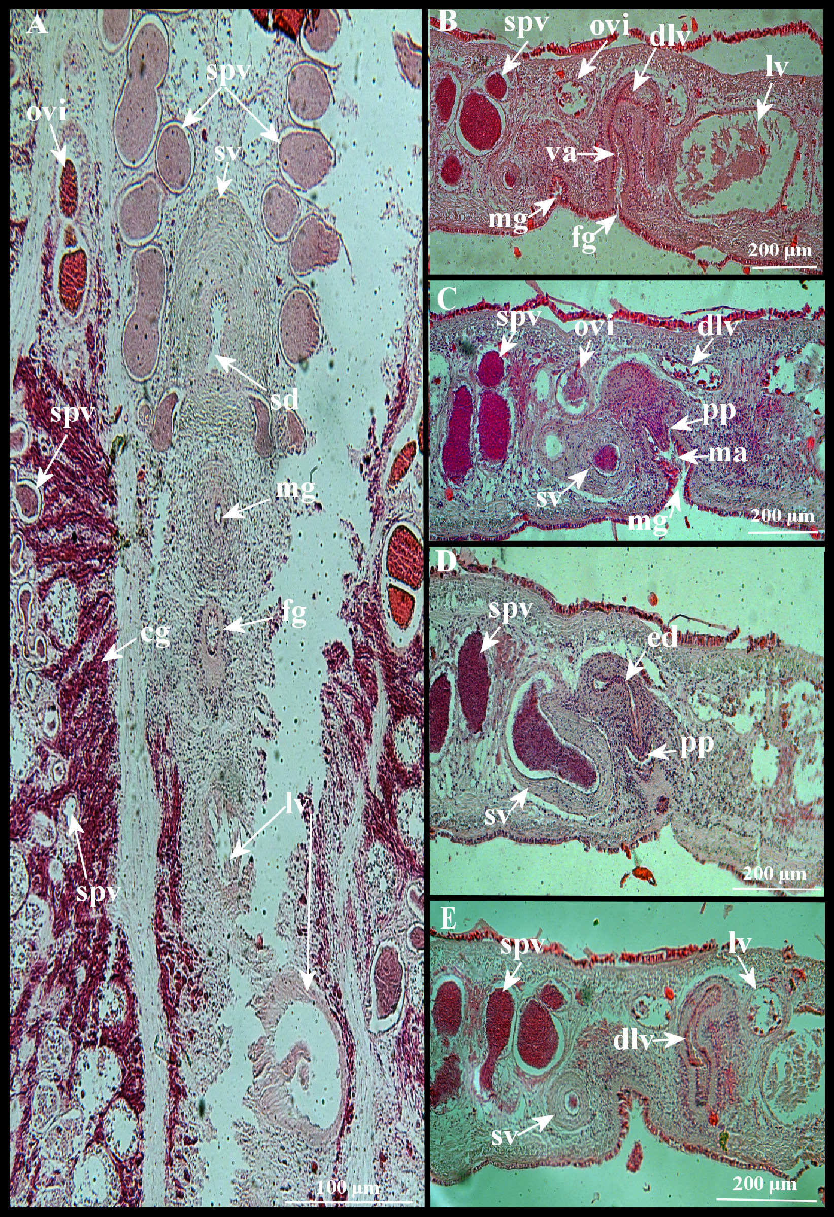
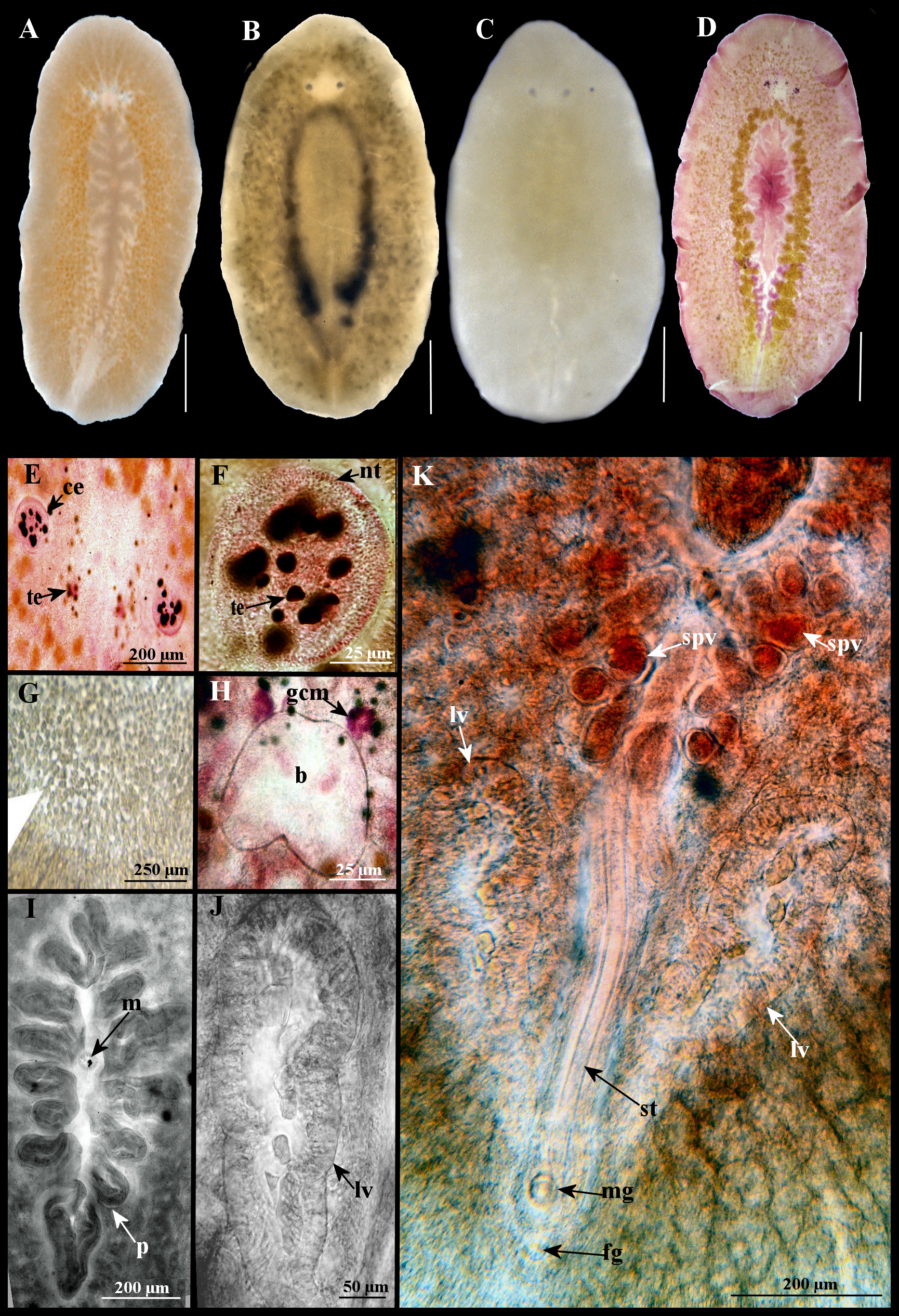
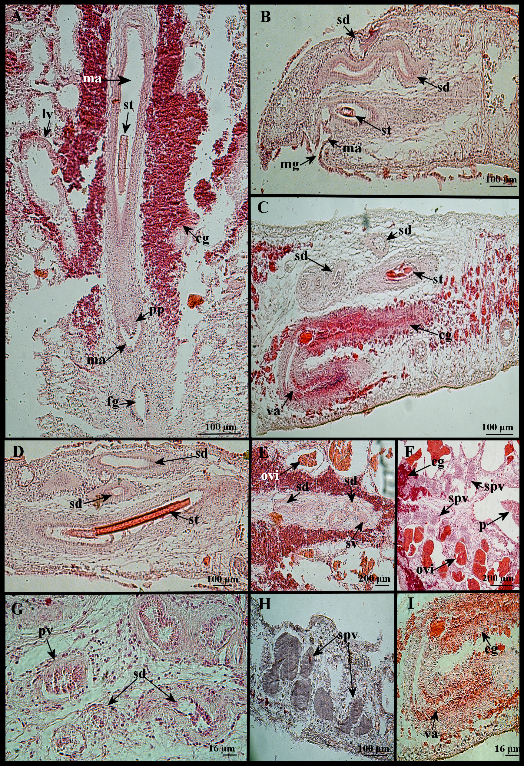
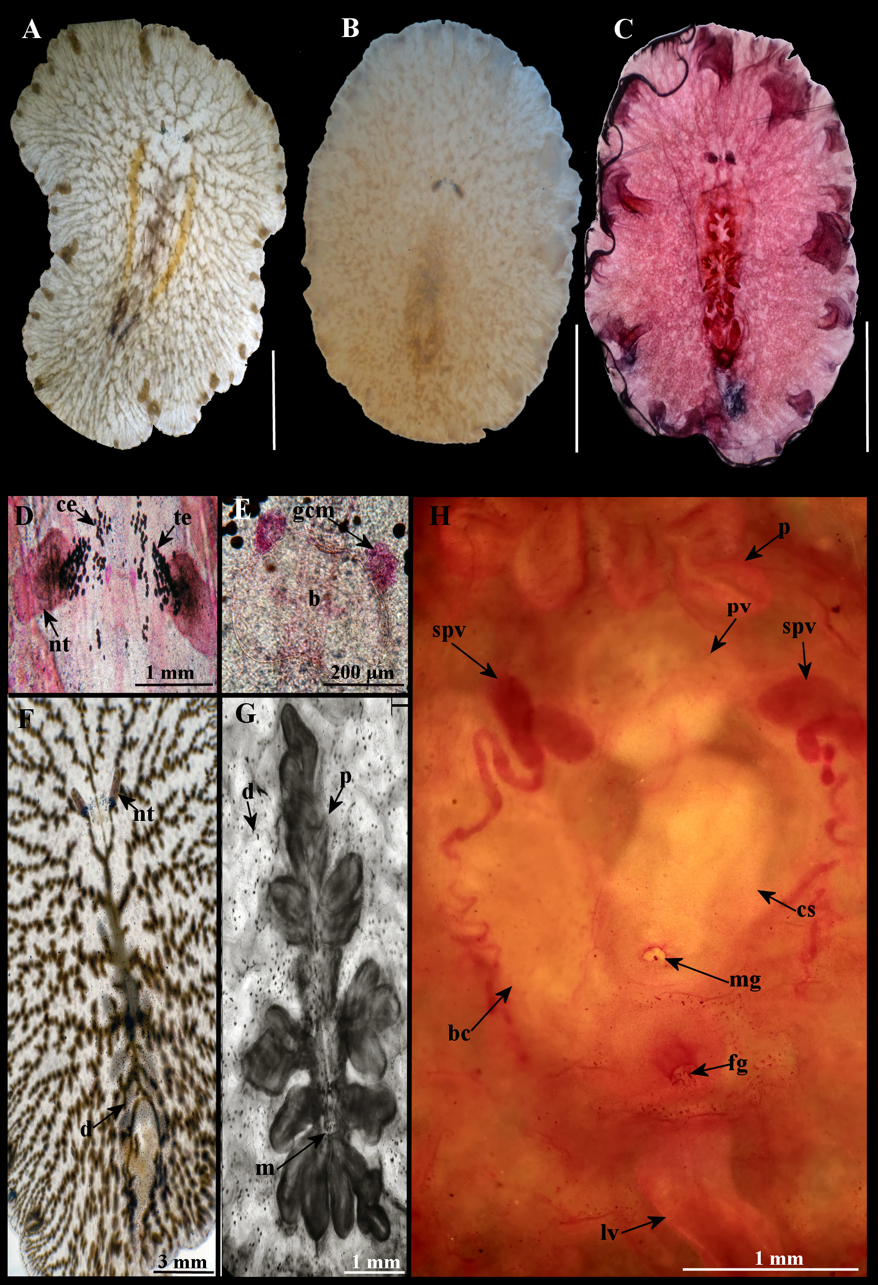
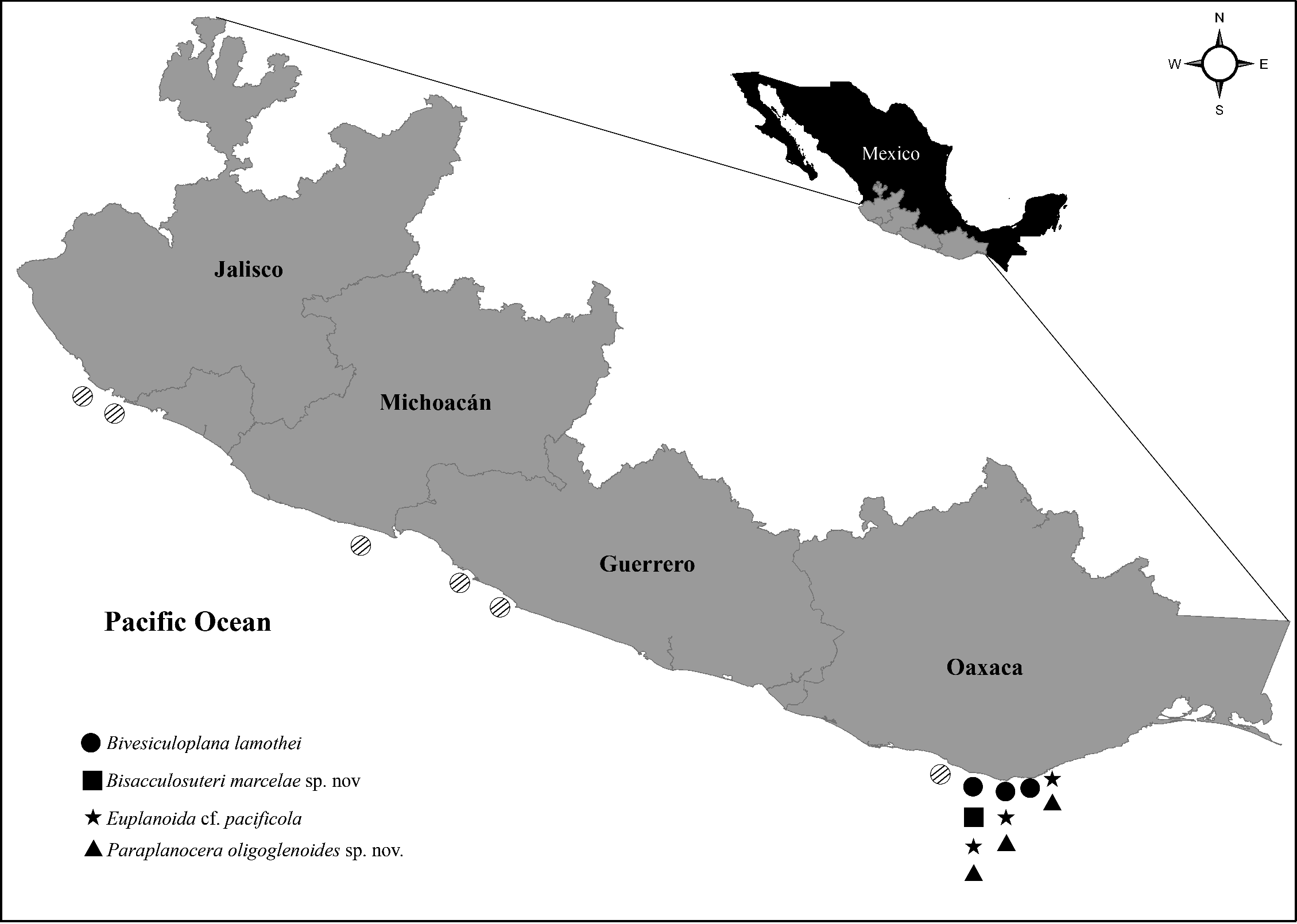
TABLE I. Comparison of the non-matching characters of Euplanoida pacificola nominal species with respect variants of the species and Euplanoida cf. pacificola.
| Taxa | Euplanoida pacificola | E. pacificola | E. pacificola | Euplanoida cf. pacificola |
|---|---|---|---|---|
| Locality | Chile | Perú | México | Oaxaca |
| References | Plehn 1896: 153–155, pl. 10, Figs 7, 9, pl. 13, Fig. 10; Bock 1913: 220 | Plehn 1896: 153–155, pl. 10, Fig. 8; Bock 1913: 220 | Hyman 1953a: 331–333, Figs 87–89 | This study: Figs 2A–K, 3A–E |
| Locality | Valparaiso, Chile (Type locality) | Peruvian coast | Gulf of California | Oaxaca (Camarón, Puerto Ángel, Estacahuite and Yerbabuena) |
| Habitat | Bottom of a boat | - | Under rocks | Under rocks |
| Cerebral and tentacular eyes distribution | Cerebral eyes distributed anterior to the brain and the tentacular eyes, without forming an arrangement of cerebral-tentacular eyes | - | Cerebral eyes distributed anterior and posterior to the brain and to the tentacular eyes, forming an arrangement of cerebral-tentacular eyes | Cerebral eyes distributed anterior and posterior to the brain and to the tentacular eyes, forming an arrangement of cerebral-tentacular eyes |
| Mouth positioned in the pharynx | In the second third of the pharynx | In the end of the last third of the pharynx | In the end of the last third of the pharynx | In the end of the last third of the pharynx |
| Gonopores | Separated | The male gonopore is so close to the female gonopore those give the appearance of being a common gonopore | The male gonopore is so close to the female gonopore that gives the appearance of being a common gonopore | Separated |
| Bursa copulatrix | “Presented” | - | - | Absent |
| Lang’s vesicle morphology | Lang’s vesicle oblong approximately the same length as the seminal vesicle | Lang’s vesicle elongate. approximately twice the length of the seminal vesicle | Lang’s vesicle elongated, approximately half of the pharynx and fourfold the length of the seminal vesicle | Lang’s vesicle elongated, approximately one third of the pharynx and fivefold the length of the seminal vesicle |
No known copyright restrictions apply. See Agosti, D., Egloff, W., 2009. Taxonomic information exchange and copyright: the Plazi approach. BMC Research Notes 2009, 2:53 for further explanation.