
Odontaspis noronhai Maul, 1955
|
publication ID |
https://doi.org/ 10.11646/zootaxa.5094.1.3 |
|
publication LSID |
lsid:zoobank.org:pub:EBD00C94-0E82-4DC9-9D3E-D1868F23F6A6 |
|
DOI |
https://doi.org/10.5281/zenodo.6301180 |
|
persistent identifier |
https://treatment.plazi.org/id/03DE5B49-FF9E-FFE6-FF1E-FD8A5B51158A |
|
treatment provided by |
Plazi |
|
scientific name |
Odontaspis noronhai Maul, 1955 |
| status |
|
Odontaspis noronhai Maul, 1955 View in CoL
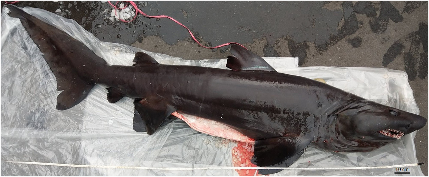
( Fig. 2 View FIGURE 2 ; Table 1 View TABLE 1 )
Material examined. EBFS 02001 , 312 cm TL, female, ca. 25°25’N, 124°10’E, 100 m, landed at NanFangAo fishing port, northeastern Taiwan, northwestern Pacific Ocean , 27 December 2019 GoogleMaps .
Description. Morphometric data is given in Table 1 View TABLE 1 . Body bulky, conical. Dorsal ridges absent from midline of the dorsal side. Caudal peduncle plump, height at anal fin insertion 4.48 % of TL. Lateral keels or ridges absent. Upper precaudal pit present, deeply depressed. Head length around 94 % of pectoral-pelvic space. Head short and moderately pointed, outline in lateral view slightly depressed dorsally above eyes; dorsal profile gently tapering. Snout moderately long and conical, preoral length 62.8 % of mouth width; snout tip rounded in dorsoventral view. Eyes large, horizontal diameter 6 % of head length, without nictitating eyelids. Eyes located closely to the dorsal surface; interorbital width 24.1 % of head length. Spiracle small, located behind and at the lower border of orbit. Five gill slits similar in height, 23.3 %–23.8 % of head length. Margins of all gill slits strongly concave. Gills opening large, gill filaments visible from outside. Nostrils with moderately large elliptical incurrent aperture, with short anterior nasal flap, and excurrent aperture small and suboval. Internarial space broad, distance between inner corners of nostrils 46 % of mouth width. Mouth arched and large, extending well behind posterior eye. Lower jaw margin concave at symphysis. Mouth length 51.7 % of width, mouth width 37.6 % of head length. Tongue large. Labial furrows absent.
Pectoral fins medium-sized, subtriangular, somewhat falcate. Anterior margin strongly curved, apices rounded; posterior margin weakly concave; free rear tip slightly pointed; inner margin straight, length of base 50.5 % of pectoral fin length. Fin origin located beneath the fifth gill slit. Pelvic fins large and triangular, not falcate. Anterior margin straight, apices rounded; posterior margin concave; free rear tip pointed; inner margin short, inner margin length 28 % of pelvic fin length; pelvic fins area larger than two dorsal fins. First dorsal fin relatively small, low and subtriangular, not falcate. Anterior margin curved, apex rounded; posterior margin weakly concave; free rear tip subangular; inner margin straight; height equal to the length of posterior margin, 72 % of dorsal fin length; origin of first dorsal posterior to the pectoral fin free rear tip, but not reaching pectoral fin insertion. Second dorsal fin small, shape very similar to the first dorsal fin; length of different margins somewhat proportional to the first dorsal fin, length 64.7 % of first dorsal fin length, length of base 48.3 % of first dorsal fin base length, length of posterior margin 76.3 % of first dorsal fin posterior margin length, height 75 % of first dorsal fin height. Origin of second dorsal fin over pelvic inner margin, insertion closer to pelvic insertion than anal fin origin. Interdorsal space 17.8 % of TL. Anal fin small, subtriangular, area slightly smaller than second dorsal fin. Shape similar to second dorsal fin, with curved anterior margin, apex bluntly rounded, posterior margin weakly concave, free rear tip more acutely pointed. Caudal fin long, broadly lobed, asymmetrical. Terminal lobe short, with large, long and triangular ventral lobe; dorsal caudal margin straight, preventral margin curved, ventral tip rounded; lower and upper postventral margin straight, posterior notch not obvious; subterminal notch shallow, subterminal margin straight, terminal margin moderately concave, posterior tip rounded.
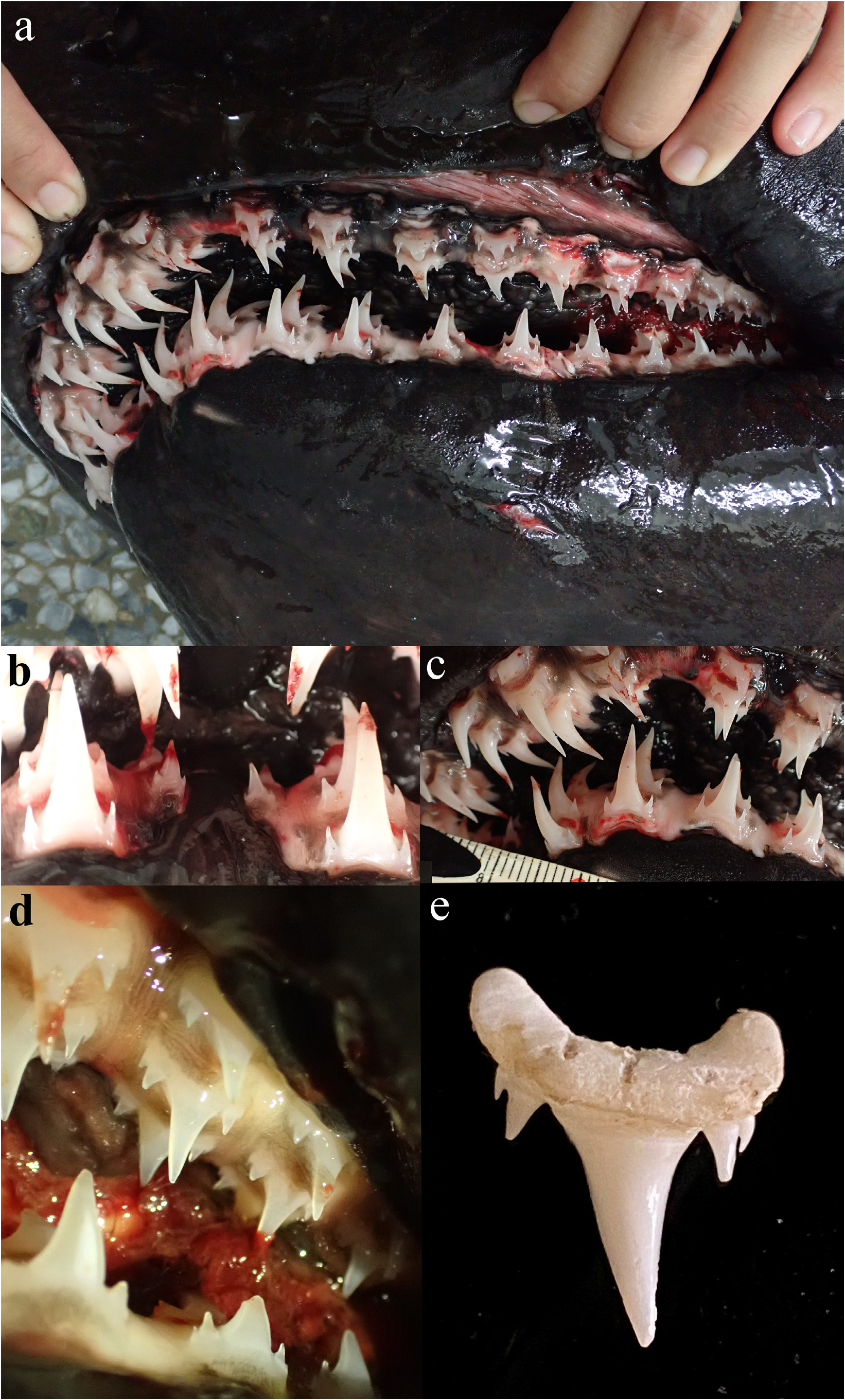
Teeth from upper and lower jaw, 29/29 rows, 58 total rows in both jaws, three to four series functional. Dental formula is given in Table 2 View TABLE 2 . Lateral view of the left jaw is shown in Fig. 3a View FIGURE 3 .
Tooth shape between the left and the right side similar. The following description was based on the left side of the jaw. Upper jaw: no symphyseal tooth, two anterior teeth and one intermediate tooth on each side; anterior teeth larger than other teeth, protruded outwards, with long and slender crown and one or two cusplets on each side of the cusps; crown height of the first anterior tooth 13.2 mm; intermediate teeth slightly smaller than anterior teeth, shape similar to anterior teeth but less protruded, with two cusplets on the left side and one cusplet on the right side of the cusp; lateral teeth less slender and protruded, gradually decrease in size from anterior teeth, with one or two cusplets on each side of cusps. The length of the inner cusplet around 1.5–2 times longer than length of outer cusplet; inner cusplets thick, hook-like or protrude outwards, outer cusplets thinner than inner cusplets, hook-like in shape. Lower jaw: one parasymphyseal tooth on each side ( Fig. 3b View FIGURE 3 ), two anterior teeth on each side ( Fig. 3c View FIGURE 3 ), no intermediate tooth; parasymphyseal tooth smaller than other teeth, with slender crown; morphological characters and sizes of anterior and lateral teeth shape mostly similar to upper jaw, except the last four lateral teeth with less slender but more triangular crown ( Fig. 3d View FIGURE 3 ). Anterior and lateral teeth with one or two cusplets on each side of cusps.
Body color uniform black, without any spots on body and blotches on fin edges. Tongue with irregular-shaped black patches. Eyes green when fresh.
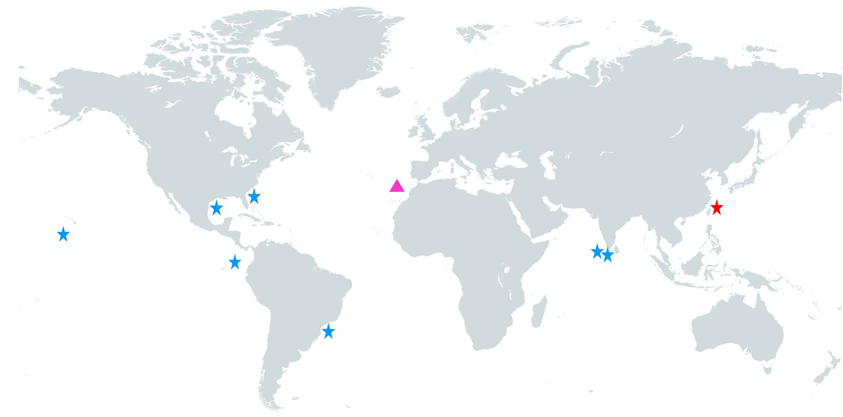
Distribution. The known distribution of O. noronhai is demonstrated in Table 3 View TABLE 3 and Fig. 4 View FIGURE 4 . This species has a wide distribution circumglobally, mainly inhabiting the mesopelagic zone at depths 100–800 m, near oceanic islands and in open oceans.
Biology. Branstetter & McEachran (1986) found cephalopods and teleosts inside the stomach of one specimen. Morón et al. (1998) found stings of stingrays inserted in the lower jaw cartilages. This species is presumably a top predator, feeding on elasmobranchs, teleosts and cephalopods.
DNA. Through comparing the COI sequence obtained in this study with other O. noronhai sequence from India ( Bineesh et al. 2017) in NCBI database (accession numbers: KF899559 View Materials , KF899560 View Materials , KF899561 View Materials ) using BLASTN ( Zhang et al. 2000), revealed the similarity were all 99.67 % respectively, indicating that our specimen belongs to O. noronhai . The newly acquired COI sequence has been uploaded to NCBI GenBank (accession number: MZ317278 View Materials ).
No known copyright restrictions apply. See Agosti, D., Egloff, W., 2009. Taxonomic information exchange and copyright: the Plazi approach. BMC Research Notes 2009, 2:53 for further explanation.
|
Kingdom |
|
|
Phylum |
|
|
Class |
|
|
Order |
|
|
Family |
|
|
Genus |