
Dipavali petri, (RUSSELL, 1964)
|
publication ID |
https://doi.org/ 10.1111/j.1096-3642.2011.00787.x |
|
persistent identifier |
https://treatment.plazi.org/id/03DE8792-FF8E-656C-FF58-FBECFD77FA42 |
|
treatment provided by |
Marcus |
|
scientific name |
Dipavali petri |
| status |
|
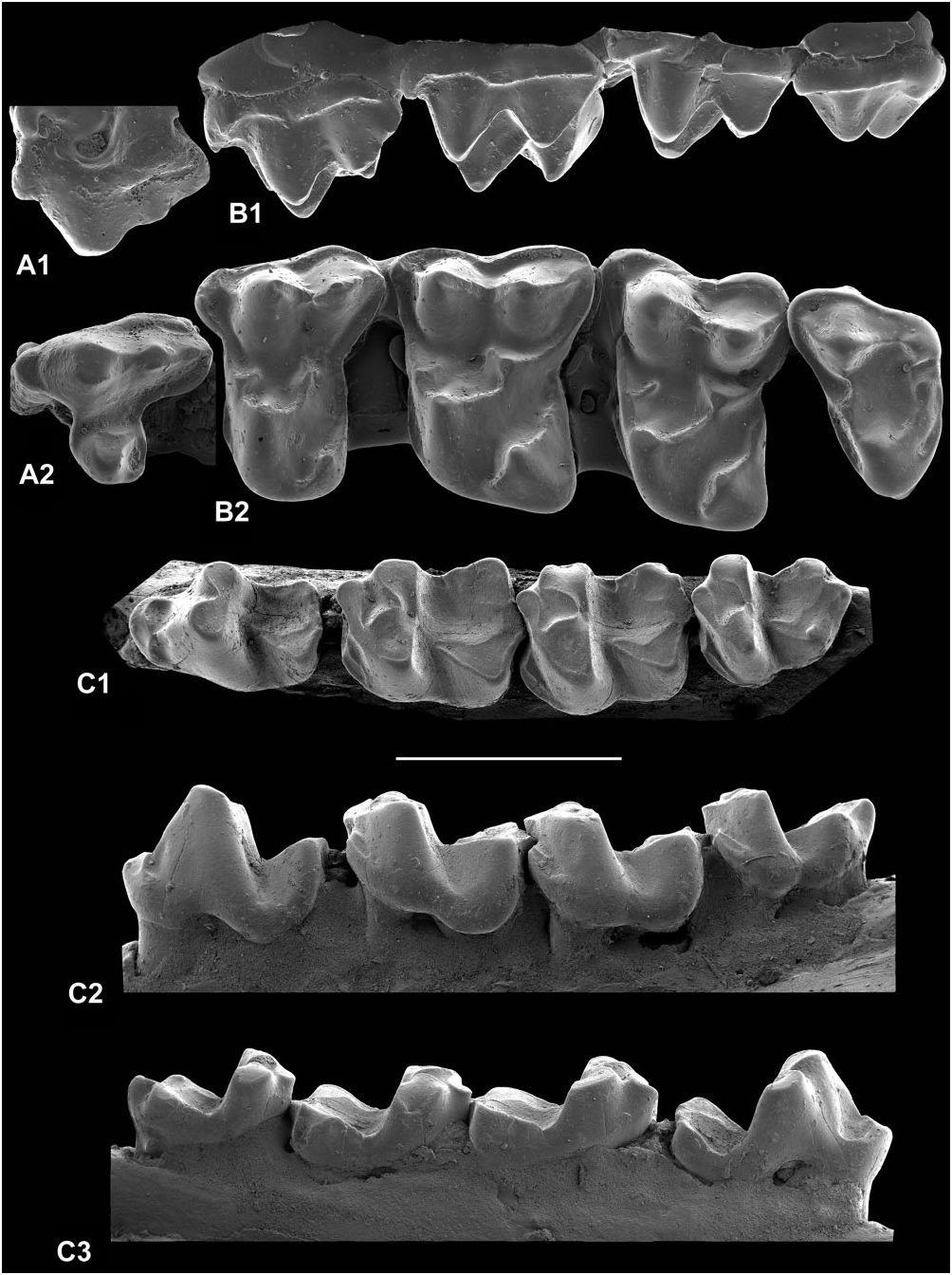
DIPAVALI PETRI ( RUSSELL, 1964) ( FIGS 15, 16 View Figure 16 , 19O)
*vp 1964 paschatherium petri Russell : pp. 239–241, 305, pl. 14, figs 3–5.
*vnon 1964 paschatherium petri Russell : p. 305 (CRL.825, 827).
v. 1964 Paschatherium sp. ; Russell, p. 242, pl. 14, fig. 6.
v. 1967 Paschatherium petri Russell ; Russell, Louis & Poirier, p. 853.
v. 1978 Dipavali petri (Russell) ; Van Valen, p. 61.
Holotype: Left dentary fragment with M 2 and alveoli for M 1, M 3, MNHN.F.CR-142, Cernay.
Paratypes: Those referred specimens also from Cernay ( Russell, 1964: 305–306, pl. 14, figs 3–5), except: the left dentary, M 2–3 (MNHN.F.BR-1198), not then identified to species; left DP 4 (MNHN.F.CR- 4397) here made paratype of Louisina marci sp. nov.; P 4 (MNHN.F.CR4400) and four M 3 s [MNHN.F.CR- 237, CR-1146, CR-1251, UCMP.62023 (CR-1104)], here made the holotype and paratypes respectively of Te. brisswalteri sp. nov.; M 2 (MNHN.F.CR-1126) here made paratype of B. pellouini gen. et sp. nov.; dentary with P 4 –M 2 (MNHN.F.CRL-825) here made paratype of W. girardi gen. et sp. nov.; and dentary with P 4 –M 3 (MNHN.F.CRL-827) here referred to Pa. dolloi (q.v.).
New material: LP 3, MNHN.F.CR-4390; three RP 3 s, MNHN.F.CR-329, CR-1137, CR-115-Bn; eight LP 4 s, MNHN.F.CR-633, CR-15938(Collier 837), CR-65-Ph, CR-885-Ph, CR-366-Pn, CR-67-Bn, CR-282-L, CR-55- MD; 15 RP 4 s, MNHN.F.CR-279, CR-1107, CR- 1176, CR-1207, CR-124-Pn, CR-359-Pn, CR-1728-Pn, CR-337-Ph, CR-338-Ph, CR-218-Bn, CR-283-L, CR-285-L, CR-286-L, CR-82-MD, CR-86-MD; five LM 1 s, MNHN.F.CR-64-Pn, CR-283-Bn, CR-696-Ph, CR-295-L, SLP.CR-404; nine RM 1 s, MNHN.F.CR-628, CR-4519, CR-15935(Collier 834), CR-86-Pn, CR-280- Bn, CR-292-L, CR-293-L, CR-294-L, CR-17481-L; five LM 2 s, MNHN.F.CR-217, CR-11897, CR-692-Ph, CR-297-L, CR-1418-Pn; 13 RM 2 s, MNHN.F.CR-218, CR-11895, CR-13775, CR-89-Ph, CR-107-Ph, CR-296-L, CR-298-L, CR-299-L, CR-300-L, CR-301-L, CR-1143-Pn, SLP.CR-319, CR-323; RM 3, MNHN.F.CR- 941; two LDP 3, MNHN.F.CR-535, CR-599; RDP 3, MNHN.F.CR-224; RDP 4, MNHN.F.CR-1438-Pn; RP 2, MNHN.F.CR-38-Pn; five LP 3 s, MNHN.F.CR-595, CR-4372, CR-101-Ph, CR-144-Ph, CR-325-Ph; eight RP 3 s, MNHN.F.CR-283, CR-303, CR-508, CR-1184, CR-13759, CR-15954(Collier 553), CR-49-Pn, CR-327- Ph; two L dentaries, P 4 -M 1, MNHN.F.CR-62-De, CR-73-MD; six LP 4 s, MNHN.F.CR-299, CR-519, CR-72-Pn, CR-689-Ph, SLP.CR-8, CR-398; six RP 4 s, MNHN.F. CR-1304, CR-4523, CR-17-Pn, CR-1462-Pn, CR-398-Ph, SLP.CR-345; ten M 1 s, MNHN.F.CR-53-Pn, CR-315-Pn, CR-317-Pn, CR-1134-Pn, CR-1711-Pn, CR-302-L, CR-303-L, CR-304-L, CR-305-L, C-418-W; eight RM 1 s, MNHN.F.CR-4514, CR-25-Pn, CR-46-Pn, CR-368-Pn, CR-1393-Pn, CR-1716-Pn, SLP.CR-316, CR-401; eight LM 2 s, MNHN.F.CR-342, CR-56-Pn, CR-1713-Pn, CR-15-Bn, CR-308-L, CR-309-L, SLP.CR- 318, CR-348; eight RM 2 s, MNHN.F.CR-86-Ph, CR-375- Ph, CR-356-Pn, CR-362-Pn, CR-1139-Pn, CR-306-L, CR-310-L, SLP.CR-322; seven LM 3 s, MNHN.F.CR-239, CR-11972, CR-324-Pn, CR-1153-Pn, CR-1732-Pn, CR-1761-Pn, CR-325-L; four RM 3 s, MNHN.F.CR-87- Pn, CR-1735-Pn, CR-326-L, CR-336-L; L dentary, DP 3–4, MNHN.F.CR-14167; LDP 3, MNHN.F.CR-590- Ph; all Cernay.
Two LP 4 s, MNHN.F.BRL-127-P, BRL-3-F; RM 1, MNHN.F.BRL-3-G; two LM 2, MNHN.F.BRL-88-P, BRL-202-P; RM 2, MNHN.F.BR-17480-L; R dentary, P 1, P 3–4, MNHN.F.BRL-9959; LP 4, MNHN.F.BRL- 9960; RM 1, MNHN.F.BRL-4531; RM 2, MNHN.F. BRL-13862; LM 3, MNHN.F.BRL-13861; all bed 4, Berru.
Associated RM 1–3, MNHN.F. BR-15695–7; three RM 1, MNHN.F.BR-2-L, BR-4-L, I-272; two LM 2 s, MNHN.F.I-587, I-690; LM 3, MNHN.F.BR-14; two LP 3 s, MNHN.F.I-668, I-672; LP 4, MNHN.F.I-671; two RP 4 s, MNHN.F.BR-1-Ph, BR-9-Bn; LM 1, MNHN. F.I-667; RM 1, MNHN.F.I-673; L dentary, M 2–3, MNHN.F.BR-1198; R dentary, M 2–3, MNHN.F.BR-101; RM 2, MNHN.F.BR-36-L; RM 3, MNHN.F.I-669; RDP 3, MNHN.F.BR-79; all bed 5, Berru.
LP 3, MNHN.F.R-202-Lass; RP 4, MNHN.F.R-1174; Cernay or Berru.
RP 4, MNHN.F.RILLY-24-Lr; RM 2, MNHN.F.RILLY- 14-Lr; RDP 4, MNHN.F.RILLY-12-L; all Rilly.
Casts in MNHN.F: LP 3, CR-8-Rob; LP 4, CR-21-Rob; two LM 2 s, CR-170-Lx, CR-87-Lass; four RM 2 s, CR-278-Bn, CR-20-Ro, CR-950-Pn, CR-1143-Pn; RP 3, CR-85; two RP 4 s, CR-271, CR-4-BW; two LM 1 s,
CR-134-Lx, CR-200-Lx; LM 2, CR-148-Lx; RM 2, CR-85; LM 3, CR-149-Lx; RM 3, CR-1060-Pn; all Cernay.
RM 2, BRL-103-CGH; bed 4, Berru.
Age and distribution: Sables de Châlons-sur-Vesle supérieurs, Cernay and Berru (beds 4, 5), Calcaire de Rilly, Rilly, France, late Thanetian, Late Palaeocene.
Emended diagnosis: As for genus, monotypic.
Description
Previously, only molars have been attributed ( Russell, 1964). P 3–4, DP 4, P 1–4, and DP 3–4 are newly recognized.
P 3–4: The outline of P 4 is broadly hourglass-shaped to triangular, with or without pre- and postflexi ( Fig. 15E, C 2 View Figure 2 ). With either shape, the buccal lobe is longer than the lingual lobe. The paracone is the dominant cusp, with smaller metacone than paracone, both somewhat bulbous, and a small hypocone may be present ( Fig. 15C, E) or absent. When present, the hypocone can be either the lingual termination of the postcingulum ( Fig. 15E) or form on the distal protocone crest ( Fig. 15C 2 View Figure 2 ). There is no postprotocrista, as on M 1. The preprotocrista joins the paracingulum and there is often a tiny paraconule. Presence and strength of an ectocingulum is very variable. Sometimes there is a tiny cusp developed in the central valley ( Fig. 15C 2 View Figure 2 ) between the metacone and hypocone (when present). MNHN.F.CR-15938 appears pathological ( Fig. 15A). The axes running between the paracone and protocone on the one hand and the paracone and metacone on the other subtend an obtuse instead of the normal right angle. There is a hypocone between the postcingulum and the protocone. In the enlarged distal space, there is a large supernumary cusp with distal crest that meets a distal cingulum and resembles a second more lingually situated metacone and postmetacrista. A distal interstitial facet distal of the more buccal (true?) metacone indicates the position of the succeeding M 1.
Five teeth that differ from P 4 in having a tiny protocone scarcely differentiated from the lingual cingulum are identified as P 3 ( Fig. 15B). Like P 4, they have a small metacone on the distal flank of the larger paracone, which is distally recurved. They occlude well with P 3.
M 1: The dominant condition is to have the large hypocone joined to the protocone by a crest and the postprotocrista lacking, leaving an isolated metaconule, whose only crest is a premetaconule crista ( Fig. 15D 2F View Figure 2 ). Variants show a thin crest joining the metaconule to the hypocone and in one case a vestige of the postprotocrista. The postcingulum and metacingulum are confluent. The distal margin is usually straight, but sometimes there is a shallow postflexus ( Fig. 15F) and in one case a distal bulge ( Fig. 15D 2 View Figure 2 ). The preprotocrista is confluent with the preparaconule crista, whether or not a paraconule is present (small if present and without postparaconule crista). The preparaconule crista may be confluent with the paracingulum or may not reach it. The precingulum is confluent with the paracingulum. The ectocingulum is usually interrupted round the paracone ( Fig. 15D, F) and often forms a tiny cingular mesostyle.
M 2: The metacone is much smaller than the paracone and the distal part of the buccal edge of the tooth is contracted lingually ( Fig. 15D). The ectocingulum is complete on most specimens. The outline is trapeziform rather than rectangular/square as on M 1. It is also relatively shorter than M 1, although the proportions are variable. The pattern of cresting is essentially as on M 1, but in some specimens there is a weak postprotocrista and no crest linking the protocone and hypocone ( Fig. 15L) and in one case both crests are lacking. Also as on M 1, occasionally there is a thin crest between the metaconule and hypocone ( Fig. 15D 2 View Figure 2 ).
M 3: One specimen (MNHN.F.BR-15697) is associated with an M 2 through matching interstitial facets, so is unequivocally identified ( Fig. 15D). The buccal margin is very oblique and the metacone is situated slightly lingual of the median plane of the tooth. The metacone is much smaller than the paracone and there is a large parastyle and a simple trigon with a paraconule and a small metaconule, both without cristae, except for the preparaconule crista that joins the paracingulum. There is a complete distal cingulum, but no precingulum.
Other less securely identified isolated M 3 s are similar, but show some precingulum development. Nevertheless, they share with MNHN.F.BR-15697 the extreme lingual position of the metacone.
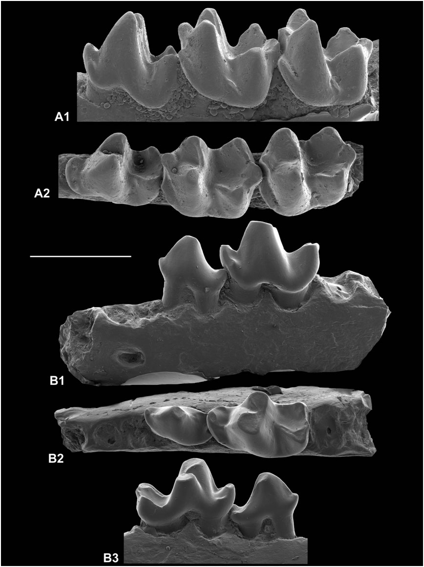
P 1–3: A right dentary (MNHN.F.BRL-9959) has P 1, P 3–4, and alveoli for a two-rooted P 2 ( Fig. 15G). P 1 is a simple tooth with a large distally recurved protoconid, a gently sloping paracristid with no paraconid, and a single small but prominent talonid cusp. An isolated lower premolar of similar morphology but with a low rounded paraconid and slightly larger size is identified as P 2 ( Fig. 15H). Its roots fit the relevant alveoli in the dentary. P 3 is similar to P 2, but larger, with a more centrally placed protoconid and with a small basined talonid, with at most only two (sometimes only one) barely differentiated cusps. The paraconid is variable too, sometimes being absent and never more than a low blunt swelling at the mesial end of the paracristid.
P 4: Unlike the other lower premolars, there is a metaconid that is variable in size, although always smaller than the protoconid, and a basined talonid wider than the trigonid, with small hypoconid and entoconid and sometimes also a tiny hypoconulid largely subsumed by the postcristid ( Fig. 15G, N). A cristid obliqua dips steeply at first mesially, then ascends the back of the protoconid. The paraconid is very low and blunt. In a few specimens there is a short precingulid ( Fig. 15N 1 View Figure 1 ), unlike the other premolars where cingula are lacking.
M 1: A paraconid is lacking, although the paracristid is present and joins mesially an evenly sloping premetacristid ( Fig. 15I, N 1 View Figure 1 ). The protoconid, metaconid, hypoconid, and entoconid are almost equal in size, the talonid being only slightly lower than the trigonid ( Fig. 15N 2 View Figure 2 , N 3 View Figure 3 ). The cristid obliqua dips fairly steeply to the base of the trigonid back wall, buccal of the midline. The protocristid is deeply notched. The trigonid is longer than the talonid. An entoconulid is usually present on the pre-entocristid ( Fig. 15I, N 2 View Figure 2 ). The postentocristid is consistently weak, gently notched, and, in occlusal view, concave distally. The hypoconulid is situated consistently buccal of the midline. The precingulid varies in how far round the protoconid it extends.
M 2: This tooth differs from M 1 in having a shorter, wider, and more open trigonid and a talonid that is shorter, narrower, and lower than the trigonid ( Fig. 15J, M, O). Cingular development is more extensive buccally, sometimes extending from the front of the paracristid almost uninterrupted to the back of the hypoconid. The entoconulid is lacking in most specimens. In other respects the two tooth types are very similar.
M 3: This differs from M 2 in having a mesially bowed paracristid and a narrow tapering talonid ( Fig. 15K, O). The hypoconulid is lingually situated close to the entoconid and may be fused to it. There is some similarity to the equivalent-sized B. pellouini , but the trigonid cusps are lower, the talonid cusps are subsumed by crests, and the paracristid recurves to join the metaconid. There is nevertheless variation in how far distally the hypoconulid projects and in the extent of the cingulum buccally, from just a short precingulid to extending as far as the hypoconid.
Deciduous premolars: DP 4 is trapeziform in outline, tapering mesially to a very large parastyle ( Fig. 15Q). The metacone is slightly lower than the paracone. The paraconule is negligible on a strong preprotocrista that joins the paracingulum. The precingulum is weak to lacking. There is a partial distal ectocingulum. The metaconule is isolated from the large hypocone, but in one specimen a crest links it to the middle of the lingually deflected centrocrista. The distal cingulum dips basally as in the other upper cheek teeth. Three teeth are identified as DP 3. They are wedge-shaped in outline, widening distally ( Fig. 15P). They have a main paracone and a smaller metacone. There is a swelling of the lingual cingulum distally but no hypocone. They are identified as deciduous because of the prominent parastyle. They vary slightly in the size of the parastyle and the distance between the paracone and metacone.
A left dentary fragment allows accurate identification of DP 3–4 ( Fig. 15R). The position of the posterior mental foramen below the middle of the more distal tooth is important in this regard (see below). DP 3 is elongate with a relatively low distally recurved protoconid and smaller but occlusally projecting paraconid and talonid. The talonid is slightly basined with a large hypoconid and small entoconid. Two main crests lie on the buccal and lingual sides of the distal wall of the protoconid, with a fainter one between. The lingual crest has a kink part way down, judged to represent a metaconid. The DP 4 is much more complex, with a talonid almost as developed as on M 1, but with a cristid obliqua meeting and ascending the trigonid back wall far buccally. There is a metaconid nearly as tall as the protoconid, with an intervening deeply notched protocristid. The trigonid is greatly elongated with a weak, gently sloping paracristid and large detached slightly basined paraconid.
Dentary: Six dentary fragments with permanent cheek teeth provide additional information. There are two mental foramina. The more anterior is preserved only in BRL-9959, where it lies below P 2 ( Fig. 15G 2 View Figure 2 ). The position of the more posterior one varies. In three specimens it is below the middle of P 4 ( Fig. 15G 2 View Figure 2 ), whereas in two it is below the distal half of P 4 and in the sixth it is between the junction of P 4 and M 1. The most distal position is associated with a nearly unworn M 2, whereas in one of the examples of the most anterior position the M 1 is heavily worn. However, it is uncertain if there is a consistent signal of anterior ontogenetic migration as the foramen is below the middle of DP 4 in the immature dentary ( Fig. 15R 3 View Figure 3 ). The position of this foramen given in the generic diagnosis reflects the median. Like Walbeckodon and Pa. dolloi , the horizontal ramus does not deepen posteriorly behind M 3 ( Russell, 1964: pl. 14, fig. 6).
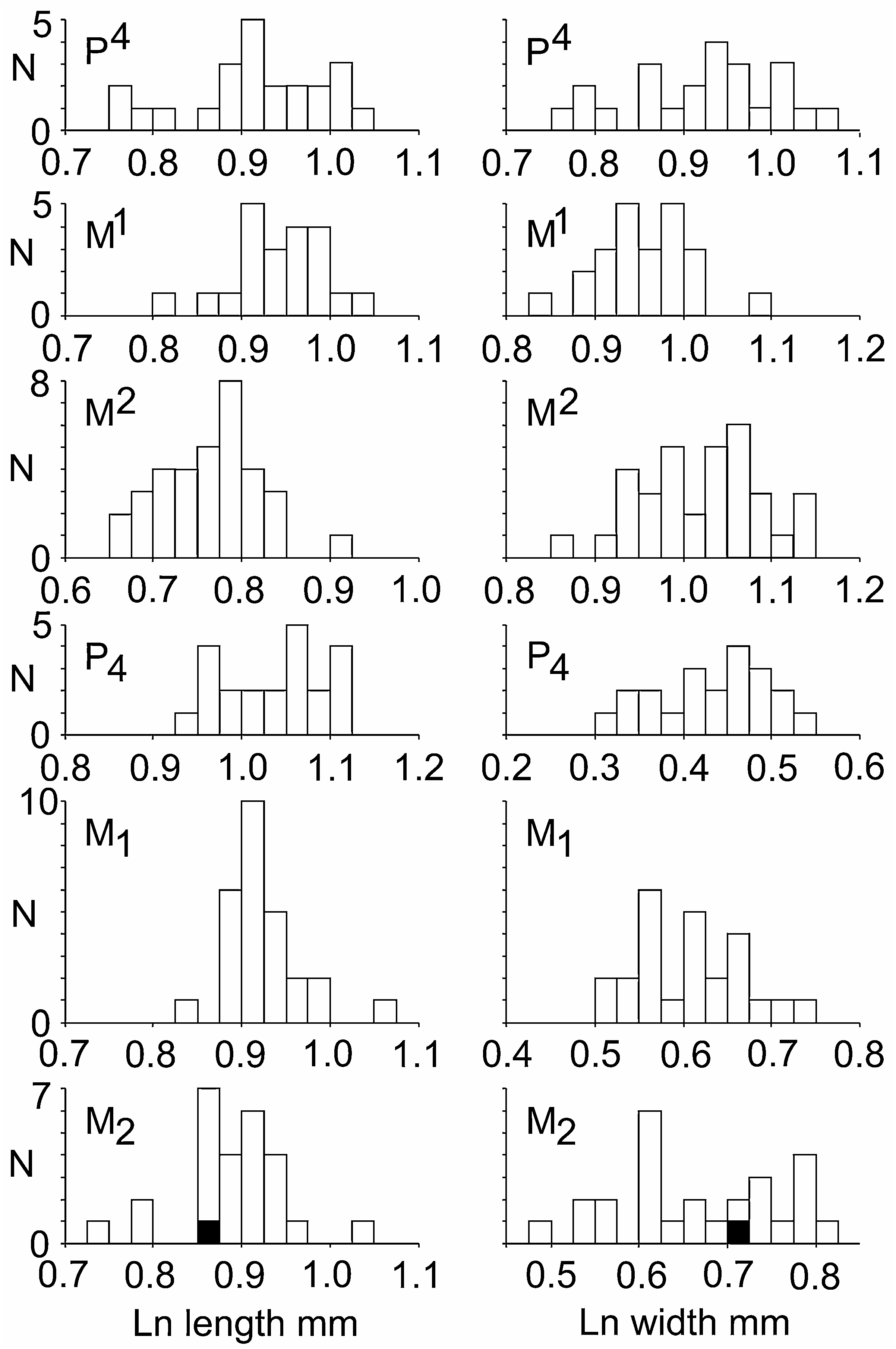
Discussion: This species is variable morphologically and dimensionally. The specimen with the largest M 2 (MNHN.F.CR-1198: Fig. 15O), although originally referred to the species, was also suggested to belong
OR, observed range; SD, standard deviation; V, coefficient of variation (SD as percentage of the mean).
to a larger but unnamed species of Paschatherium ( Russell, 1964: 242, pl. 14, fig. 6), the genus to which D. petri was then referred. Despite the apparently large size variation, the length and width dimensions of at least the first and second molars produce unimodal histograms ( Fig. 16 View Figure 16 ) and the length and width dimensions of the first and second molars and P 4 produce coefficients of variation indicative of a single species ( Table 5). Only P 4 has larger coefficients of variation. These are comparable to those found in the large sample of P 4 of Te. reisi by Tabuce et al. (2006).

| MNHN |
Museum National d'Histoire Naturelle |
No known copyright restrictions apply. See Agosti, D., Egloff, W., 2009. Taxonomic information exchange and copyright: the Plazi approach. BMC Research Notes 2009, 2:53 for further explanation.