
Kodaianella mua, Constant & Pham, 2024
|
publication ID |
https://doi.org/10.5852/ejt.2024.919.2407 |
|
publication LSID |
lsid:zoobank.org:pub:72B7907F-F901-4DDD-B2DD-63D03253837E |
|
DOI |
https://doi.org/10.5281/zenodo.10568746 |
|
persistent identifier |
https://treatment.plazi.org/id/5BECEAE4-B7C2-4138-BC82-F16E0D4A0B07 |
|
taxon LSID |
lsid:zoobank.org:act:5BECEAE4-B7C2-4138-BC82-F16E0D4A0B07 |
|
treatment provided by |
Plazi |
|
scientific name |
Kodaianella mua |
| status |
sp. nov. |
Kodaianella mua sp. nov.
urn:lsid:zoobank.org:act:
Figs 1 View Fig , 2A View Fig , 24–25 View Fig View Fig
Diagnosis
Kodaianella mua sp. nov. can be recognized by
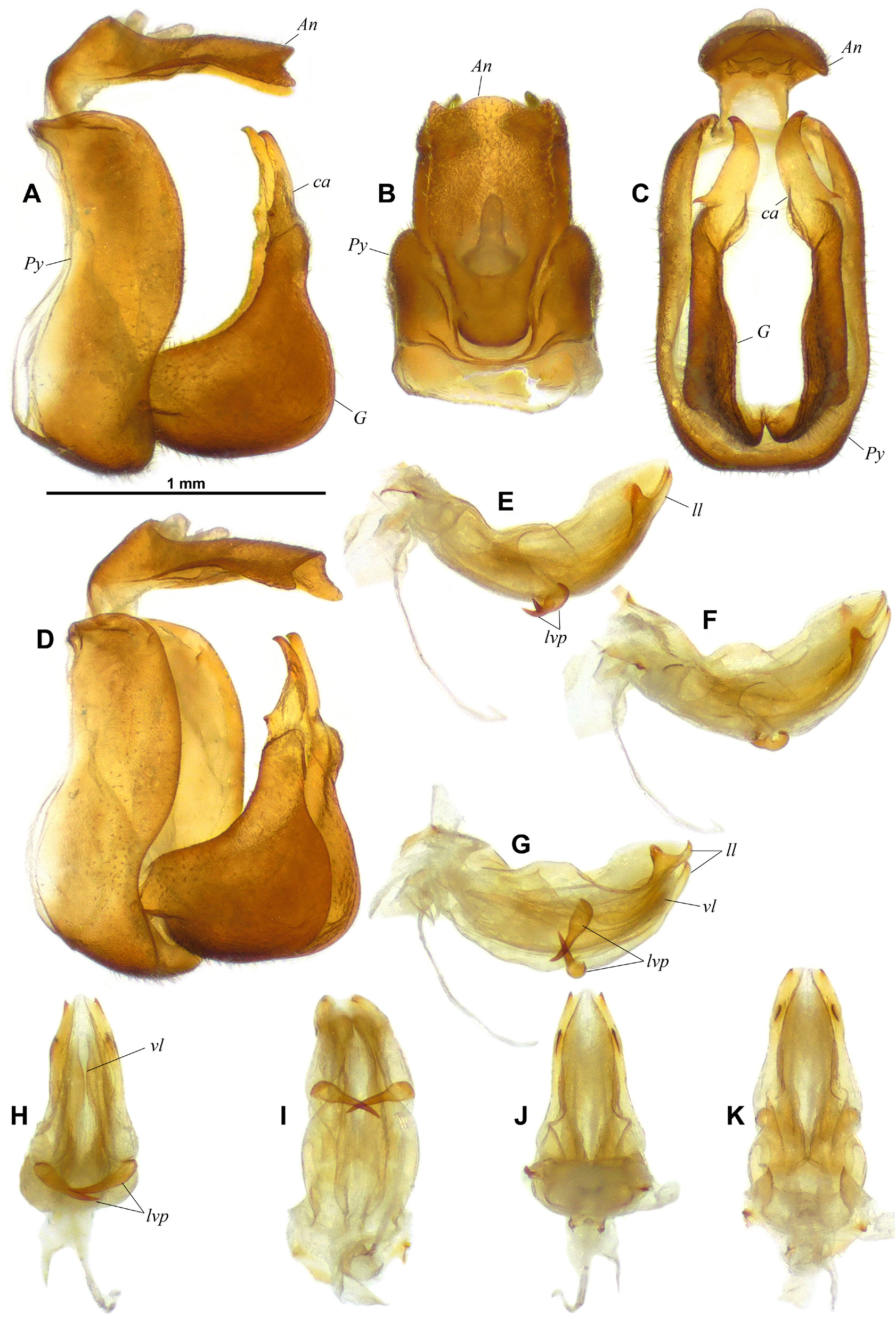
(1) the short gonostyli, mostly developed vertically (about 1.7 times as high as long in lateral view) and without triangular projection on anterodorsal margin at base of capitulum in lateral view ( G, ca – Fig. 25A View Fig );
(2) the aedeagus with pair of lateroventral processes shaped as rather short ventral hooks directed mesoanteriorly, rather robust basally then tapering into acute point after half-length ( lvp – Fig. 25G– I View Fig );
(3) the lateral lobes of periandrium with an apical hook pointing dorsad and an anteapical strong triangular tooth directed dorsad ( ll – Fig. 25E–G View Fig ).
Differential diagnosis
Only one other species, K. furcata , shares the gonostyli without triangular projection on anterodorsal margin at base of capitulum in lateral view but it can be easily separated from K. mua sp. nov. by the ventral hooks of the aedeagus which are very elongate and furcate in K. furcata ( Chang et al. 2020: figs 12–13) as opposed to the short simple hooks in K. mua .
Etymology
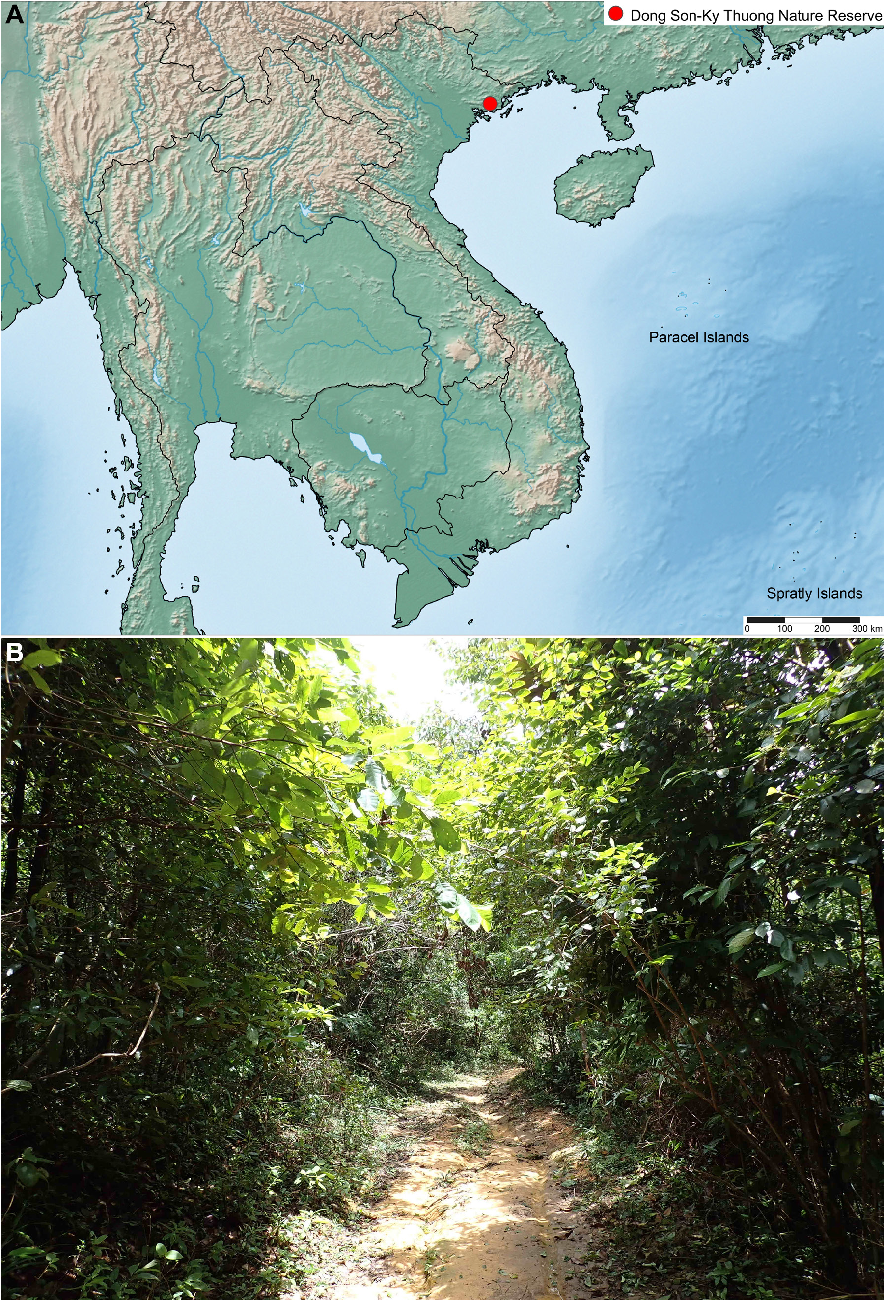
The species epithet ‘mua’ is the Vietnamese word for ‘rain’ and refers to the heavy rains which prevented our team from collecting in Dong Son-Ky Thuong Nature Reserve for two days. It is used as a noun in apposition.
Type material
Holotype
VIETNAM • ♂ (dissected – Figs 2A View Fig , 24–25 View Fig View Fig ); Quang Ninh Province, Dong Son-Ky Thuong Nature Reserve ; 21°08′29″ N, 107°04′53″ E; 27 Aug. 2022; 550 m a.s.l.; secondary forest; GTI Project; J. Constant, J. Bresseel and L. Semeraro leg.; I.G.: 34.518; RBINS.
GoogleMapsDescription
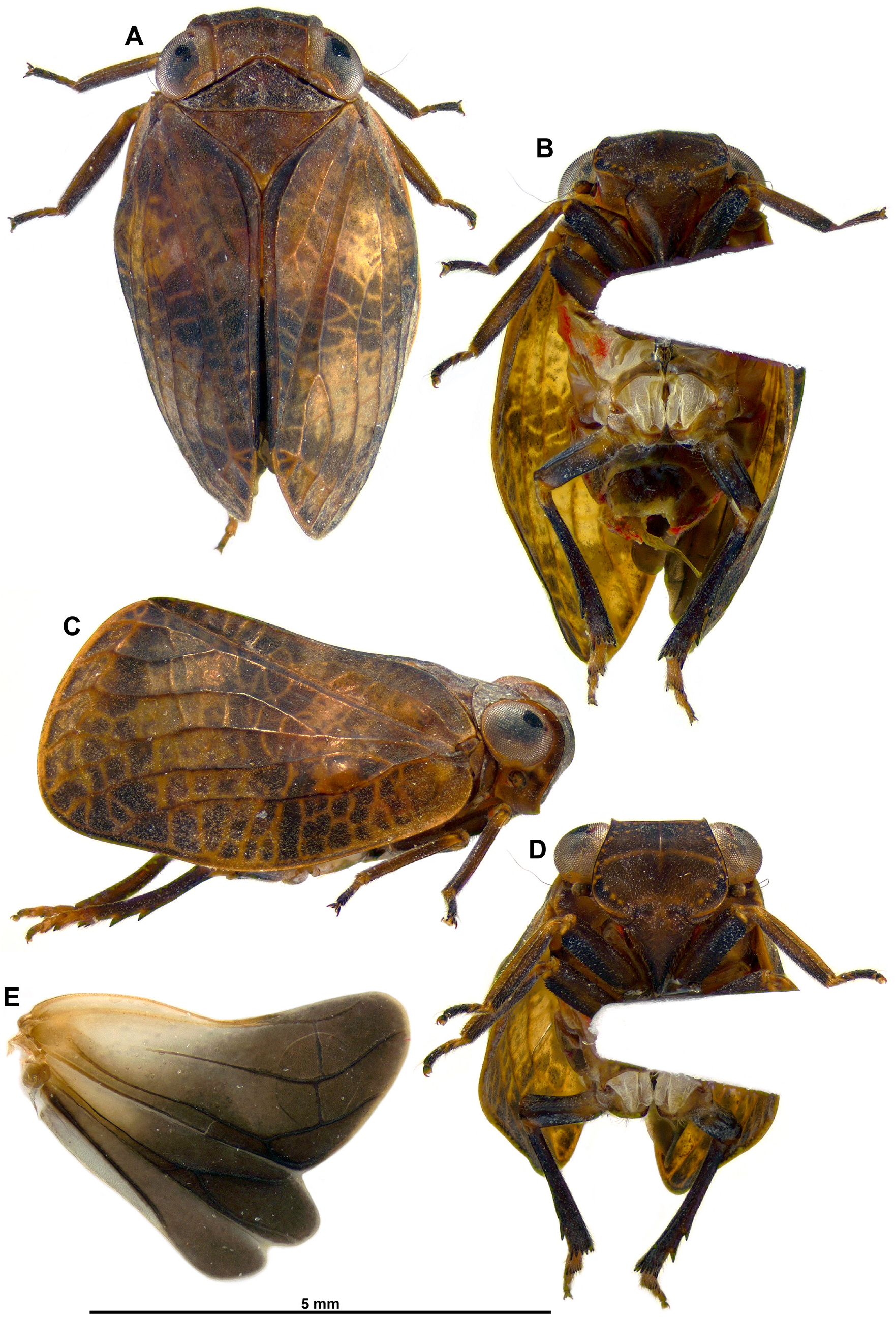
MEASUREMENTS AND RATIOS. LT: ♂ (n = 1): 5.7 mm; LT/BB = 1.7; LTg/BTg = 1.60; LW/BW = 1.36; BV/ LV = 2.5; LF/BF = 0.64.
HEAD ( Fig. 24A–D View Fig ). Vertex distinctly broader than long in midline but slightly narrower in middle than on sides, brown with margins slightly carinate; anterior margin convex, posterior one concave and lateral subparallel; disc shallowly concave. Side of head brown with pale yellowish line at mid-height from eye to anterior margin. Frons about 1.6 times as wide as long, brown with curved transverse yellowish narrow line, and a darker area along lateral margins, wider along lateroventral angle; row of small yellowish tubercles parallel to lateral margins, two rows along dorsal margin; strong median carina in dorsal half; ventrolateral angle carinate and projecting anteriorly, forming angular projection in lateral view. Clypeus brown, darker apically and elevated medially but not carinate. Labium brown with last segment longer than broad, and shorter than penultimate. Scape short, ring-shaped, black; pedicel bulbous, brown.
THORAX ( Fig. 24A, C View Fig ). Brown. Pronotum rather short, 0.6 times as long as mesonotum in midline; median carina obsolete, with one small, impressed point on each side and some faint tubercles along posterior margin; anterior and posterior margins weakly carinate, disc weakly concave; posterior margin bisinuate; paranotal lobes brown turning to yellowish behind eye. Mesonotum smooth, with weak carina parallel to anterior margin; posterior portion of disc and scutellum slightly elevated. Tegulae yellowish brown.
TEGMINA ( Fig. 24A–C View Fig ). Moderately convex; brown with three darker area: one near base, broad transverse band at midlength, and in distal portion; widest in distal portion with apical margin obliquely truncate and broadly rounded; costal margin slightly reflexed in basal half; distinct hump in fork ScP+RA–RP. Venation: ScP+R forked near base; MP forked slightly beyond basal ⅓; CuA very long, forked at about ¾ of tegmen length; CuP reaching apicosutural angle; Pcu weakly visible in distal portion, fused with A 1 at about midlength of clavus; A 1 well marked; Pcu+A 1 moderately keeled; continuous row of aligned cross-veins joining main veins, parallel to apical margin.
HIND WINGS ( Fig. 24E View Fig ). Dark brown with basicostal portion paler, trilobed, with veins darker, slightly shorter than tegmina. ScP-R-MP-Cu lobe more than twice as broad as Pcu-A 1, even broader than combined lobe Pcu-A 1 + lobe A 2; A 2 lobe with anterior and posterior margins subparallel and distinctly surpassing half-length of Pcu-A1 lobe; Pcu single, subdistally anastomising with 2-branched A 1; A 2 simple.
LEGS ( Fig. 24A–D View Fig ). Moderately elongate and slender, brown with femora and metatibiae darker, base of metatibiae shortly yellowish. Metatibiae with 2 lateral spines in apical ⅓ and 7 apical spines. Metatibiotarsal formula: (2) 7 / 9 / 2.
MALE TERMINALIA. Pygofer ( Py – Fig. 25A–D View Fig ) in lateral view about 3.1 times as high as long and with anterior and posterior margins subparallel, moderately curved in dorsal half, sinuate in ventral half; in caudal view subrectangular, about 1.7 times as high as wide. Gonostyli ( G – Fig. 25A, C–D View Fig ) about 1.7 times as high as long in lateral view, with anterodorsal margin roundly, evenly emarginate, posterior margin moderately sinuate, broadly rounded in ventral portion, and ventral margin broadly rounded; mostly vertical in caudal view with capitulum projecting dorsomesad; capitulum ( ca) elongate and with neck rather wide in lateral view, distal portion anteroposteriorly laminate and subcrescent-shaped in caudal view ( Fig. 25C View Fig ). Anal tube ( An – Fig. 25A–D View Fig ) elongate and dorsoventrally flattened; in dorsal view 1.9 times as long in midline, as wide, with lateral margins diverging from base to basal ⅓, then distinctly bisinuate; apical margin distinctly bisinuate with lateral angles acute; anal opening in basal ¼; in lateral view moderately curved posteroventrad and with posterior margin distinctly emarginate. Aedeagus ( Fig. 25E–K View Fig ) moderately curved posterodorsad, moderately tapering towards apex in dorsal view and with pair of lateroventral processes ( lvp) as rather short ventral hooks directed mesoanteriorly, rather robust basally then tapering into acute point after half-length; ventral lobe of periandrium ( vl) broad, abruptly narrowing at level of ventral hooks then slowly tapering to narrowly rounded apex; lateral lobes of periandrium ( ll) with triangular subapical tooth directed posterodorsad and apical tooth curved dorsad.
Biology
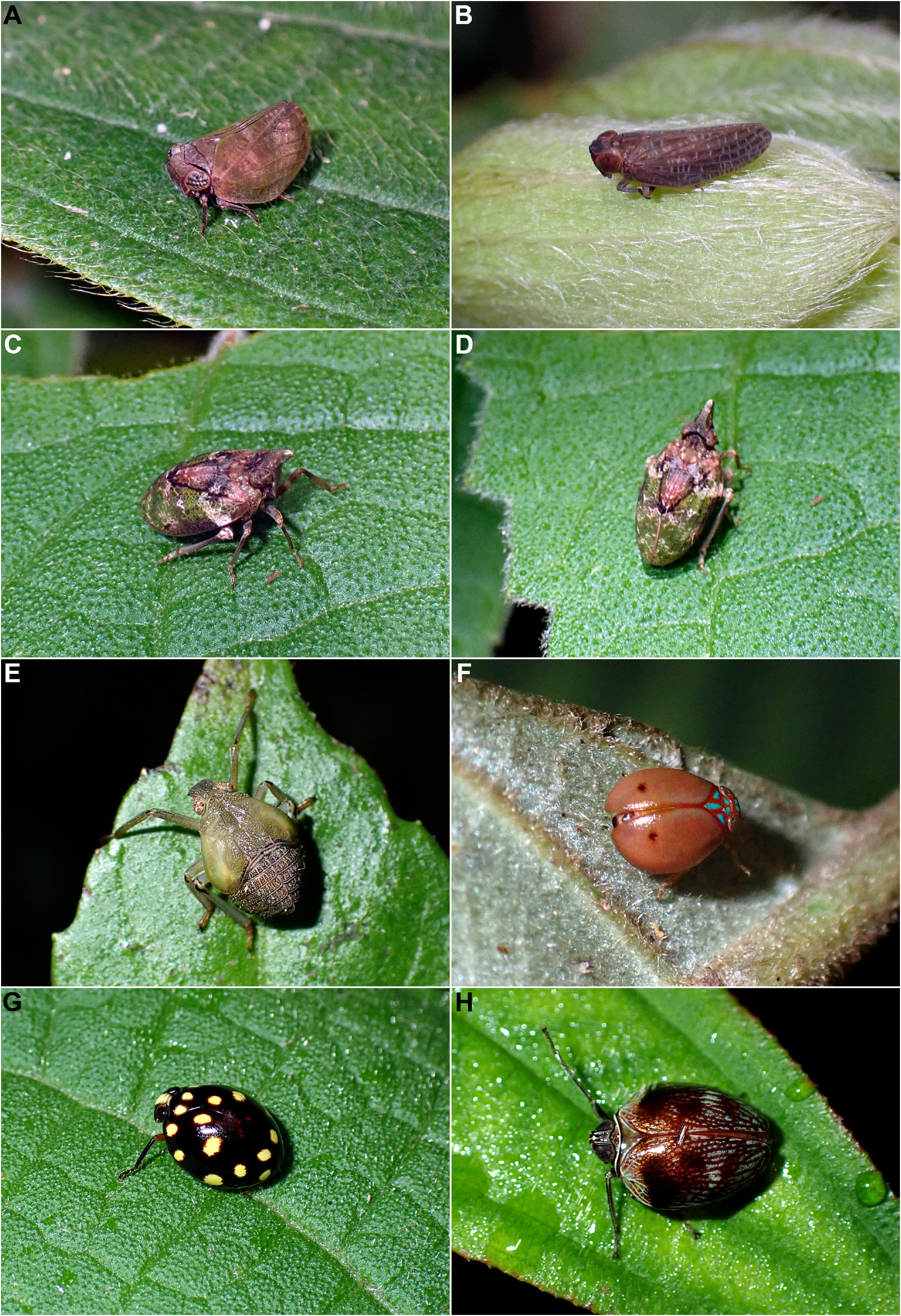
Kodaianella mua sp. nov. was collected in August on lower vegetation and bushes, in moist evergreen tropical forest at about 550 m in altitude ( Fig. 1B View Fig , 2A View Fig ).
Distribution
Vietnam: Quang Ninh Province, Dong Son-Ky Thuong Nature Reserve ( Fig. 1A View Fig ).
| RBINS |
Royal Belgian Institute of Natural Sciences |
No known copyright restrictions apply. See Agosti, D., Egloff, W., 2009. Taxonomic information exchange and copyright: the Plazi approach. BMC Research Notes 2009, 2:53 for further explanation.
|
Kingdom |
|
|
Phylum |
|
|
Class |
|
|
Order |
|
|
SubOrder |
Auchenorrhyncha |
|
InfraOrder |
Fulgoromorpha |
|
SuperFamily |
Fulgoroidea |
|
Family |
|
|
SubFamily |
Issinae |
|
Tribe |
Kodaianellini |
|
Genus |