
Otocelis erinae, Hooge, Matthew D. & Rocha, Carlos E. F., 2006
|
publication ID |
https://doi.org/ 10.5281/zenodo.174287 |
|
DOI |
https://doi.org/10.5281/zenodo.6263459 |
|
persistent identifier |
https://treatment.plazi.org/id/03DE87D4-E609-FF94-FE84-12EFFC7BFB9B |
|
treatment provided by |
Plazi |
|
scientific name |
Otocelis erinae |
| status |
sp. nov. |
Otocelis erinae View in CoL sp. nov.
( Figs. 25–26 View FIGURE 25 View FIGURE 26 )
Type material. Holotype. MZUSP PL. 193, one set of 2-µm-thick serial sagittal sections of epoxy-embedded specimen stained with toluidine blue. Paratypes. MZUSP PL. 194, one set of 2-µm-thick serial frontal sections of epoxy-embedded specimens stained with toluidine blue, and MZUSP PL. 195, epoxy-embedded whole mount.
Type locality. As Ilhas, São Sebastião, São Paulo, Brazil, from subtidal fine-grained sand (23°47’16.0”S, 45°42’31.2”W).
Other material examined. Living specimens in squeeze preparations from Pedro do Sino beach (23°44’49.8”S, 45°20’52.9”W) and Praia de Feiticeira (23°50’45.2”S, 45°24’33.8”W), Ilhabela, from intertidal and subtidal medium-grained sand; whole mounts for fluorescence imaging of musculature; photographs of living specimens in squeeze preparations.
Etymology. Species name in honor of Ms. Erin Suhr of Portland, Oregon, USA.
Description. Examined specimens 400 to 490 µm long and 120 to 130 µm wide ( Figs. 25 View FIGURE 25 A, B). Anterior and posterior ends rounded. Body cylindrical. Epidermis completely ciliated. Rhabdoid glands in distinct rows ( Fig. 25 View FIGURE 25 A). Body colorless in transmitted light, but digestive syncytium with green coloration. Frontal organ present, and seen especially well in living specimens in regions flanking statocyst ( Fig. 25 View FIGURE 25 B). Mouth opening on ventral surface, middle of body. Digestive central syncytium extends from frontal glands posteriorly to male copulatory organ.
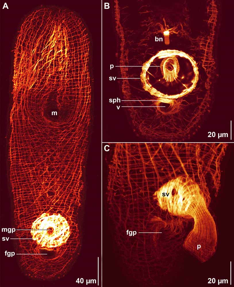
Body-wall musculature with circular muscles that encircle the body along entire length of animal; straight longitudinal muscles present between frontal organ and anterior edge of mouth; longitudinal muscles with a longitudinal orientation anteriorly that bend medially to cross diagonally over the body (longitudinal-cross-over fibers) present in dorsal and ventral body walls; longitudinal muscles in the anterior half of body that wrap around the posterior rim of the mouth (U-shaped muscles) present in ventral body wall ( Fig. 26 View FIGURE 26 A).
Ovary unpaired, ventral; extends from level of mouth posteriorly to bursal nozzle ( Figs. 25 View FIGURE 25 A, C). Testes paired, lateral to eggs. Testes extend from frontal gland posteriorly to level of the bursal nozzle ( Fig. 25 View FIGURE 25 B).
Male gonopore opens ventrally, anterior to female gonopore ( Figs. 25 View FIGURE 25 C, 26). Gonopore opens directly to well-developed, muscular and glandular penis. Penis musculature composed of outer non-anastomosing longitudinal muscles and inner circular muscles ( Figs. 26 View FIGURE 26 B, C). Lumen of penis filled with glandular secretions ( Fig. 25 View FIGURE 25 C). Penis invaginated into muscular seminal vesicle filled with sperm and small granules that abut the posterior side of penis.
Female gonopore opens ventrally, posterior to male gonopore and seminal vesicle ( Figs. 25 View FIGURE 25 C, 26). Gonopore opens to a short ciliated antrum that leads to vagina ( Fig. 25 View FIGURE 25 C). Immediately distal to antrum, vagina surrounded by muscular sphincter ( Fig. 26 View FIGURE 26 B). Vagina passes dorsally to seminal vesicle; leads to seminal bursa with well-developed sclerotized bursal nozzle ( Figs. 25 View FIGURE 25 A, B, 26B).
Remarks. Within the genus Otocelis , O. erinae differs from all other described species in having an unpaired ovary. O. erinae is most similar to O. phycophilus Ehlers and Dörjes, 1979 and O. westbladi Ax, 1959 , which also have vaginal sphincters and separate male and female gonopores. In contrast to these two species, O. erinae has a slightly curved penis, not a hooked penis, and is without ocelli.

| MZUSP |
Museu de Zoologia da Universidade de Sao Paulo |
No known copyright restrictions apply. See Agosti, D., Egloff, W., 2009. Taxonomic information exchange and copyright: the Plazi approach. BMC Research Notes 2009, 2:53 for further explanation.