
Archaphanostoma marcusi, Hooge, Matthew D. & Rocha, Carlos E. F., 2006
|
publication ID |
https://doi.org/ 10.5281/zenodo.174287 |
|
DOI |
https://doi.org/10.5281/zenodo.6263431 |
|
persistent identifier |
https://treatment.plazi.org/id/03DE87D4-E638-FFAA-FE84-15BFFB7CFE63 |
|
treatment provided by |
Plazi |
|
scientific name |
Archaphanostoma marcusi |
| status |
sp. nov. |
Archaphanostoma marcusi View in CoL sp. nov.
( Figs. 13–14 View FIGURE 13 View FIGURE 14 )
Type material. Holotype. MZUSP PL. 188, one set of 2-µm-thick serial sagittal sections of epoxy-embedded specimen stained with toluidine blue.
Type locality. Praia de Feiticeira, Ilhabela, São Paulo, Brazil, from subtidal coarsegrained sand (23°50’45.2”S, 45°24’33.8”W).
Other material examined. Three sets of 2-µm-thick serial sagittal sections of epoxyembedded immature specimens stained with toluidine blue; whole mount for fluorescence imaging of musculature; photographs of living specimen in squeeze preparations.
Etymology. Species name in honor of Prof. Ernst Marcus, in recognition of his significant contribution to our understanding of the Acoela .
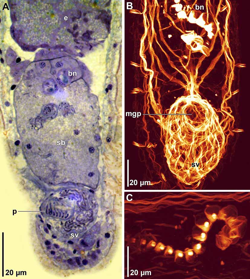
Description. Living specimens in squeeze-preparation ~470 µm long and ~135 µm wide ( Fig. 13 View FIGURE 13 A). Anterior and posterior ends rounded. Epidermis completely ciliated. Large rhabdoid glands in longitudinal rows ( Fig. 13 View FIGURE 13 A). Body colorless in transmitted light. Frontal glands well developed, extend from frontal pore posteriorly to level behind statocyst. Mouth opening on ventral surface, middle of body. Digestive central syncytium extends from position behind statocyst posteriorly to level of seminal bursa.
Ovary unpaired, extends from level of mouth posteriorly to seminal bursa ( Figs.13 View FIGURE 13 A, 14A). Testes paired, follicular; extend from frontal glands posteriorly to seminal vesicle ( Figs. 13 View FIGURE 13 B).
Single gonopore present; opens directly to male copulatory organ. Unclear from histological sections whether gonopore also opens to seminal bursa ( Fig. 14 View FIGURE 14 A)
Seminal bursa a dense syncytial mass surrounding sperm and bursal nozzle ( Figs. 13 View FIGURE 13 B, 14A). Bursa capped anteriorly with several cells that separate bursa from developing eggs ( Fig. 14 View FIGURE 14 A). Bursal nozzle S-shaped; composed of many (~17) disjunct, actin-rich nozzle components, through which sperm pass on way to ovary ( Figs. 13 View FIGURE 13 B, 14A–C)
Isodiametrid-type male copulatory organ composed of a muscular penis (~13 µm long) invaginated into a muscular seminal vesicle ( Figs. 13 View FIGURE 13 B, 14A, B). Space within seminal vesicle filled with sperm that surrounds penis ( Fig. 14 View FIGURE 14 A).
Remarks. This species was very fast moving and fairly uncommon in our samples. Due to a lack of available specimens, only a single mature animal was fixed for histological sectioning. Unfortunately, this single specimen is slightly damaged around the gonopore, and as such, it is not possible to determine whether it is solely a male gonopore, or a common gonopore that also opens to the seminal bursa.
Our species is placed in the genus Archaphanostoma due to its possession of the diagnostic character—a syncytial seminal bursa. A. marcusi differs most substantially from the other three known species of the genus by its possession of a bursal nozzle. A disjunct nozzle is an uncommon occurrence among members of the Isodiametridae , but is present in Aphanostoma bruscai Hooge and Tyler, 2003 (see also Petrov et al. 2006).

| MZUSP |
Museu de Zoologia da Universidade de Sao Paulo |
No known copyright restrictions apply. See Agosti, D., Egloff, W., 2009. Taxonomic information exchange and copyright: the Plazi approach. BMC Research Notes 2009, 2:53 for further explanation.