
Notofairchildia motacuensis Ježek, Oboňa & Le Pont, 2018
|
publication ID |
https://doi.org/ 10.11646/zootaxa.4442.3.8 |
|
publication LSID |
lsid:zoobank.org:pub:99AEA819-E5EE-42D8-B8CE-7714D4FC8217 |
|
DOI |
https://doi.org/10.5281/zenodo.5952074 |
|
persistent identifier |
https://treatment.plazi.org/id/03DF173E-FFCA-D350-FF1D-2D1D041C4DAA |
|
treatment provided by |
Plazi |
|
scientific name |
Notofairchildia motacuensis Ježek, Oboňa & Le Pont |
| status |
sp. nov. |
Notofairchildia motacuensis Ježek, Oboňa & Le Pont View in CoL sp. nov.
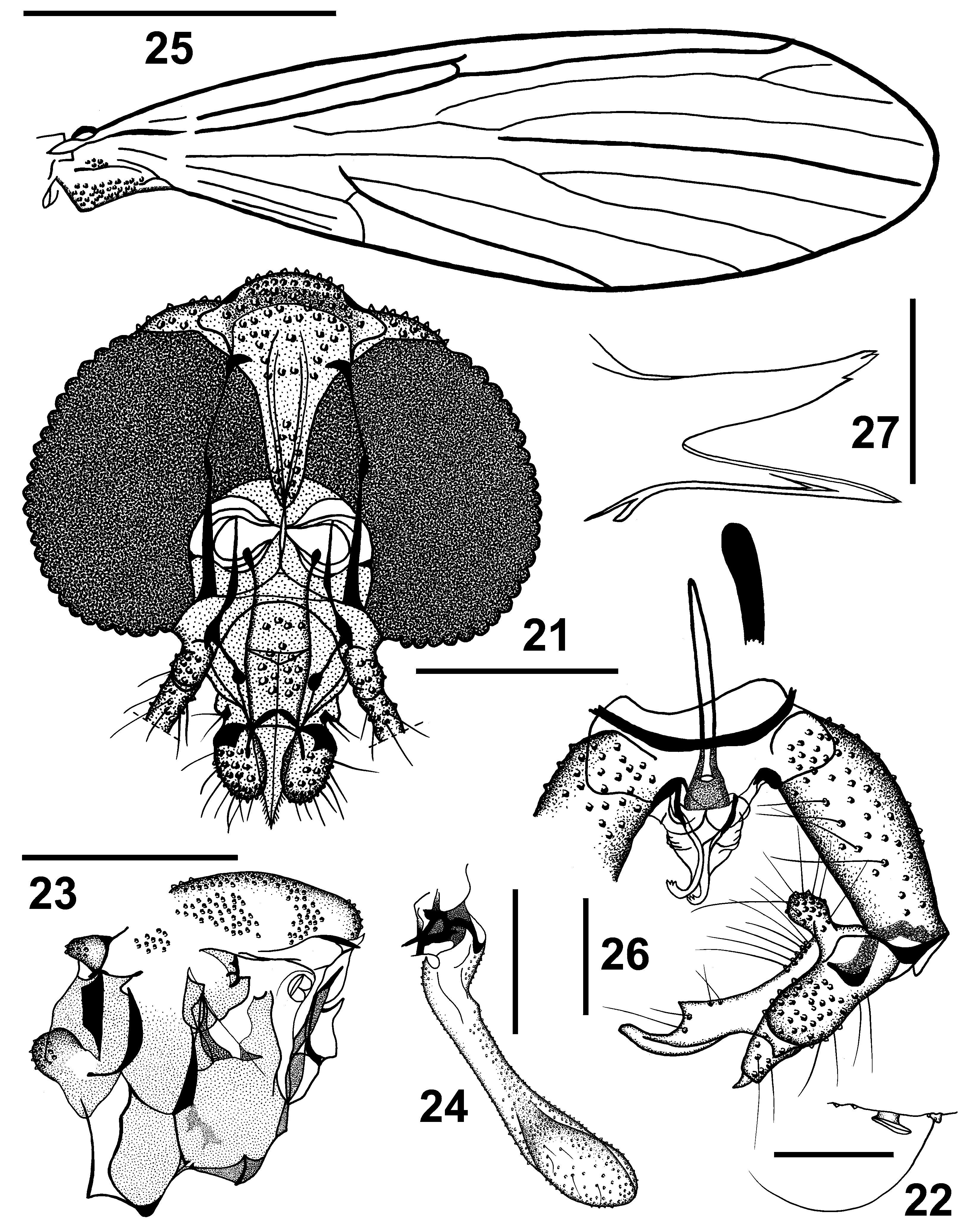
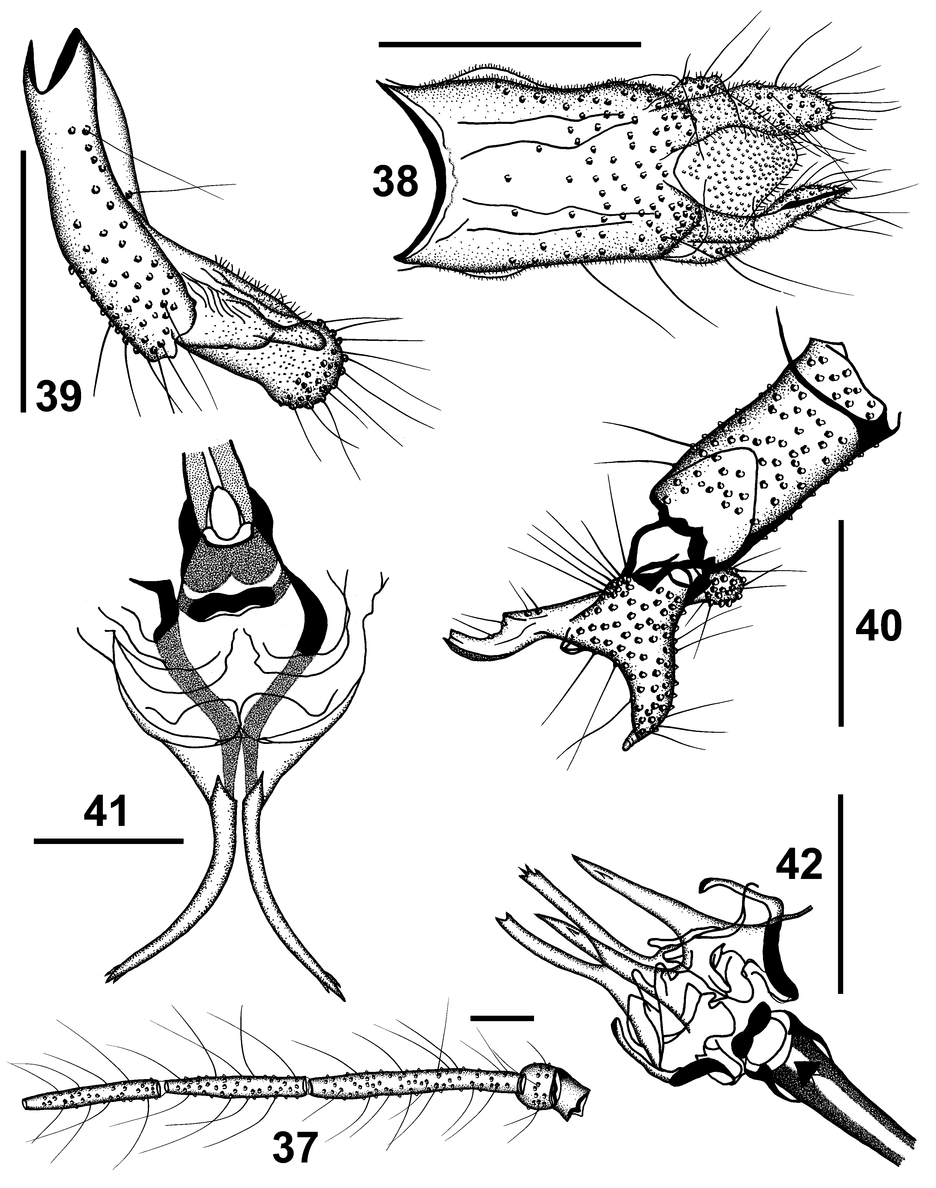
( Figs. 21–42 View FIGURES 21–27 View FIGURES 28–36 View FIGURES 37–42 )
Description. Head ( Fig. 21 View FIGURES 21–27 ) with slightly protuberant dome-shaped vertex. Eyes separated by one facet diameter ( Fig. 28 View FIGURES 28–36 ); frons with cluster of approximately 13 hair scars at anterior angle of eye, cluster separated from other scars on frons and vertex. Supraocular enlarged bristles on dorsal margins of eyes missing. The fold of eye on Fig. 21 View FIGURES 21–27 from caudal view is not figured (but exist). Antenna 16-segmented, with flagellomeres elongate ( Figs. 29 View FIGURES 28–36 , 37 View FIGURES 37–42 ), cylindrical, covered with abundant needle-shaped soft sensilla, with paired mushroom- or platter-shaped ascoids ( Fig. 22 View FIGURES 21–27 ). Terminal flagellomere with digit-shaped apiculus. Scape stump-shaped, cut, pedicel subsphaerical. Ratio of lengths of segments of palpus maxillaris 1.0:3.2:3.4:4.9:14.6, maxila as long as the second palpal segment ( Fig. 30 View FIGURES 28–36 ). Third segment not conspicuousl enlarged, Newstead’s scales not evident. Mouthparts extending over mid length of palpus segment 3. Labial lobes ( Fig. 31 View FIGURES 28–36 ) prolonged with a little swollen sides, parallel inner lines of small microsetae are not visible. Cibarium ( Figs. 21 View FIGURES 21–27 , 32 View FIGURES 28–36 ) longer than broad, well sclerotized, without chitinous arch.
Thorax. Insertions of setae on mesothorax as in Fig. 23 View FIGURES 21–27 , anepisternum without setose patch, thoracic spiracle inside slot-shaped. Wings ( Fig. 25 View FIGURES 21–27 ) translucent, 3.4 times longer than wide. Wing length 3.1 (holotype) - 3.0 mm (paratype), Sc bifurcated near middle of wing and attached to R1 (not to C). Radial fork before ending of R1 and conspicuously distal to medial fork, which is proximally to end of CuA1; r-r and r-m missing.M3joined to CuA1at base, a connection of M1+2 and CuA1 missing. Vein sector of CuA1 and CuA2 developed. A1and their fold ending free in the wing basis. Following veins strengthened: Sc, R1, R5 and CuA1. The greater part of veins is quite clearly attached to the costa, but notfully sclerotized distally near their end in wing apex (5 veins). There are additionally several invisible connections of veins in central area of wing (see Fig. 25 View FIGURES 21–27 ). Halteres ( Fig. 24 View FIGURES 21–27 ) almost ovoid with a prolonged stem and many microsetae mainly in distal part. Ratio of maximum length of halteres to their maximum width hardly 5.0:1. Ratios of lengths of femora, tibiae and first tarsal segments: P1 1.4:2.0:1.0, P2 1.5:2.1:1.1, P3 1.7:2.5:1.2. Paired tarsal claws of P1 haired as in Fig. 33 View FIGURES 28–36 , widened in the middle, only pointed tip is bare and bent. Areas of insertion points of elongate erectile setae of medial abdominal segments (tergal chaetotaxy) see on Fig. 34 View FIGURES 28–36 .
Terminalia. Hypandrium ( Fig. 26 View FIGURES 21–27 ) narrow, inconspicuously bilobed in the middle, approximately as wide as ejaculatory apodeme from dorsal view. Epandrium ( Figs. 38, 39 View FIGURES 37–42 ) quadrangular in dorsal view, about 1.9 times as long as wide, almostfour times longer in lateral view. Ventral plate ( Fig. 38 View FIGURES 37–42 ) reduced to hardly visible ligaments laterally on both sides of epandrium. Hypoproct largely triangular, conspicuousl broadened at its base, tongueshaped, haired; epiproct membraneous and the shape is hardly detected from dorsal and lateral view ( Figs 38, 39 View FIGURES 37–42 ). Cerci cylindrical from dorsal and lateral views, one surstylus is ina diagonal position on the slide (paratype, Cat. No. 34714), pointed, one of the cerci was partially folded or rolled ( Figs. 38, 39 View FIGURES 37–42 ). Gonocoxite cylindrical, unmodified ( Figs. 26 View FIGURES 21–27 , 35 View FIGURES 28–36 , 40 View FIGURES 37–42 ), with numerous conspicuous setae alveoli dorsally and laterally, with several elongate setiform sensilla inserted medially. Gonostylus complex ( Figs. 26 View FIGURES 21–27 , 35 View FIGURES 28–36 , 40 View FIGURES 37–42 ) approximatelly as long as gonocoxite, with four strong radial protuberances rounded or more or less pointed from different views ( Figs. 35 View FIGURES 28–36 , 40 View FIGURES 37–42 ). Main protuberance of gonostylus is prolonged by bent arm, similarity point of spanner- or bottle-opener, with upright arranged tooth basally ( Figs. 26 View FIGURES 21–27 , 35 View FIGURES 28–36 , 40 View FIGURES 37–42 ). Terminl area of main protuberance is characteristic, rounded or cut hooked tip from different views, tapering to a point from diagonal view ( Fig. 35 View FIGURES 28–36 ). Parameres bifid, with fused arms basally and two-fourteeth apically ( Figs. 27 View FIGURES 21–27 , 36 View FIGURES 28–36 , 42 View FIGURES 37–42 ), inconspicuouslysclerotized. Ejaculatory apodeme ( Fig. 26 View FIGURES 21–27 ) narrow, straight, stick-shaped, pointed proximally in dorsal view, rounded from lateral one. Ratio of length of ejaculatory apodeme to gonocoxite 1:1.3. Aedeagal complex symmetrical ( Figs. 26 View FIGURES 21–27 , 41 View FIGURES 37–42 ), terminating in backward curved apex, distiphallus composed from two divergent tapering arms with three spines apically.
Differential diagnosis. Similar to Notofairchildia acaenohybos ( Quate & Alexander, 2000) witch is characterized by Sc distally free in basal wing area; r-r and r-m developed; M3 arising from M1+2; vein sector of CuA1 and CuA2 missing; veins of wing uniformly strengthened, clearly attached to the costa; arms of main protuberance of gonostylus divided by a deep rounded cleft (not upright arranged); parameres a pair of outward curved, tapered spikes lying below aedeagus, membranous and inconspicuous. Contrasting to this, Notofairchildia motacuensis Ježek, Oboňa & Le Pont sp. nov. has Sc bifurcated near middle of wing and connected with R1 (not with C); r-r and r-m missing ( Fig. 25 View FIGURES 21–27 ); M3 arising from CuA1 basally; vein sector of CuA1 and CuA2 developed; veins Sc, R1, R5 and CuA1 stronger (more infuscated); some veins apices not quite clearly attached to the costa (not fully sclerotized); main protuberance of gonostylus prolonged by bent arm with upright arranged tooth basally, similarity point of spanner- or bottle-opener ( Figs. 26 View FIGURES 21–27 , 35 View FIGURES 28–36 , 40 View FIGURES 37–42 ); parameres bifid, fused basally, with two-four teeth apically ( Figs. 27 View FIGURES 21–27 , 36 View FIGURES 28–36 , 42 View FIGURES 37–42 ).
Type locality. Bolivian dry tropical semideciduous forest in the southern side of the Santiago serrania, Motacu study site, near the town of Santiago de Chiquitos.
Type material. Holotype: male ( NMPC, dissected, slide, red label), Bolivia: Motacu , iii.-iv.2008 , CDC miniature light-trap, Le Pont leg., Cat . No. 34713, Inv. No. 24167. Paratype: male ( NMPC, dissected, yellow label), same data, Cat. No. 34714, Inv. No. 24168.
Etymology. This species is named after the type locality.
Distribution. This species is currently known from a single locality in Bolivia.

No known copyright restrictions apply. See Agosti, D., Egloff, W., 2009. Taxonomic information exchange and copyright: the Plazi approach. BMC Research Notes 2009, 2:53 for further explanation.