
Uariniensis
|
publication ID |
https://doi.org/10.11646/zootaxa.5129.1.8 |
|
publication LSID |
lsid:zoobank.org:pub:F4B01EE2-AA90-408A-8D10-D52373C9880E |
|
DOI |
https://doi.org/10.5281/zenodo.6502559 |
|
persistent identifier |
https://treatment.plazi.org/id/03DFCB05-FFD0-801B-FF50-FB042FA1FE93 |
|
treatment provided by |
Plazi |
|
scientific name |
Uariniensis |
| status |
|
Species Group Uariniensis
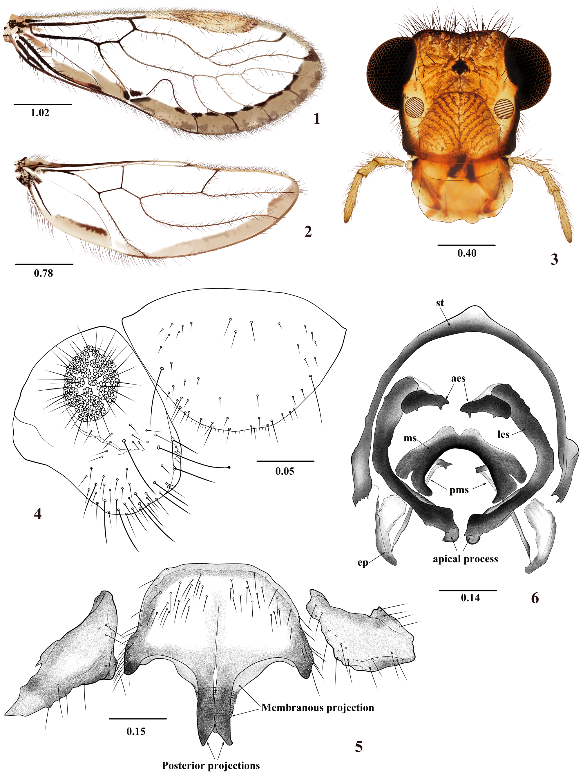
Diagnosis. Forewings with a pigmented marginal band from R4+5 to cu1 and cu2 cells ( Fig. 1 View FIGURES 1–6 ). Pterostigma rounded, not angulate nor extended towards Rs. Hindwings with a pigmented marginal band from R4+5 to cu1 and cu2 cells ( Fig. 2 View FIGURES 1–6 ). Hypandrium of three sclerites, central one large, with two median, close together, sclerotized, acuminate or distally widened posterior projections, located ventrally to a distally acuminate membranous projection ( Fig. 5 View FIGURES 1–6 ). Phallosome with three pairs of endophallic sclerites ( Figs 6 View FIGURES 1–6 and 11 View FIGURES 7–11 ), mesal endophallic sclerite fused, inverted U-shaped, with short, lateral laminar processes, postero-mesal sclerites elongate, narrow and laminar ( Figs 12–15 View FIGURES 12–15 ), basally fused to lateral process, apically acuminate and connected to the external parameres. Anterior sclerites small, transverse, lateral sclerites bow-shaped, apically directed mesally and with variable processes. Species included: E. atlantica Silva-Neto, 2021 , E. macuxi Pereira et al., 2022 , E. uariniensis Silva-Neto et al., 2019 , and two more species described below.
Key to the species of the group Uariniensis (males)
1. Central sclerite of the hypandrium with the two posterior median processes distally wide and curved slightly inward, each arm machete shaped (see Fig. 6 View FIGURES 1–6 in Pereira et al. 2022)......................................... E. macuxi Pereira et al.
- Central sclerite of the hypandrium with the two posterior median processes distally acuminate ( Figs 5 View FIGURES 1–6 and 10 View FIGURES 7–11 )........... 2
2. Mesal endophallic sclerite with anterior margin aligned with the anterior margin of the lateral processes, appearing as a substraight anterior margin; anterior sclerites smaller than in the other three species........... E. uariniensis Silva-Neto et al. View in CoL
- Mesal endophallic sclerite with anterior margin convex, or medially concave, lateral processes not forming a sub-straight line with the anterior margin of the sclerite ( Figs 6 View FIGURES 1–6 and 11 View FIGURES 7–11 )....................................................... 3
3. Lateral endophallic sclerites with concave laminar process apically, clearly widened, mesal endophallic sclerite with rounded anterior margin, not concave medially ( Fig. 11 View FIGURES 7–11 )............................................................. 4
- Lateral endophallic sclerites apically with process not widened or developed as in the previous ones, mesal endophallic sclerite usually with medially concave anterior margin ( Figs 6 View FIGURES 1–6 , 12, 14, 15 View FIGURES 12–15 )........................... E. guticortesorum n. sp.
4. Posterior processes of the central sclerite of the hypandrium distally narrow; mesal endophallic sclerite basally narrow, apical process of lateral endophallic sclerite posteriorly wide, and distally curved outward (see Fig. 7 View FIGURES 7–11 in Silva-Neto 2021).......................................................................................... E. atlantica Silva-Neto View in CoL
- Posterior processes of the central sclerite of the hypandrium distally more widened than in the previous species ( Fig. 10 View FIGURES 7–11 ); mesal endophallic sclerite basally broader than in the previous species, apical process of the lateral endophallic sclerites with concave laminar and crescent-shaped process ( Fig. 11 View FIGURES 7–11 )................................................. E. wilsoni n. sp.


No known copyright restrictions apply. See Agosti, D., Egloff, W., 2009. Taxonomic information exchange and copyright: the Plazi approach. BMC Research Notes 2009, 2:53 for further explanation.