
Hopperia beaglense Chen & Vincx, 1998
|
publication ID |
https://doi.org/ 10.5852/ejt.2012.24 |
|
publication LSID |
lsid:zoobank.org:pub:F8ED2AA9-83C1-4CB8-8327-58C501B6C42A |
|
DOI |
https://doi.org/10.5281/zenodo.3858989 |
|
persistent identifier |
https://treatment.plazi.org/id/03E0332F-F817-FFEB-FDA7-FC9DFB5DFDE3 |
|
treatment provided by |
Valdenar |
|
scientific name |
Hopperia beaglense Chen & Vincx, 1998 |
| status |
|
Hopperia beaglense Chen & Vincx, 1998
Figs 15-17 View Fig View Fig View Fig , Table 5
Material examined
5 ƋƋ and 4 ♀♀, collected 5 May 2010, Kaikoura Canyon axis (1061 m water depth), 42.5081°S, 173.6325°E (NIC 84450).
Description
Males
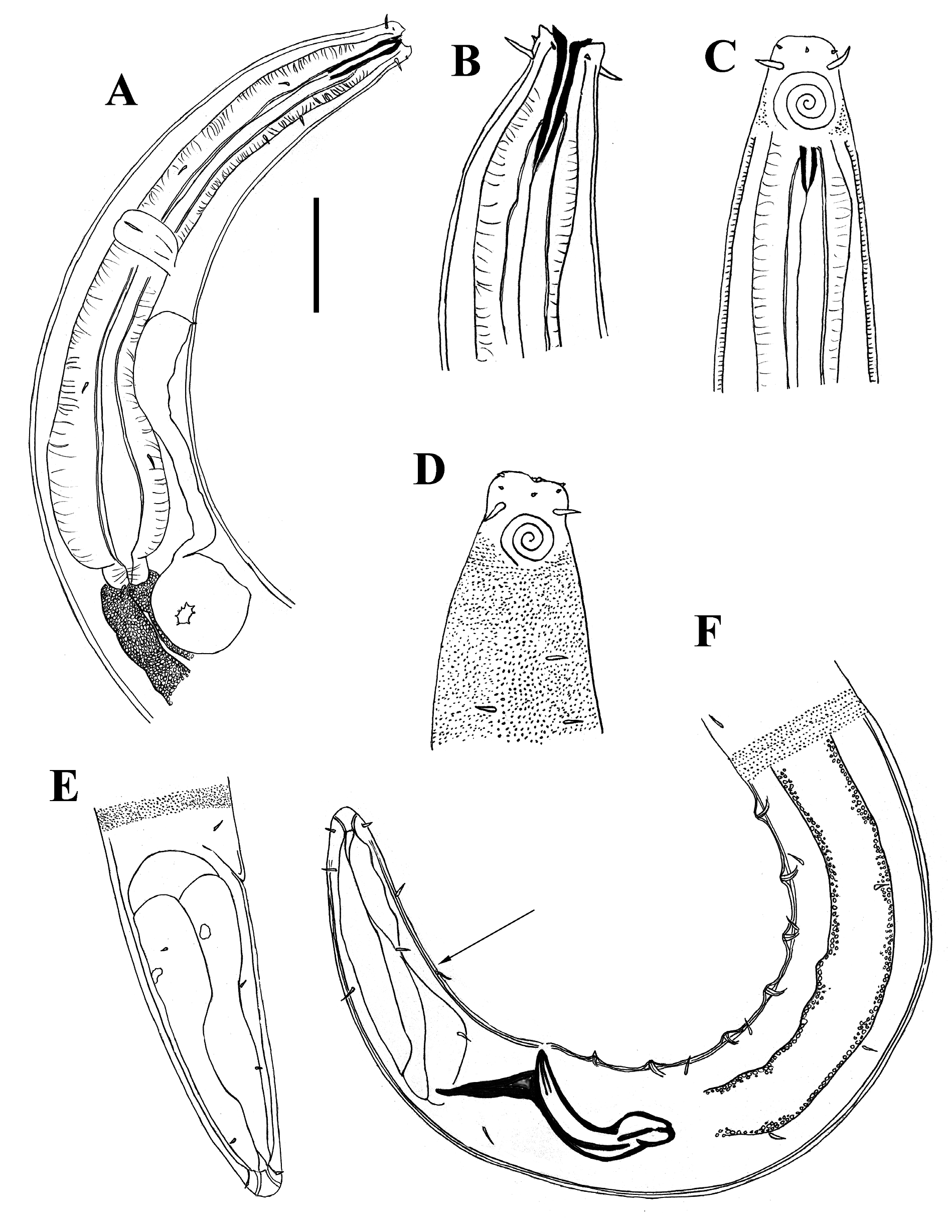
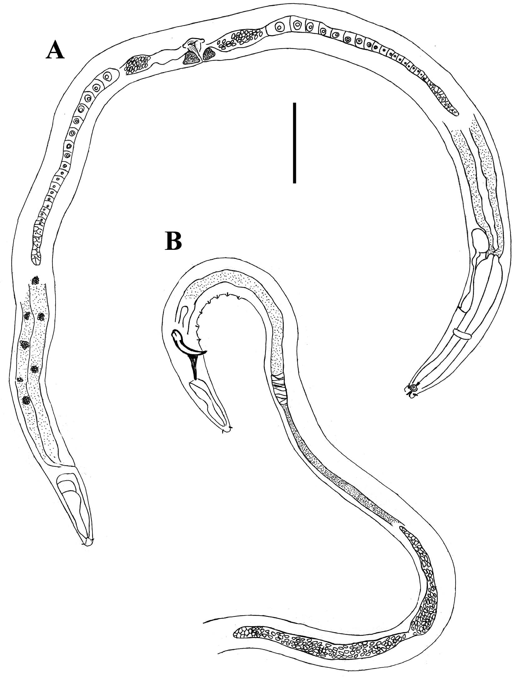
Body cylindrical, tapering slightly towards anterior extremity. Cuticle punctate from level of amphid to near tail tip, with lateral differentiation consisting of larger, irregularly-spaced dots. Somatic setae short and sparse, in four dorso- and ventro-lateral longitudinal rows. Six inner labial papillae, six outer labial papillae, and four cephalic setae in three distinct circles. Anterior portion of buccal cavity cupshaped. Posterior portion of buccal cavity cylindrical, strongly cuticularised, 24-26 μm deep, extending into three strongly cuticularised teeth at border to anterior portion. Teeth everted in some specimens. Cuticularisation of posterior portion of buccal cavity extends posterior to junction of marginal tubes with pharyngeal lumen ( Fig. 15B View Fig ). Amphideal fovea spiral, 3.0-3.5 turns, located immediately posterior to cephalic setae. Pharynx with oval posterior bulb. Cardia short. Nerve ring near middle of pharynx, situated anteriorly to secretory-excretory pore. Cellular body of ventral gland large (up to 37 x 20 μm), situated just posteriorly to cardia. Intestine wall with numerous small, clear granules, and small clusters of round, clear inclusions, sometimes with smaller orange-brown granules ( Fig. 17B View Fig ).
Reproductive system diorchic, opposed, outstretched. Anterior testis to left of intestine and posterior testis to right of intestine. Spicules paired, equal, arcuate, strongly cuticularised, 1.3-1.6 abd long, with swollen proximal end and internal cuticularise projection (lamella) extending one fourth of spicule length from proximal end, no velum. Gubernaculum with straight dorso-caudal apophyses, tapering distally. One pre-cloacal seta and 7-8 conspicuous, cup-shaped pre-cloacal supplements. Tail short, cylindrical, with rounded tip, several short caudal setae and no terminal setae. Three large caudal glands and well-developed spinneret ( Fig. 17C View Fig ).
Females
Similar to males, but slightly greater maximum body diameter and smaller amphids, 3.0 turns. Reproductive system didelphic, opposed, outstretched, with anterior branch to left of intestine and
posterior branch to right of intestine, except for one individual with opposite arrangement. Vulva slightly post median. Granular vaginal glands present, vagina uterina surrounded by constrictor muscle.
Discussion
My specimens closely resemble the description given by Chen & Vincx (1998) based on specimens from the Beagle Channel, Chile (100-110 m water depth). My specimens, however, have slightly lower a (range: 29-36 vs. 30-41; mean: 34.2 vs. 39.1) and c values (range: 14-19 vs. 19-22; mean: 15.8 vs. 20.6), have 7-8 supplements instead of 6-7, and lack the supplement-like structure situated halfway down the ventral side of the tail ( Fig. 15F View Fig ). This is the first record of this species outside the type locality.
Within the Dorylaimopsinae , cuticularisation of the posterior buccal cavity does not usually extend into the pharyngeal lumen (i.e., cuticularisation stops before marginal tubes begin) (fig. 2E in Jensen 1979). My specimens are unusual in that the cuticularisation of the buccal cavity extends into the anteriormost portion of the pharyngeal lumen (see Fig. 15B View Fig ). It is unclear whether this is also a feature of the type specimens, however, because Chen & Vincx (1998) did not describe the marginal tubes.

No known copyright restrictions apply. See Agosti, D., Egloff, W., 2009. Taxonomic information exchange and copyright: the Plazi approach. BMC Research Notes 2009, 2:53 for further explanation.