
Echinoderes obtuspinosus, Sørensen & Rho & Min & Kim & Chang, 2012
|
publication ID |
https://doi.org/ 10.11646/zootaxa.3368.1.8 |
|
DOI |
https://doi.org/10.5281/zenodo.5253996 |
|
persistent identifier |
https://treatment.plazi.org/id/03E08792-E21A-FFB4-FF36-FA59FEEEFE47 |
|
treatment provided by |
Felipe |
|
scientific name |
Echinoderes obtuspinosus |
| status |
sp. nov. |
Echinoderes obtuspinosus View in CoL sp. nov.
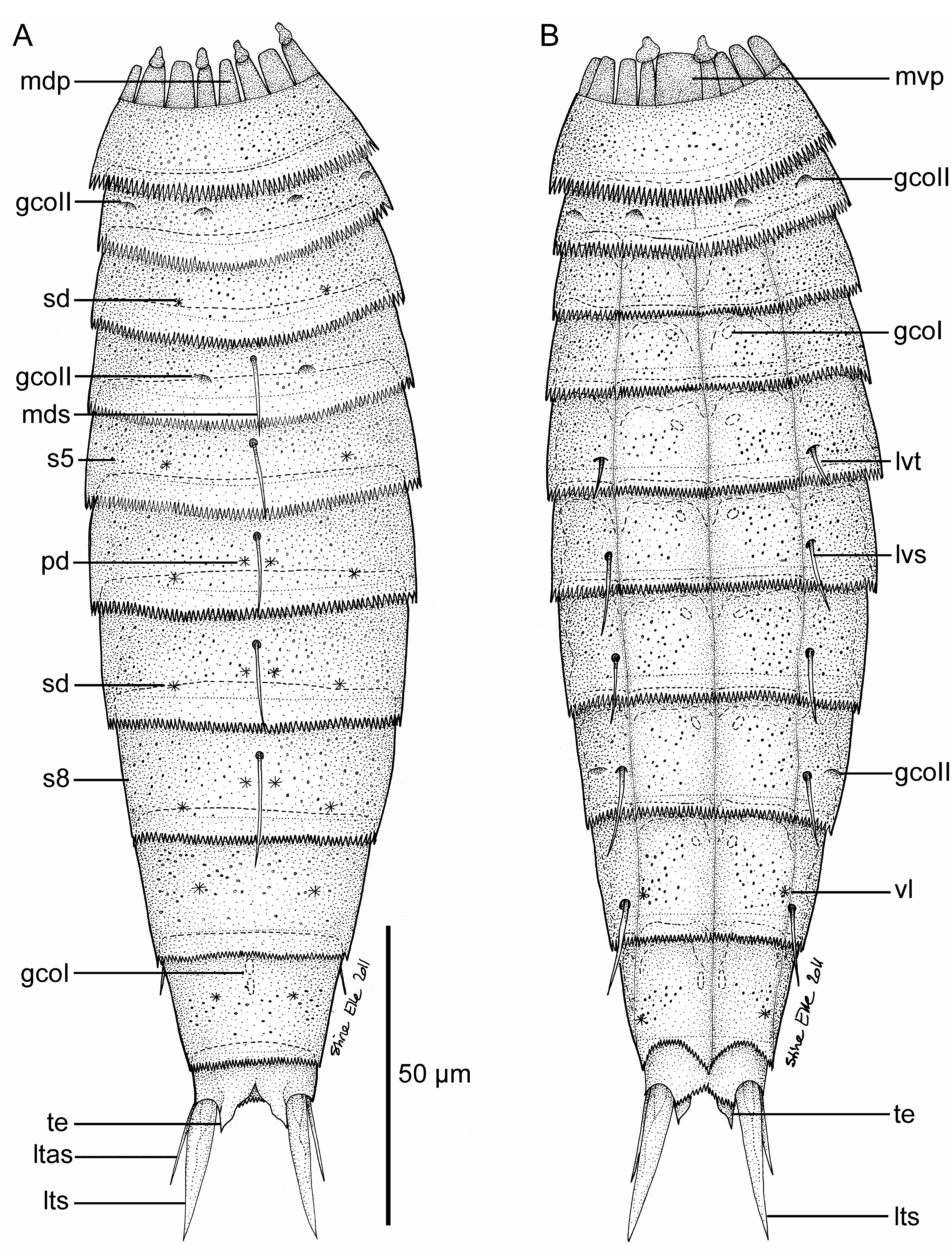
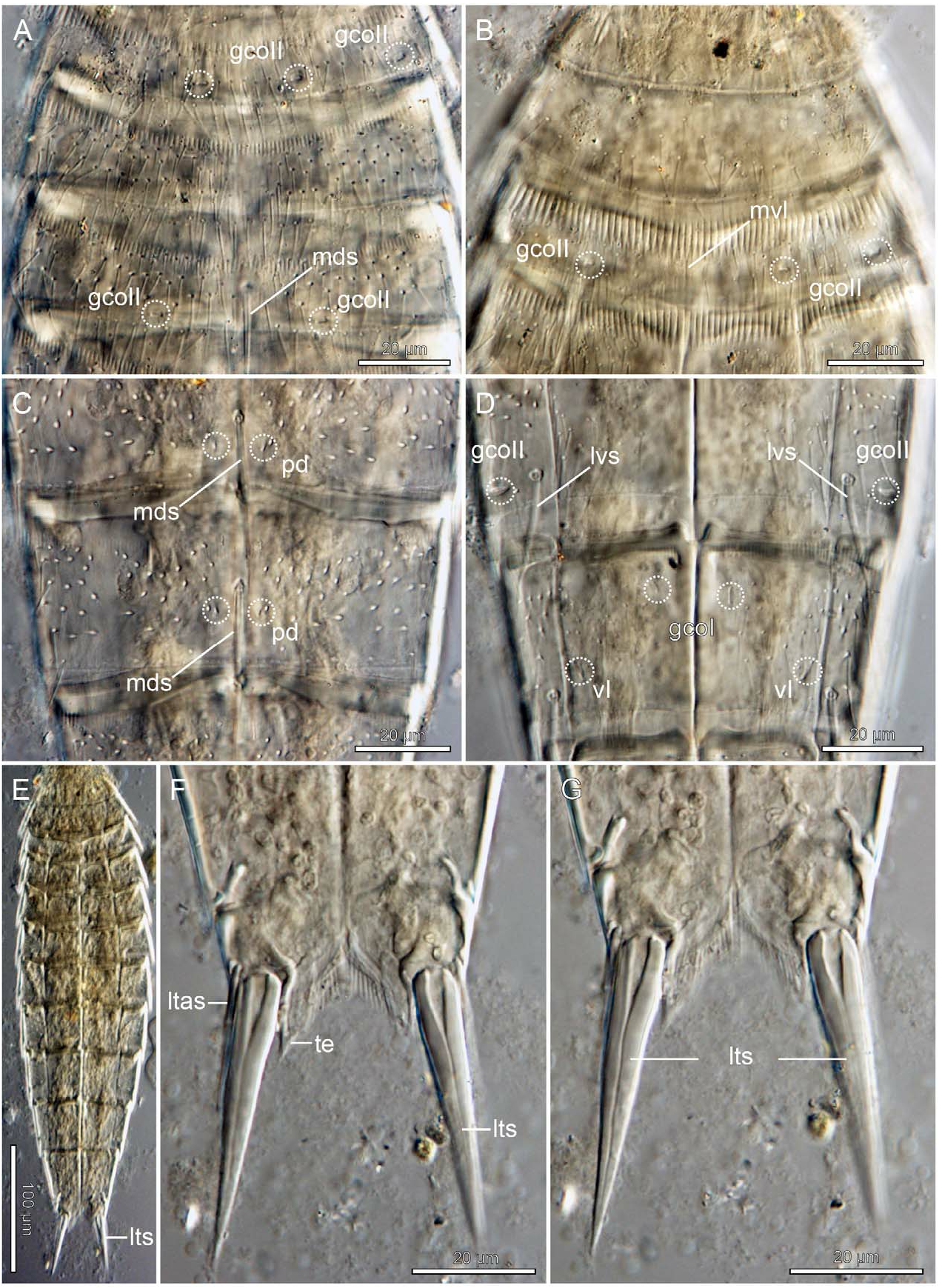
( Figures 11–12 View FIGURE 11 View FIGURE 12 , Tables 7–8)
Diagnosis. Echinoderes with middorsal spines on segments 4–8; lateroventral tubules on segment 5 and lateroventral spines on segments 6–9. Lateral terminal spines very short and stout, about 14% of trunk length. Glandular cell outlets type 2 present in subdorsal, laterodorsal, sublateral and ventrolateral positions on segment 2, in subdorsal positions on segment 4, and in sublateral position on segment 8. Segment 2 consisting of a closed cuticular ring, but with a weak indication of a midventral line.
Type material. Holotype: adult female, collected on 26 February 1999 at station CYC-26, Munseum Islet at the south coast of Jeju Island, ( Fig. 1A View FIGURE 1 ), 33 o 13.52’N 126 o 33.92’E, Korea, found among intertidal algae, mounted in glycerine, deposited at deposited at NIBR under accession number INBRIV0000245084. No allotype designated GoogleMaps . Paratypes: two female specimens (one rather damaged), collected on 5 November 1999 at station CYC-03, in the harbor of Geumjin on the Korean east coast, about 18 km southeast of Gangneung ( Fig. 1A View FIGURE 1 ), 37 o 39,13’N 126 o 03,00’E, found among material washed off from a hermit crab, mounted in glycerine, deposited at NHMD GoogleMaps under accession numbers ZMUC KIN-549 and KIN-550 .
Etymology. The species name is composed of the Latin obtusi - (thick) and - spina (spine), meaning the one with thick spines, with reference to the stout lateral terminal spines.
Description. Adult specimens consist of a head, a neck and eleven trunk segments ( Figs 11A–B View FIGURE 11 , 12E View FIGURE 12 ). Measurements and dimensions are given in Table 7. A summary of sensory spot, spine, tubule and glandular cell outlet positions is provided in Table 8. No specimens were available for SEM examinations, and some minor cuticular structures, especially sensory spots, could not be identified with light microscope. Hence, no mention of sensory spots in the description should not be seen as a positive confirmation of their absence.
The head consists of a retractable mouth cone and an introvert. Nine outer oral styles composed of two subunits are present. Inner armature and scalid distribution could not be examined in detail.
The neck consists of 16 placids, all measuring 15 µm in length and 9 µm in width at bases ( Fig. 11A View FIGURE 11 ), except midventral placid that measures 12 µm in width ( Fig. 11B View FIGURE 11 ). Placids number 2 and 16 (counting clockwise from midventral placid) with broad trichoscalid plate and attached trichoscalid. Smaller trichoscalid plates with trichoscalids on placids number 6, 8, 10, and 12.
Segment 1 consists of one complete cuticular ring ( Fig. 12B View FIGURE 12 ). Sensory spots or pore fields could not be identified with certainty. Cuticular hairs emerge through rounded perforation sites, and are scattered all over the dorsal side of the segment, whereas those on the ventral side tend to be concentrated in a median belt that only extends towards the anterior margin in the midventral and lateroventral regions. Posterior margin with regular pectinate fringe; fringe tips clearly longest and strongest on ventral side.
Segment 2 consists of one complete cuticular ring, but with a weak indication of a midventral line ( Fig. 12B View FIGURE 12 ). Pairs of glandular cell outlets type 2 present in subdorsal, laterodorsal, sublateral and ventrolateral positions ( Figs 11A–B View FIGURE 11 , 12A–B View FIGURE 12 ). Cuticular hairs are distributed in a median belt around the segment, limited posteriorly by the IJline. Posterior segment margin as on preceding segment.
Segment 3 and following eight segments consist of one tergal and two sternal plates. On all segments with this composition, pachycycli are well-developed along the anterior segment margins, and along anterior 1/3 of tergosternal and midsternal junctions. Segment with sensory spots, at least in subdorsal positions ( Fig. 11A View FIGURE 11 ). Bracteate cuticular hairs distributed in a broad belt around the tergal plate, and in the ventrolateral and ventromedial parts of the sternal plates. Posterior segment margin with regular, well-developed pectinate fringe; fringe tips appear uniform around the segment, except in the paraventral areas where they are conspicuously shorter and thinner.
Segment 4 with middorsal spine ( Fig. 11A View FIGURE 11 ). Cuticular markings indicate the presence of glandular cell outlets type 2 in subdorsal position ( Fig. 12A View FIGURE 12 ). Sensory spots could not be observed, but paired pore fields (gco1) are present in ventromedial positions ( Fig. 11B View FIGURE 11 ). Cuticular hairs and pectinate fringe as on preceding segment.
Segment 5 with middorsal spine and lateroventral tubules. Pairs of subdorsal sensory spots and ventromedial pore fields (gco1) present ( Fig. 11A View FIGURE 11 ). Cuticular hairs and pectinate fringe as on preceding segment, except for differentiation of fringe tips in the paraventral regions that no longer appear shorter.
Segments 6 and 7 with middorsal and lateroventral spines. Sensory spots present at least in paradorsal and subdorsal positions ( Figs 11A View FIGURE 11 , 12C View FIGURE 12 ). Paired pore fields (gco1) present in ventromedial positions. Cuticular hairs and pectinate fringe as on preceding segment.
Segment 8 with middorsal and lateroventral spines. Paired glandular cell outlets type 2 present in sublateral positions ( Figs 11B View FIGURE 11 , 12D View FIGURE 12 ). Sensory spots present at least in paradorsal and subdorsal positions ( Figs 11A View FIGURE 11 , 12C View FIGURE 12 ). Paired pore fields (gco1) present in paraventral positions. Cuticular hairs and pectinate fringe as on preceding segment.
Segment 9 with lateroventral spines. Pairs of sensory spots present in subdorsal and ventrolateral positions ( Fig. 12D View FIGURE 12 ), and pore fields (gco1) in paraventral positions. Sieve plates present in lateral accessory positions. Cuticular hairs and pectinate fringe as on preceding segment.
Segment 10 without acicular spines. Sensory spots present in the subdorsal and ventrolateral positions ( Figs 11A–B View FIGURE 11 ). Two middorsal glandular pore fields (gco1) present on the anterior part of the segment; paired pore fields furthermore present in paraventral positions. Cuticular hairs in a middorsal patch, and a pair of lateral patches that extend into narrow, median areas on the sternal plates. Posterior segment margin of tergal plate straight, with weak pectinate fringes; sternal plates with deep ventromedial notches and a stronger fringe tips.
Segment 11 with short and conspicuously stout lateral terminal spines ( Figs 11A–B View FIGURE 11 , 12F–G View FIGURE 12 ). Females furthermore with much thinner lateral terminal accessory spines ( Fig. 12F View FIGURE 12 ). No males were available for examination of sexually dimorphic male characters. No sensory spots or pore fields were observed. Actual cuticular hairs emerging through perforation sites are not present. Tergal extensions are narrow, and with interrupted mesial margins ( Figs 11A View FIGURE 11 , 12G View FIGURE 12 ); sternal extensions short, oblique with dense fringes ( Fig. 11B View FIGURE 11 ).
Notes on diagnostic features. Echinoderes obtuspinosus sp. nov. can be distinguished from most other congeners by its characteristically short, but very stout lateral terminal spines. Other species with such spines include only E. abbreviatus Higgins, 1983 , E. brevicaudatus , E. cavernus and E. ulsanensis Adrianov, 1999 in Adrianov & Malakhov, 1999. Especially the latter is relevant in regard to E. obtuspinosus sp. nov., because it is described from the Korean southeast coast (see Adrianov & Malakhov 1999), and the two species may very well have overlapping distributions. They also resemble each other in many ways, but can be distinguished by the lateroventral spines on segment 9 that are present in E. obtuspinosus sp. nov. but lack in E. ulsanensis . The new species is much more easily distinguished from E. abbreviatus that has only three middorsal spines (see Higgins 1983) opposed to five in E. obtuspinosus sp. nov.
The two remaining species, E. cavernus and E. brevicaudatus , both have conspicuously short middorsal spines that never project beyond the posterior segment margin. Echinoderes brevicaudatus furthermore has a characteristically strong pectinate fringe on the ventral side of segment 1, and a rather dense covering of cuticular hairs. Besides its distinct difference in length of middorsal spines, E. cavernus can be distinguished from E. obtuspinosus sp. nov. by the presence of relatively long midlateral tubules on segment 10 (mistakenly referred to as “genital setae” by Sørensen et al. 2000) and by a different distribution of glandular cell outlets type 2 (gco2). In E. cavernus gco2 are present in sublateral positions on segment 2, subdorsal positions on segments 4 to 6, and in midlateral position on segment 9 (referred to as “muscle scars”, “cuticular scars” and “mucous glands” by Sørensen et al. 2000).

No known copyright restrictions apply. See Agosti, D., Egloff, W., 2009. Taxonomic information exchange and copyright: the Plazi approach. BMC Research Notes 2009, 2:53 for further explanation.
|
Kingdom |
|
|
Phylum |
|
|
Class |
|
|
Order |
|
|
Family |
|
|
Genus |