
Acanthobothrium ningdense, Yang, Chaopin, Sun, Yuan, Zhi, Tingting, Iwaki, Takashi, Reyda, Florian B. & Yang, Tingbao, 2016
|
publication ID |
https://doi.org/10.11646/zootaxa.4169.2.3 |
|
publication LSID |
lsid:zoobank.org:pub:49268DE3-C5D5-4899-9DC8-8A32F6BB5D4A |
|
DOI |
https://doi.org/10.5281/zenodo.6071925 |
|
persistent identifier |
https://treatment.plazi.org/id/03E087F2-622F-B27F-15D0-F8DEFD3BA3AF |
|
treatment provided by |
Plazi |
|
scientific name |
Acanthobothrium ningdense |
| status |
sp. nov. |
Acanthobothrium ningdense n. sp.
( Figs 1 – 14 View FIGURES 1 – 7 View FIGURES 8 – 14 )
Type host. Dasyatis akajei (Müller & Henle) .
Site of infection. Spiral intestine.
Type locality. Fuhai aquatic market ( 26°66′N, 119°53′E), Ningde , Fujian Province. GoogleMaps
Additional localities. Off Wanjichi aquatic wholesale market ( 26°68′N, 121°44′E), Taizhou , Zhejiang Province GoogleMaps ; the 8th Seafood Market ( 24°46′N, 118°08′E), Xiamen , Fujian Province GoogleMaps ; Guanghai Port ( 21°95′N, 112°80′E), Taishan , Guangdong Province GoogleMaps ; Sanya Fishing Port ( 18°24′N, 109°50′E), Sanya , Hainan Province GoogleMaps .
Deposited Specimens. Holotype (MPM 21226) and 2 paratypes (MPM 21227 and 21228), 30 paratypes (SYSU 20121113 -1-3 and 20141002-1-27).
Etymology. This specific name refers to one of the collecting locations, Ningde, Fujian Province, China.
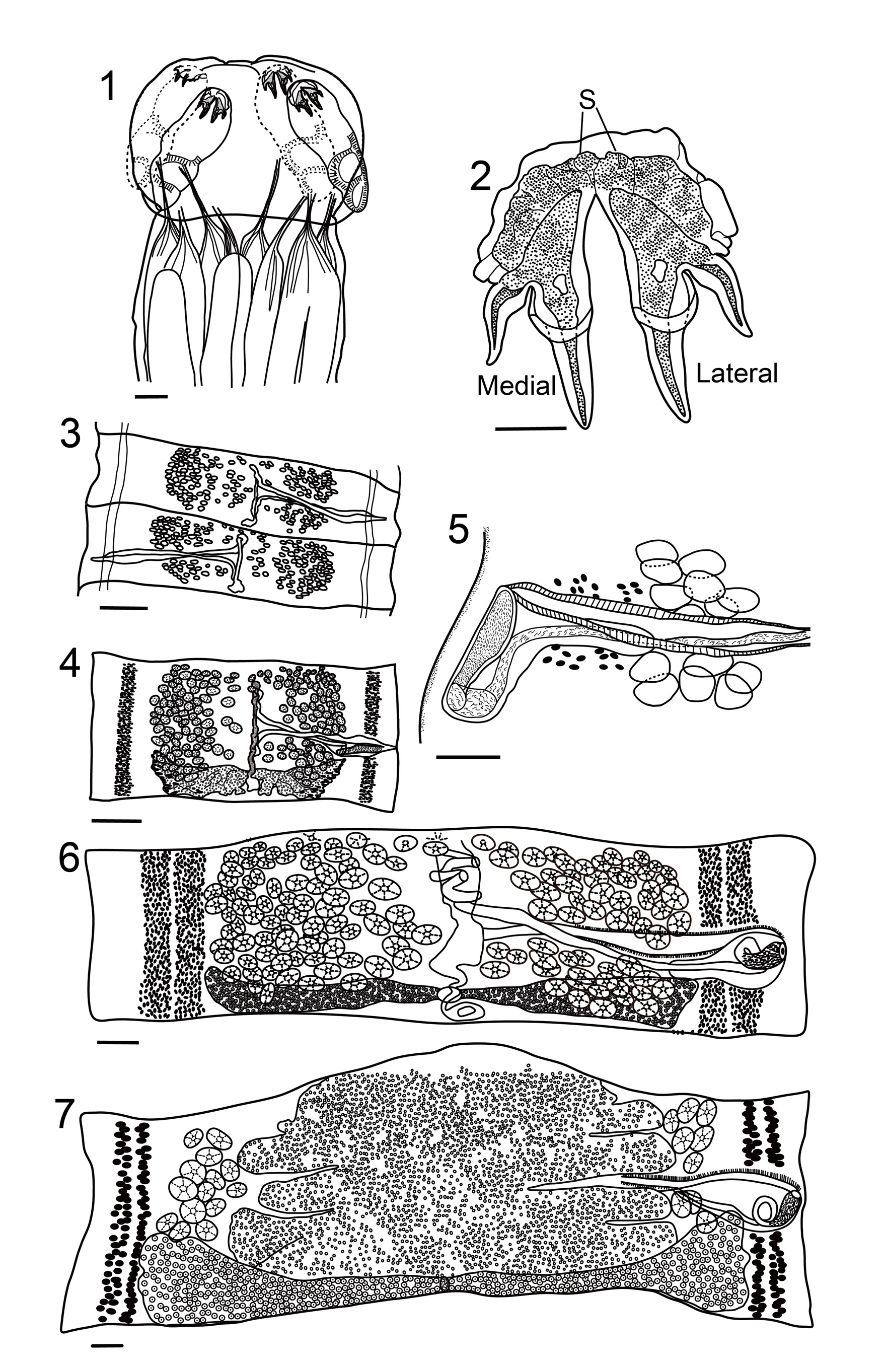
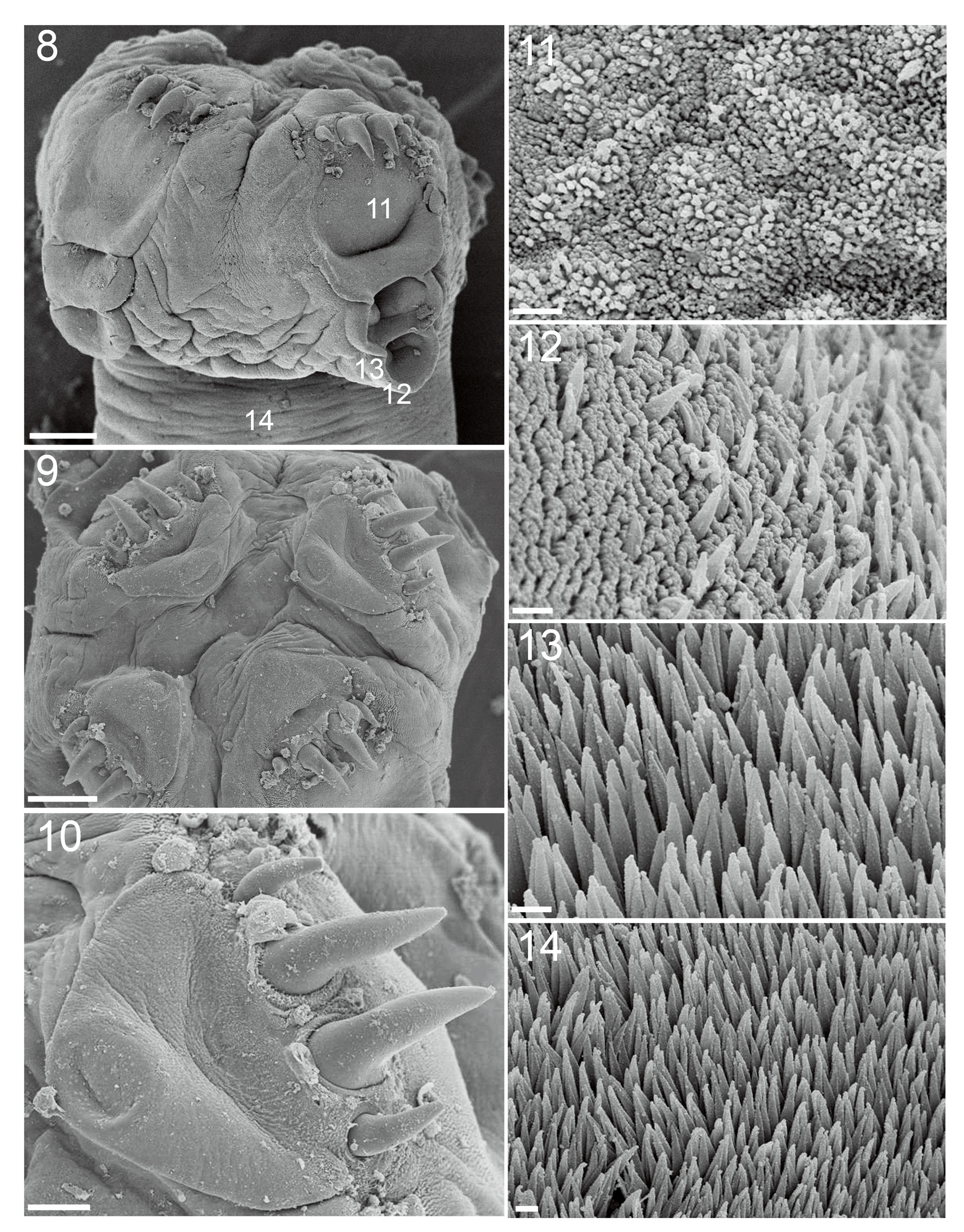
Morphological description. [Based on 38 whole mounts (33 mature and 5 immature) and 2 worms examined with SEM]: Worms anapolytic, 5.1 – 24.1 cm (13.06 ± 5.00; 22) long; greatest width at level of terminal proglottid; with 171 – 499 (254 ± 67; 20) proglottids per worm, excluding some immature proglottids too small to count clearly; genital pores marginal, irregularly alternating, 42 – 56% (48 ± 4; 19) of proglottids length from posterior end. Scolex consisting of scolex proper and conspicuous cephalic peduncle. Scolex proper with 4 linguiform bothridia, bothridia 601 – 1028 (790 ± 111; 19) long by 286 – 405 (326 ± 29; 19) wide; not free posteriorly. Each bothridium with 3 loculi, muscular pad and apical sucker; muscular pad and apical sucker conspicuous with SEM ( Figs. 8 – 10 View FIGURES 8 – 14 ). Muscular pad triangular, 167 – 193 (180 ± 18; 2) long by 132 – 140 (136 ± 5; 2) wide. Apical sucker oval, 42 – 45 (44 ± 2; 2) in diameter. Anterior loculus 297 – 548 (444 ± 64; 19) long, middle loculus 108 – 245 (179 ± 38; 19) long, and posterior loculus 111 – 255 (169 ± 34; 19) long; loculi length ratio 1:0.36 – 0.45:0.37 – 0.47 (1:0.40:0.38 ± 0.14:0.09:0.08; n=19); maximum width of bothridia at the level of anterior loculus. Velum absent. Hooks bipronged, hollow, internal channel smooth; each hook with single conspicuous tubercle on proximal part of axial prong; axial prongs longer than abaxial prongs. Axial prong fenestrated in proximal part. Hooks on each bothridium closely linked in proximal portion, with large irregular-shaped sclerite attached ( Fig. 2 View FIGURES 1 – 7 ). Lateral and medial hooks approximately equal in size. Lateral hook measurements (n=23): Hook base length 66 – 103 (86 ± 10; 23), axial prong length 90 – 122 (107 ± 9; 23), abaxial prong length 44 – 64 (53 ± 6; 23), total lateral hook length 145 – 195 (177 ± 14; 23). Medial hook measurements (n=23): Hook base length 66 – 101 (84 ± 11; 23), axial prong length 91 – 125 (107 ± 9; 23), abaxial prong length 45 – 65 (54 ± 5; 23), total medial hook length 148 – 202 (177 ± 15; 23). Ratio of total hook length (both medial hook and lateral hook) to bothridial length: 1:4.00 – 5.09 (4.48 ± 0.37; 19). Cephalic peduncle 3202 – 8692 (5829 ± 1806; 16) long by 913 – 1607 (1222 ± 191; 16) wide.
Distal bothridial surfaces covered with acicular and capilliform filitriches ( Fig. 11 View FIGURES 8 – 14 ). Bothridial surfaces at lateral margin coated with gladiate spinitriches interspersed with capilliform filitriches, beginning at mid-level of posterior loculus ( Fig. 12 View FIGURES 8 – 14 ). Proximal portion with densely arranged gladiate spinitriches ( Fig. 13 View FIGURES 8 – 14 ). Surfaces of cephalic peduncle covered with gladiate spinitriches only ( Fig. 14 View FIGURES 8 – 14 ).
Proglottids slightly craspedote. Immature proglottids 94 – 338 (156 ± 51; 22) in number; mature proglottids 21 – 73 (53 ± 13; 20) in number, 225 – 1507 long by 704 – 2077 wide and with length-to-width ratio 0.16 – 1.85 (0.6 ± 0.46; 25):1; gravid proglottids 0 – 103 (45 ± 33; 19) in number; 718 – 2395 long by 1011 – 2974 wide; terminal gravid proglottids length-to-width ratio 0.48 – 1.74 (1.0 ± 0.40; 18):1. Testes irregularly oval in dorsoventral view, 37 – 96 long by 25 – 75 wide, arranged irregularly, from level of ovarian isthmus, overlapping ovary, to anterior margin of proglottid; testicular field one or two layers deep. Testes 117 – 174 (145 ± 20; 18) in total number in mature proglottid, 29 – 66 (46 ± 9; 18) pre-vaginal; 17 – 37 (28 ± 5; 18) post-vaginal; 53 – 94 (76 ± 11, 18) aporal. In gravid proglottids, testes more or less atrophied and situated dorsal to uterus. Vas deferens extensive, coiled, filling intervitelline region anterior to cirrus sac. Cirrus sac straight ( Fig. 5 View FIGURES 1 – 7 ), post-equatorial, 214 – 415 (300 ± 61, 18) long by 96 – 196 (144 ± 29, 18) wide, containing coiled cirrus; cirrus expanded at base; proximal and part of distal portion with spinitriches. Vagina opening into genital atrium anterior to cirrus, thick walled, extending from ootype along medial line of proglottid anteriorly, then turning towards genital pore, passing anterior margin of cirrus sac to common genital atrium; vaginal sphincter not observed; seminal receptacle not seen. Ovary located at or near posterior end of proglottid, each lobe consisting of numerous small and distally rounded acini, maximum width 139 – 374 (250 ± 67; 19), bilobed in dorsal or ventral view, essentially symmetrical; poral lobe 359 – 823 (561 ± 139; 18) long and aporal lobe 341 – 731 (547 ± 113; 18) long, extending to (or slightly overlapping) cirrus sac; ovarian isthmus located at approximate mid-point of ovary. Mehlis’ gland posterior to ovarian isthmus. Vitellarium follicular, including 2 lateral bands, each further comprising 2 columns; extending from near anterior margin of proglottid to near posterior margin, interrupted by vagina and cirrus sac dorsally and ventrally, not interrupted by ovary. Uterus in gravid proglottids full of eggs; extending full length, or nearly full length of proglottid. Eggs spherical, conspicuous in gravid proglottid, 10 – 14 (12 ± 1; 16) in diameter.
Remarks. Acanthobothrium ningdense n. sp. belongs to species category 4 of Acanthobothrium suggested by Ghoshroy & Caira (2001) because of its characteristics of total length ≥ 15 mm, ≥ 50 proglottids, ≥ 80 testes, and with a symmetrical ovary. Currently, 38 species in category 4 have been reported ( Pramanik & Manna 2010; Fyler & Caira 2010; this paper), including six species from D. akajei , the only known host for A. ningdense . Acanthobothrium ningdense n. sp. is readily distinguished from the 31 species in category 4 by the number of the testes, 117 – 174, which is substantially more than A. adlardi Campbell & Beveridge, 2002 (83 – 103), A. cribbi Campbell & Beveridge, 2002 (72 – 96), A. karachiense Bilqees, 1980 (74 – 98), A. microcephalum Alexander, 1953 (90 – 100), A. myliomaculata Srivastav, Shweta & Noopur, 1995 (60 – 98), A. robustum Alexander, 1953 (99 – 101), A. satyanarayanaraoi Sanaka, Vijaya Lakshmi & Hanumantha Rao, 1993 (80 – 90), and A. wedli Robinson, 1959 (80 – 100), but significantly fewer than A. magnum Euzet, 1959 (220) and A. polytesticularis Wang & Yang, 2001 (208 – 228). Acanthobothrium ningdense n. sp. has more post-vaginal testes (17 – 37) than A. zugeinense Yang & Lin, 1994 (9 – 14), A. ramiroi Ivanov, 2005 (5 – 11), and fewer testes on the anti-poral side (53 – 94) than A. hanumantharaoi Rao, 1977 (97 – 108). Acanthobothrium ningdense n. sp. can be distinguished from A. triacis Yamaguti 1952 and A. terezae Rego & Dias, 1976 by possessing symmetrical hooks (medial and lateral hooks nearly identical) rather than asymmetrical hooks as in the latter two. The new species can be distinguished from A. barusi Pramannik & Manna, 2010 , A. majumdari Pramannik & Manna, 2010 , and A. paramanandai Pramannik & Manna, 2010 , by having shorter bothridia (601 – 1028 vs. 1200 – 1300, 1700 – 2300, 1100 – 1900 in length, respectively). The new species is also different from 8 other category 4 species ( A. bajaense Appy & Dailey, 1973 , A. coronatum (Rudolphi, 1819) Blanchard, 1848 , A. dighaense Srivastava & Capoor, 1980 , A. heterodonti Drummond, 1937 , A. intermedium Perrenoud, 1931 , A. musculosum (Baer, 1948) Yamaguti, 1959 , A. rhynchobatidis Subhapradha, 1955 , A. cestraciontis Yamaguti, 1934 ) by possessing a conspicuously sclerotised plate connecting two hooks ( Fig. 2 View FIGURES 1 – 7 ). Acanthobothrium ningdense n. sp. differs from A. pichelinae Campbell & Beveridge, 2002 by lacking conspicuous lateral spurs on the proximal portion of abaxial prongs, and from A. bengalense Baer & Euzet, 1962 by hook shape (axial prong length much longer than abaxial prong length in A. ningdense vs. axial and abaxial prong in same length in A. bengalense ). Acanthobothrium ningdense n. sp. differs from A. matttaylori Fyler & Caira, 2010 , A. cannoni Campbell & Beveridge, 2002 and A. dysbiotos (MacCallum, 1921) Williams, 1969 in that it is anapolytic.
Acanthobothrium ningdense n. sp. differs from six of the seven species of Acanthobothrium reported from the same host, D. akajei View in CoL , as follows: the bothridia of A. ningdense n. sp. are oriented laterally ( Fig. 1 View FIGURES 1 – 7 ), whereas the bothridia of A. ijimai , A. grandiceps and A. macrocephalum are oriented more apically (forward), so that the scolex appears quadrangle in apical view (Fig. 69 in Yamaguti, 1952 for A. grandiceps ), a type of configuration termed ‘clover-leaf’ by some authors ( Yoshida 1917; Fyler & Caira 2010). Acanthobothrium ningdense n. sp. further differs from A. ijimai and A. grandiceps by lacking a conspicuous nodular swelling of the septum between the anterior and middle loculi on the bothridia. Acanthobothrium ningdense n. sp. can be differentiated from A. dasybati by having longer hooks (145 – 195 for lateral hooks and 148 – 202 for medial hooks), rather than about 130 for all hooks of A. dasybati . Acanthobothrium ningdense n. sp. possesses a sclerite ( Fig. 2 View FIGURES 1 – 7 , see also Figs. 4, 5, 6 View FIGURES 1 – 7 , in Campbell & Beveridge, 2002) at the base of the hooks, whereas there is no such structure in A. dasybati .
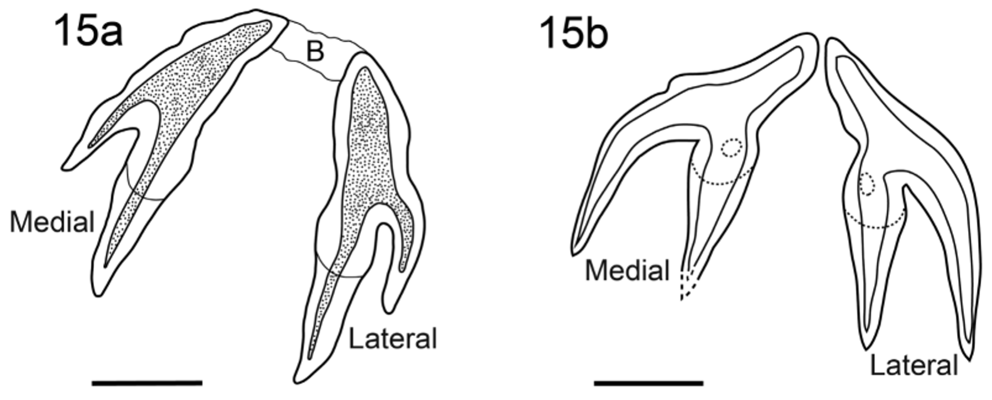
Among the species previously described from D. akajei View in CoL , A. latum and A. micracantha are most similar to A. ningdense n. sp.. Careful observations of the paratypes (MPM: 2 2635 and 22636) of A. micracantha confirmed that there is a long connecting bar between the bases of medial and lateral hooks ( Fig. 15a View FIGURES 15 a – 15 b. 15 a ), which was not indicated in the original description (see Fig. 61 in Yamaguti, 1952). The connecting bar in A. micracantha distinguishes it from A. ningdense n. sp. which does not have such a structure, but possesses a large irregular shaped sclerite ( Fig. 2 View FIGURES 1 – 7 ). In addition, A. ningdense n. sp. has distinctively larger hooks than those of A. micracantha 145 – 202 vs. 75 – 90 for total hook length, 90 – 125 vs. 40 – 50 for axial prong length and 44 – 65 vs. 20 – 25 for abaxial prong length. The hook drawing of A. latum in Yamaguti’s original paper is problematic because the angle is such that it seems that it was observed en face (See Fig. 63 in Yamaguti 1952). Examination of the holotype of A. latum Yamaguti, 1952 revealed that the author chose one of two pairs of hooks to illustrate in his original description, drawing the hook pair at an unusual angle. In this paper the hooks of the holotype of A. latum were redrawn (see Fig. 15 View FIGURES 15 a – 15 b. 15 a b). Acanthobothrium ningdense n. sp. can be differentiated from A. latum by its much longer axial prong than the abaxial one (90 – 125 vs. 44 – 65; see Fig. 2 View FIGURES 1 – 7 ), rather than the axial prong as long as the abaxial one in A. latum (about 80, Fig. 15 View FIGURES 15 a – 15 b. 15 a b, in order to facilitate comparison). The seventh species of Acanthobothrium previously reported from D. akajei View in CoL , A. tsingtaoense was considered either incertae sedis ( Goldstein 1967), or not possible to categorize ( Fyler & Caira 2006). Nonetheless, the information in the original description of A. tsingtaoense is sufficient to distinguish it from A. ningdense n. sp.. The bothridia of A. ningdense n. sp. are much narrower than those of A. tsingtaoense (286 – 405 vs. 940 – 1280).
No known copyright restrictions apply. See Agosti, D., Egloff, W., 2009. Taxonomic information exchange and copyright: the Plazi approach. BMC Research Notes 2009, 2:53 for further explanation.
|
Kingdom |
|
|
Phylum |
|
|
Class |
|
|
Order |
|
|
Family |
|
|
Genus |
Acanthobothrium ningdense
| Yang, Chaopin, Sun, Yuan, Zhi, Tingting, Iwaki, Takashi, Reyda, Florian B. & Yang, Tingbao 2016 |
A. latum
| Yamaguti 1952 |