
Paraleptophlebia falcula Traver 1934
|
publication ID |
https://doi.org/10.11646/zootaxa.4098.2.9 |
|
publication LSID |
lsid:zoobank.org:pub:1EDE074A-2C2A-4BB0-8A35-7C4C2DFA208D |
|
DOI |
https://doi.org/10.5281/zenodo.6075464 |
|
persistent identifier |
https://treatment.plazi.org/id/03E08C6B-FFB5-FFD1-74E6-FB6936432E8C |
|
treatment provided by |
Plazi |
|
scientific name |
Paraleptophlebia falcula Traver 1934 |
| status |
|
Paraleptophlebia falcula Traver 1934 View in CoL
( Figs 1–37 View FIGURES 1 – 13 View FIGURES 14 – 15 View FIGURES 16 – 22 View FIGURES 23 – 25 View FIGURES 26 – 30 View FIGURES 31 – 32 View FIGURES 33 – 37 )
Paraleptophlebia falcula Traver 1934: 190 View in CoL (male imago); Needham et al. 1935: 520 (male imago); Harper & Harper 1986: 1465 (male imago).
Material. RUSSIAN FEDERATION: AMURSKAYA OBLAST’: Zeya River basin: Bolshaya Makcha river, bridge at highway Tygda – Zeya, 28.VI.2013, Coll. T. Tiunova: 1 ♂ im., 1 2♀im. (reared from larvae); same place, 28.VI.2015, coll. T. Tiunova: 1 ♂ im., 3 ♀im. (reared from larvae), 3 larvae; Allinga river (river system Allinga- Tynda-Dep-Zeya), low bridge at highway Zeya – Snezhnogorsk, 14.VII.2012, coll. T. Tiunova: 1 ♂ im., 1 ♀im. (reared from larvae), 8 larvae; same place, 24.VII.2014, coll. T. Tiunova: 10 larvae; Allinga river near mouth, 29– 30.VI.2015, coll. T. Tiunova: 5 ♂ im., 3 ♀im. (reared from larvae), 11 larvae; Amur river basin, Talali river, upper reaches, about 5 km above Gonzha village, 25.VIII.2004, coll. T. Tiunova: 2 ♂ im., 3 ♀im ( IBSS). TRANSBAIKAL KRAY, Chasovinka river (tributary of Shilka river) about 300 m above mouth, 24.VII.2005, coll. T. Tiunova: 2 ♂ im., 3 ♀im. (reared from larvae), 2 L ( IBSS). SAKHA YAKUTIA, Chulman river basin, Amnunnykta river, about 500 m upper mouth, 28.VII.2010, coll. T. Tiunova: 1 ♂ subim., 1 larva ( IBSS). KOMI REPUBLIC, Vorkuta: Ayach Yaga river (tributary of Vorkuta river), 4.VIII.1958, coll. Chernykh: 3 ♂ im.; Sabrey- Yaga river, coll. Iordansky: 1 ♂ im. (only styliger on slide made by O. Chernova) ( ZIN).
Description. Mature larva. Length (mm): body 5.3–6.8; cerci 4.6–8.2. Head: Brown with occipit light brown and pale spots around lateral ocelli and anterior to median ocellus (cuticular coloration). Antennae white. Labrum dark brown; dorsal surface covered with long hair-like setae; anterior margin with shallow emargination and several rows of stout setae ( Fig. 1 View FIGURES 1 – 13 ). Mandibles dark brown, with broad band extending parallel to outer edge ( Figs 2–3 View FIGURES 1 – 13 ). Left mandible with incisor terminated by 4 denticles and kinetodontium terminated by 3 denticles ( Fig. 4 View FIGURES 1 – 13 ), right mandible with incisor terminated by 3 denticles and kinetodontium terminated by 3 denticles ( Fig. 5 View FIGURES 1 – 13 ). Both mandibles with extensive patches of setae on dorsal surface in middle area. Right mandible with row of long hairlike setae proximal of molar surface ( Fig. 3 View FIGURES 1 – 13 ). 1st segment of maxillary palp with several spine-like setae along outer margin and small thin setae along inner margin; 2nd segment with long hair-like setae on both margins; 3rd segment almost completely covered with very long, thin hair-like setae ( Fig. 6 View FIGURES 1 – 13 ). Maxillary palp with 2nd and 3rd segments subequal in length, together 1.55 times longer than 1st segment. Labium as in Fig. 7 View FIGURES 1 – 13 ; apical part of dorsal surface of glossae densely covered with stout hair-like setae; among them small stout setae located nearer to inner edge; dorsal surface of paraglossae covered with small stout setae and long hair-like lateral setae ( Fig. 7 View FIGURES 1 – 13 ); 3rd segment of labial palp with rows of stout variably spaced setae ( Fig. 8 View FIGURES 1 – 13 ). Labial palp with 2nd segment shorter than 3rd segment, 2nd and 3rd segments together longer than 1st segment. Hypopharynx slightly emarginated apically ( Fig. 9 View FIGURES 1 – 13 ). Thorax: Pronotum brown with white lateral margins and wide pale stripe along posterior margin; mesonotum brown with diffuse pale spots anteriorly and between protoptera (cuticular coloration) ( Fig. 14 View FIGURES 14 – 15 ). Legs grayish. Forefemora with row of stout setae on inner edge; anterior side of forefemora covered with stout setae of identical form and size ( Fig. 10 View FIGURES 1 – 13 ). Claw with single row of denticles, reaching middle of claw; apex of claw elongate ( Fig. 11 View FIGURES 1 – 13 ). Ratio of length femur/tibia and tibia/tarsus: fore and middle legs 1.3–1.2 and 1.4–1.5; hind leg 1.4–1.2 and 1.4–1.7. Abdomen: Terga brown with light spots; lateral edges pale; terga II–III with small triangular light spot on median part of posterior margin; terga IV–VI with a toothed light spot occupying about half of tergum length, on terga VII–IX reaching anterior margin; tergum X light with brown lateral edges (cuticular coloration) ( Fig. 14 View FIGURES 14 – 15 ). Abdominal terga III–IX with regular row of triangular stout spines on posterior margin and irregular rows of small spines anteriad of it ( Fig. 12 View FIGURES 1 – 13 ). Posterolateral spines on abdominal segment IX longer than on segment VIII. Abdominal sterna light brown with brown band on anterior margin, lateral edges pale; sterna VIII–IX pale, yellowish; sternum X pale, yellowish with brown lateral angles (cuticular coloration) ( Fig. 15 View FIGURES 14 – 15 ). Sterna I–VII with ganglion spots. Sternum IX of female with postero-median emargination ( Fig. 13 View FIGURES 1 – 13 ). All tergalii hairy ( Figs 16–22 View FIGURES 16 – 22 ); hairs very thin, pale, and break off easily; well visible only on a dark background. Cerci pale, grayish, darker at base.
Male imago (in alcohol). Length (mm): body 6.0–8.5; forewings 6.8–8.5; cerci 7.7–9.5. Head: General color dark brown to dark. Antennae brown; ocelli whitish apically and black basally. Upper portion of compound eyes light brown to yellowish or grayish-brown; low portion dark ( Figs 23–24 View FIGURES 23 – 25 ); compound eyes contiguous ( Fig. 24 View FIGURES 23 – 25 ). Thorax: General color dark brown. Medioscutum and submedioscutum dark brown ( Fig. 24 View FIGURES 23 – 25 ); posterior scutal protuberance dark with small light spot on lateral sides; sublateroscutum brown to light brown. Wings pale, hyaline; all veins pale, almost invisible; veins C and Sc slightly darker and visible; pterostigma milky ( Fig. 23 View FIGURES 23 – 25 ). Femora and tibia of forelegs whitish or yellowish, joints brown; tarsal segments pale or whitish. Middle and hind legs grayish, femur slightly darker. Ratio of length of femur/tibia 0.8–0.9; ratio of tarsal segments length 0.95:1.5:2.8:3.3. Abdomen: Terga with contrasting pattern ( Fig. 24 View FIGURES 23 – 25 ): tergum I dark brown; tergum II light brown or brownish; terga III–VI dirty yellowish, translucent, posterior margins darker; lateral sides with dark tracheal trunk; tergum VII dirty brownish with pair of darker stripes in middle area, with posterior margin dark brown; terga VIII– IX dark. Abdominal sterna with contrasting pattern: sternum I dark brown; sternum II brownish; sterna II–VI yellowish, translucent; sternum VIII dirty gray or dark brown; sternum IX dark brown with light central area. Nerve ganglia brown, visible on segments II–VII ( Fig. 25 View FIGURES 23 – 25 ). Cerci white. Genitalia: Styliger brown, somewhat lighter in posterior area, gonostyli yellowish. Styliger deeply concave medially, with U-shaped incision and two long slightly curved processes reaching bases of ventral processes of penis ( Figs 26, 29, 30 View FIGURES 26 – 30 ). Gonostylus with 1st+2nd segment widest at middle, bent ventrally near base; 3rd segment slightly widened apically; 4th segment smallest ( Fig. 26 View FIGURES 26 – 30 ). Penis lobes separated by U-shaped cleft reaching middle of penis length; each penis lobe with triangular widening, with apex pointed and curved inwards, ventrally with long crescent-shaped pointed process directed proximally and reaching proximal 1/3 of penis ( Figs 27, 30 View FIGURES 26 – 30 ).
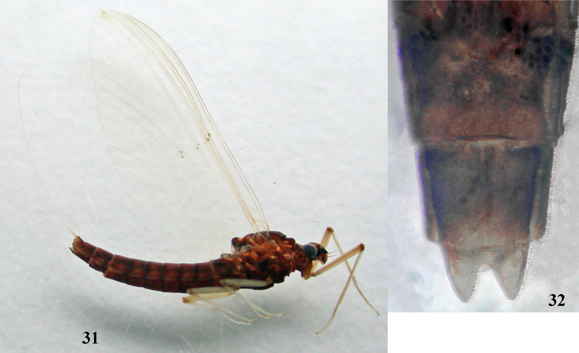
Female imago (in alcohol). Length (mm): body 7.3–8.1; forewing 8.0–9.0; cerci 7.5–8.5. Head: General color dark brown. Antennae brown; ocelli whitish apically, black basally. Thorax: General color dark brown ( Fig. 31 View FIGURES 31 – 32 ). Medioscutum and submedioscutum from brown; posterior scutal protuberance dark; sublateroscutum brown with white maculation; scuto-scutellar impression dirty brownish or dirty whitish. Wings pale, hyaline; longitudinal veins of forewing light brown, visible; hind wing colorless; all cross veins colorless, poorly visible. Legs light brown, femur slightly darker. Ratio of length femur/tibia and tibia/tarsus on forelegs 1.0–0.9 and 1.3–1.4. Abdomen: Terga and sterna dark brown ( Fig. 31 View FIGURES 31 – 32 ). Subanal plate with deep V-shaped incision ( Fig. 32 View FIGURES 31 – 32 ). Cerci yellowish or whitish.
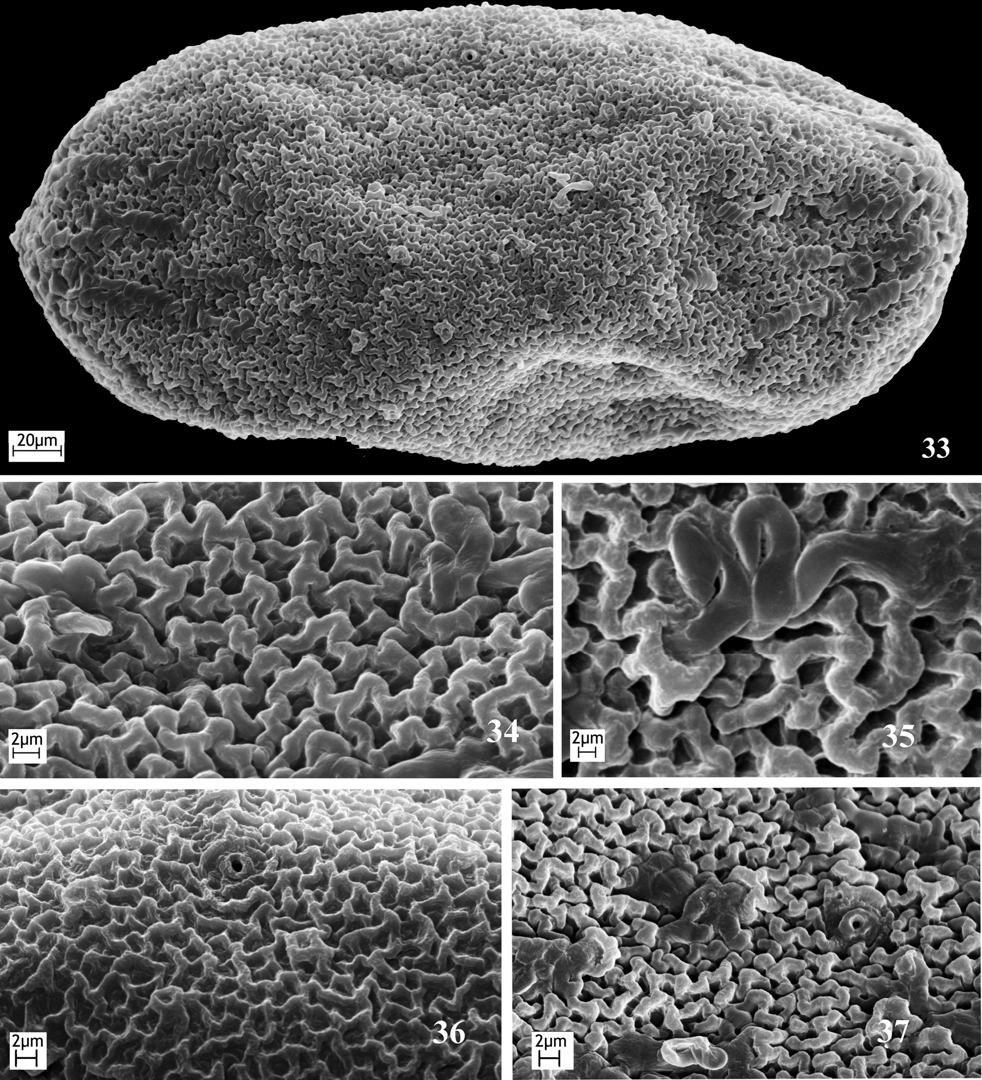
Eggs. General form oval 190–214 µm length and 85–118 µm width ( Fig. 33 View FIGURES 33 – 37 ). Chorion rugose, with broad and deep folds ( Fig. 34 View FIGURES 33 – 37 ), with small flower-shape KCT sparsely arranged ( Figs 33, 35 View FIGURES 33 – 37 ). Two or three small round micropyles in middle area ( Fig. 33 View FIGURES 33 – 37 ). Sperm guide absent ( Figs 36–37 View FIGURES 33 – 37 ).
Comparison. Male imago of P. falcula differs from all other species by presence of long processes on styliger ( Figs 26, 29, 30 View FIGURES 26 – 30 ).
Among species of Paraleptophlebia with available larval descriptions, P. falcula differs from Nearctic species P. bicornuta ( McDunnough 1926) , P. helena Day 1952 , P. packii (Needham 1927) and P. z a y a nt e Day 1952 by absence of tusk-like elongation of mandibular incisor; from P. altana Kilgore & Allen 1973 and P. cachea Day 1954 it differs by non-widened lobes of tergalii without dark lateral branches of main trachea. The same is known for the following species of Paraleptophlebia : P. calcarica Rowbotham & Allen 1988 ( Jacobus & McCafferty 2004) , P. c i nc t a ( Retzius 1783) ( Macan 1952; Landa 1969), P. debilis ( Walker 1853) ( Ide 1930; Gordon 1933), P. guttata ( McDunnough 1924a) ( Ide 1930; Gordon 1933), P. jeanae Berner 1955 ( Randolph & McCafferty 1996) , P. lacustris Ikonomov 1962 ( Ikonomov 1962) , P. moerens ( McDunnough 1924a) ( Gordon 1933; Burks 1953), P. ontario ( McDunnough 1926) ( Needham, Traver & Hsu 1935; Burks 1953), P. praepedita (Eaton 1884) ( Gordon 1933; Burks 1953), P. quisquilia Day 1952 ( Day 1952) , P. ruffoi Biancheri 1956 ( Belfiore & Giangrande 1979) , P. strandii ( Eaton 1901) ( Tiensuu 1939) , P. strigula McDunnough 1932 ( McDunnough 1932) , P. submarginata ( Stephens 1835) ( Komarek 1921; Macan 1952; Landa 1969; Belfiore & Giangrande 1979), P. volitans ( McDunnough 1924a) ( Ide 1930; Gordon 1933), P. w e r n e r i Ulmer 1920 ( Landa 1969) and P. w e s t on i Imanishi 1937 ( Gose 1979 –1980).
Mature larvae of P. falcula can be distinguished by the following features: thorax is not hairy (in contrast to P. volitans ); hypopharynx is slightly emarginated apically ( Fig. 9 View FIGURES 1 – 13 ) ( P. strandii has medioapical cleft); maxillary palp segments 2 and 3 are subequal in length, and together 1.55 times longer than segment 1 ( Fig. 6 View FIGURES 1 – 13 ) (in P. de b i l i s and P. moerens segments 2 and 3 together are equal to segment 1); labial palp segments 2 is shorter than segment 3, and together are longer than segment 1 ( Figs 7, 8 View FIGURES 1 – 13 ) (in P. calcarica , P. lacustris and P. strandii segments 2 and 3 together are shorter or equal to segment 1); legs have no brown band ( Figs 14, 15 View FIGURES 14 – 15 ) (in contrast to P. debilis and P. jeanae ); row of denticles reaches middle of claw ( Fig. 11 View FIGURES 1 – 13 ) (in P. lacustri s, P. submarginata , P. r uf f o i, P. w er n e r i and P. westoni row of denticles occupies more than half of claw); abdominal segment 8 with posterolateral spines, which are shorter than spines on segment 9 ( Fig. 15 View FIGURES 14 – 15 ) (in P. gu t t a t a and P. strigula lateral spines are present on segment 9 only).
Habitat. Larvae and adults of P. falcula were collected in the Trans-Baikal Region, Sakha Yakutia and Amurskiy Region at the end of June – beginning of July. The larvae were collected in small rivers (width 10–20 m, average depth 0.15–0.80 m in mean water). The water temperature in this period did not exceed 13ºC. The bottom substratum was rocky and gravel. The rivers of the Bolshaya Makcha and Amnunnykta have aquatic vegetation ( Figs 38–39 View FIGURES 38 – 39 ). The larvae inhabited gravel substratum with detritus in the slow flow.
Distribution. Range of P. falcula , as revealed by this study, well agrees with the concept of Amphipacific sector of Holarctic as defined by Levanidova (1982). Area of distribution of P. falcula includes Eastern Palaearctic (known from certain localities in Transbaikal Kray, Sakha Yakutia and Amurskaya Oblast') and West Nearctic (known from certain localities in states Washington, Oregon and Idaho of the USA). As well as areas of some other amphipacific species, area of P. f a l c ul a is stretched to the west at the north, so that includes north of the Urals.


| ZIN |
Russian Academy of Sciences, Zoological Institute, Zoological Museum |
No known copyright restrictions apply. See Agosti, D., Egloff, W., 2009. Taxonomic information exchange and copyright: the Plazi approach. BMC Research Notes 2009, 2:53 for further explanation.
|
Kingdom |
|
|
Phylum |
|
|
Class |
|
|
Order |
|
|
Family |
|
|
Genus |
Paraleptophlebia falcula Traver 1934
| Tiunova, Tatiana M. & Kluge, Nikita 2016 |
Paraleptophlebia falcula
| Harper 1986: 1465 |
| Needham 1935: 520 |
| Traver 1934: 190 |