
Leptocerina pauliani (Ross, 1957)
|
publication ID |
https://doi.org/10.11646/zootaxa.4981.1.10 |
|
publication LSID |
lsid:zoobank.org:pub:66C49072-2A0E-4DD4-A440-F156ADD8F639 |
|
persistent identifier |
https://treatment.plazi.org/id/03E187CD-FFA0-FFBC-77AF-3D0B4A15F8CC |
|
treatment provided by |
Plazi |
|
scientific name |
Leptocerina pauliani |
| status |
|
Description of last instar larva View in CoL
The last instar larva of L. pauliani inhabits a case that is triangular in cross-section, made of three pieces of leaf attached by their longitudinal edges ( Fig. 2A View FIGURE 2 ). [Early instars build a cylindrical case made of sand and diverse plant particles ( Fig. 2B View FIGURE 2 )].
Body: Last instar larva length ~ 9.5 mm.
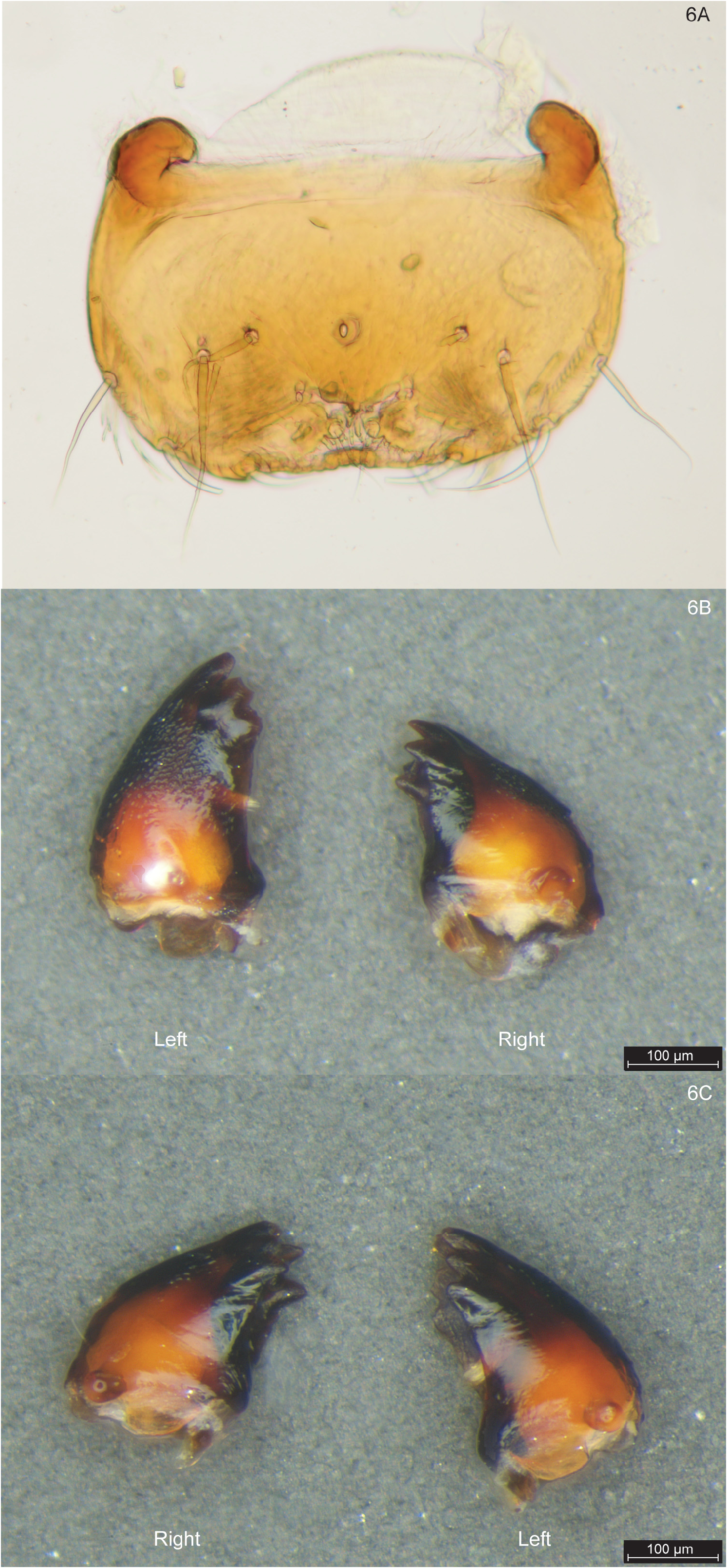
Head: Colouration uniformly brown with light-coloured muscle scars and pale circular areas around eyes ( Figs 3A–3C View FIGURE 3 ). Head elongate, tapering anteriorly, with antennae longer that half of width of frontoclypeal apotome, but not extending beyond anterior edge of labrum ( Figs 3B, 3C View FIGURE 3 ). Fronto-clypeal apotome narrowly triangular, constricted near mid-length, with pointed posterior apex joining short, asymmetrically curved coronal suture, convex to left ( Fig. 3B View FIGURE 3 ). Eyes large, dark, simple, surrounded by large, pale, circular areas ( Figs 3A, 3B View FIGURE 3 ). Pair of subocular ecdysial lines present on head capsule, running under eyes, horizontally reaching posterior part of head and dividing genae into upper and lower parts ( Fig. 3A View FIGURE 3 ). Labrum yellowish, rectangular, medially indented on anterior edge ( Figs 3B View FIGURE 3 , 6A View FIGURE 6 ). Mandibles brown, short, shredder-type; left mandible with median short brush, slightly larger than right mandible ( Figs 6B, 6C View FIGURE 6 ). Anterior ventral apotome forming pear-shaped elongate triangle nearly reaching posteriorly to two small posterior ventral sclerites ( Fig. 3C View FIGURE 3 ).
Thorax: Pronotum brown, concolorous with head capsule, medially divided and with anterolateral corners delimited by pair of slanting ecdysial lines, each starting from middle of lateral edge to reach submedial one-fifth of anterior edge of respective half sclerite ( Figs 3D, 3E View FIGURE 3 ). Foretrochantins flat, elongate, and finger-shaped ( Figs 3A, 3D View FIGURE 3 ). Sclerotised mesonotum medially divided, yellowish with brownish muscle scars and pair of conspicuous dark mesonotal bars characteristic of Athripsodini larvae extending from posterolateral corners to midlength; these mesonotal bars nearly parallel, each branching anteriorly to form fork ( Fig. 3E View FIGURE 3 ). Metanotum membranous and bearing pair of setal area 1 ( sa 1) setae submesally (best seen in profile in Figs 3D View FIGURE 3 , 4D View FIGURE 4 ); pair of small and ill-defined sclerites, each bearing one long and three small setae, present on setal area 3 ( sa 3, Figs 3E View FIGURE 3 , 4F View FIGURE 4 ). Pair of dark, transverse intersegmental sclerites between mesosternum and metasternum. Metasternum bearing three pairs of setae, two pairs of sa 3 setae laterally and one pair of sa 2 seta submesally ( Fig. 3F View FIGURE 3 , with right submesal seta broken).
Forelegs short and stocky; femora broad and with numerous long setae and row of pale strong spine-like setae on ventral margin; these strong setae also present on tibiae and tarsi; tarsal claws simple, short, curved, with prominent basal spine ( Fig. 4A View FIGURE 4 ). Midlegs longer than forelegs and also with rows of strong spine-like setae on femora, tibiae, and tarsi, and prominent basal spine on each claw ( Fig. 4B View FIGURE 4 ). Hind legs long and projected forward; rows of strong setae present on only tibiae ( Fig. 4C View FIGURE 4 , black arrow) and tarsi; prominent basal spine present on each claw. Hind femora, tibiae, and tarsi each with two rows of dense, long fringes of swimming setae, one row on each of dorsal and ventral margins ( Fig. 4C View FIGURE 4 ).
Abdomen: Segment I bearing one dorsal and two lateral humps ( Fig. 4D View FIGURE 4 ). Pair of long setae located on each side of dorsal hump ( Fig. 3E View FIGURE 3 , black arrows). Each lateral hump with patch of very short brownish spines and two long setae anteriorly and darker, slightly sinuous, longitudinal, sclerotized bar posteriorly ( Fig. 4E View FIGURE 4 ), similar to patch and bar of larvae of other Athripsodini species.
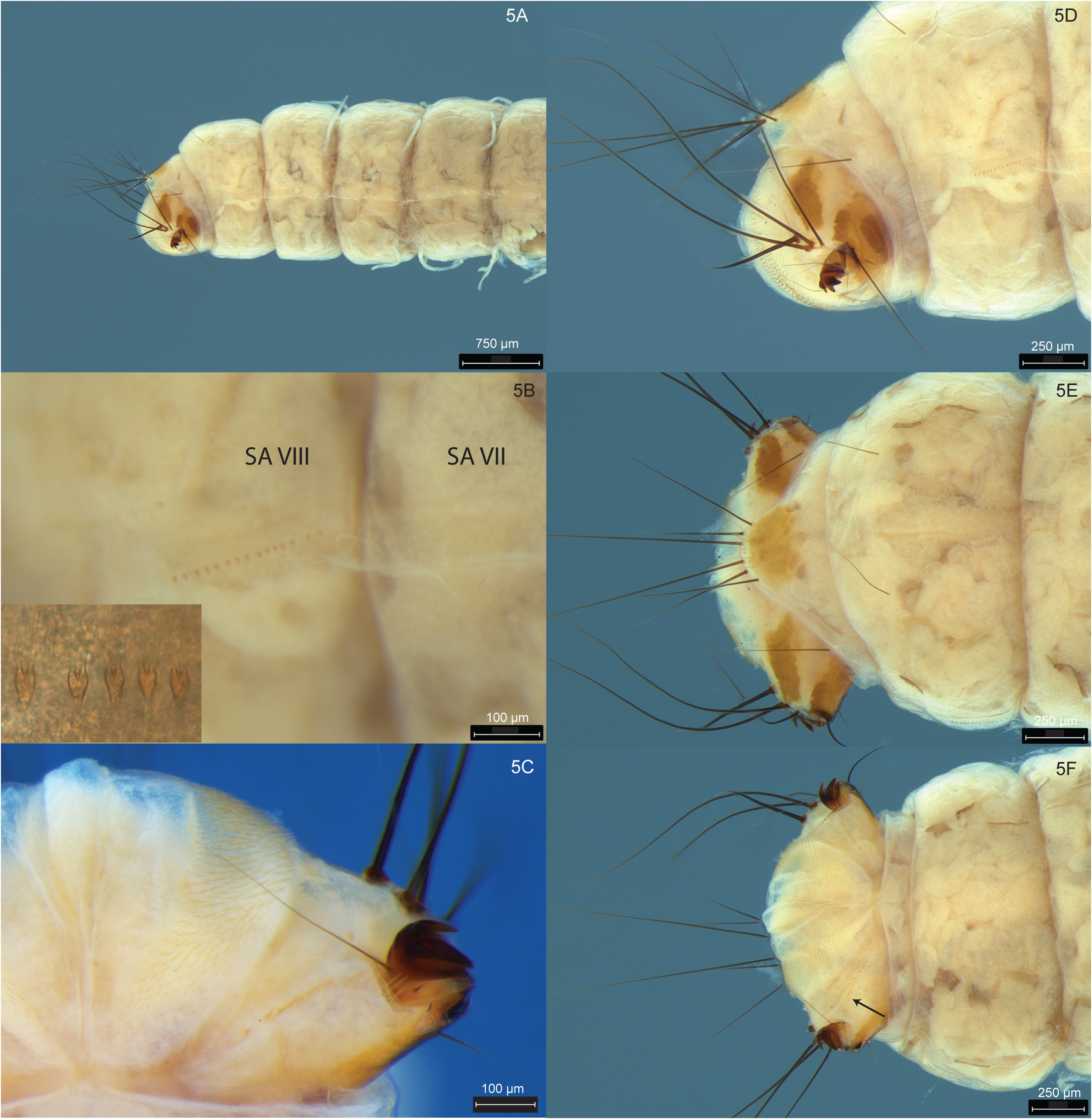
Gills simple ( Fig. 4D View FIGURE 4 ) and present on abdominal segments II–VII (only two dorsal gills on abdominal segment VII, see Fig. 5A View FIGURE 5 ).
Lateral fringe composed of dense, long, and fine setae from abdominal segment III to end of segment VII ( Fig. 5A View FIGURE 5 ). Continuing along line of lateral fringe on each side, row of short lateral tubercles present on abdominal segment VIII ( Fig. 5B View FIGURE 5 ).
Two long, dorsomesal upright setae present on abdominal tergite VIII ( Fig. 5E View FIGURE 5 ). Abdominal tergite IX bearing one dorsomesal sclerite with three pairs of large setae posteriorly; inner pair of setae longer than others and intermediate pair of setae shortest ( Figs 5D, 5E View FIGURE 5 ); row of two pairs of short, fine, lighter setae inserted between the row of these three pairs of long black setae and posterior edge of this sclerite. Two short lateral setae present on abdominal sternite IX (slightly visible in profile in Fig. 5D View FIGURE 5 ).
Anal prolegs short, each with smaller dorsal sclerite and larger sole plate in contact with claw ( Figs 5D, 5E View FIGURE 5 ). At base of claw insertion, on sole plate, one small and one long seta projecting ventrad ( Fig. 5D View FIGURE 5 ). Each basal tuft inserted just above claw consisting of four thick and long black setae, mesal seta shorter than other three ( Fig. 5D View FIGURE 5 ). In ventral view, oblique row of about fifteen upright spines extending from base of claw insertion almost to abdominal sternum IX ( Figs 5C, 5F View FIGURE 5 , black arrow). Ventral side of each anal proleg on each side of anus covered with many tight transverse rows of short, flattened spines ( Figs 5C, 5F View FIGURE 5 ). Anal claws strongly curved and armed apically with three accessory hooks ( Figs 5C, 5D View FIGURE 5 ).


No known copyright restrictions apply. See Agosti, D., Egloff, W., 2009. Taxonomic information exchange and copyright: the Plazi approach. BMC Research Notes 2009, 2:53 for further explanation.
|
Kingdom |
|
|
Phylum |
|
|
Class |
|
|
Order |
|
|
Family |
|
|
Genus |