
Chlamydolecanium Goux, 1933
|
publication ID |
https://doi.org/ 10.5281/zenodo.207983 |
|
DOI |
https://doi.org/10.5281/zenodo.5692131 |
|
persistent identifier |
https://treatment.plazi.org/id/03E187F5-FFDB-FF96-53CA-FA54FAB2FE37 |
|
treatment provided by |
Plazi |
|
scientific name |
Chlamydolecanium Goux, 1933 |
| status |
|
Chlamydolecanium Goux, 1933 View in CoL
Chlamydolecanium Goux 1933: 119 View in CoL . Type species Chlamydolecanium conchioides Goux View in CoL by original designation.
Generic diagnosis. Adult female
Dorsum. Dorsal setae entirely absent. Dorsal tubercles and tubular ducts absent. Preopercular pores present in a single longitudinal line medially, extending from anterior to anal plates to head. Simple pores and dorsal microducts present among preopercular pores and marginally. Anal plates each subtriangular and elongate, bearing strong spinose setae near apex.
Margin. Marginal setae spinose and sharply conical. Stigmatic setae not differentiated. Stigmatic clefts absent. Simple pores and minute microducts present among marginal setae.
Venter. Antennae short, 8 segmented. Legs well developed, without a tibio-tarsal articulatory sclerosis. Claw with a denticle. Claw digitules slightly knobbed. Stigmatic furrow with 5-loculi spiracular disc-pores. Tubular ducts present in a submarginal band and also medially on head and thorax. Pregenital disc-pores, each with 5-loculi, present on either side of anal cleft, pores with more than 5 loculi absent. Abdominal and inter-antennal setae short.
continued.
2 2 2 2 2 2 2 2 2 2 3 3 3 3 3 3 3 3
0 1 2 3 4 5 6 7 8 9 0 1 2 3 4 5 6 7 Antandroya View in CoL _ euphorbiae View in CoL 1 1 0 1 0 1 0 1 0 0 0 1 0 0 3 - 2 0 Cardiococcus View in CoL _umbonatus ?? 1? 0 1 0 1 0 1 0 1 2 1 0 0 0 0 Dicyphococcus View in CoL _ bigibbus View in CoL 1 1 0 1 0 1 0 1 0 1 0 1 1 1 1 0 0 0 Drepanococcus View in CoL _ cajani View in CoL 1 1 1 1 1 1 2 1 0 0 0 1 2 1 1 1 1 0 Edwallia View in CoL _rugosa 0 2 2 0 0 1 1 1 0 0 0 1 2? 0 0 0 0 Inglisia View in CoL _ patella View in CoL 0 1 1 0 0 1 0 0 0 0 0 0 2 1 0 0 0 0 Lecanochiton View in CoL _ metrosideri View in CoL 1 1 1 1 0 0 2 0 0 0 0 0 0 0 0 0 0 0
continued next page 2 2 2 2 2 2 2 2 2 2 3 3 3 3 3 3 3 3
0 1 2 3 4 5 6 7 8 9 0 1 2 3 4 5 6 7 Mitrococcus View in CoL _ celsus View in CoL ???? 1 1 0 0 0 1 0 0 2 1??? 0 Neolecanochiton View in CoL _grevilleae 0 0 1 1 0 1 0 1 0 0 0 0 3 1 0 0 1 0 Platinglisia View in CoL _ noacki View in CoL 0 0 1 1 0 1 0 0 0 0 0 0 0? 0 0 0 0 Pseudokermes View in CoL _ nitens View in CoL 1 1 1 1 0 1 2 1 0 0 0 0 0 1 0 0 0 0 Schizochlamidia View in CoL _mexicana 1 1 1 0 0 1 1 1 0 0 0 0 3 1 0 0 0 0 Chlamydolecanium View in CoL _ conchioides View in CoL 1 1 0 0 0 1 1 0 0 0 0 1 2 1 0 0 0 0 Cryptinglisia View in CoL _ lounsburyi View in CoL 1 1 1 1 0 1 1 1 0 0 0 1 2 1 0 0 0 0 Vitrococcus View in CoL _conchiformis 1 1 1 0 1 1 0 1 0 0 0 0 1 1 0 0 0 0 Coccus View in CoL _ hesperidum 1 0 1 0 0 0 1 1 1 0 1 1 0 0 0 1 1 1 Paralecanium View in CoL _ frenchii 1 0 1 1 0 0 0 1 0 0 1 1 0 0 1 1 1 0 Kenima View in CoL _ galilit View in CoL 1 1 1 1 0 0 2 0 0 1 0 1 1? 0 0 0 0 Parafairmairia View in CoL _ bipartita View in CoL 0 0 2 1 0 1 2 1 0 0 0 1 0 0 1 1 1 0 Ceroplastodes View in CoL _ dugesii View in CoL 1 1 1 0 0 1 0 1 0 1 0 1 3 0 1 1 1 0 Paracardiococcus View in CoL _actinodaphnis 1 0 0 1 0 1 2 0 0 1 0 1 2 1 3 - 2 1 Aphenochiton View in CoL _ inconspicuus View in CoL 1 0 1 0 0 1 1 1 0 0 1 1 2 0 1 0 1 0 Crystallotesta View in CoL _fagi 1 1 1 0 0 1 0 1 0 0 0 1 0 0 1 1 1 0 Ctenochiton View in CoL _viridis 1 0 1 0 0 1 1 1 0 0 1 1 2 0 1 1 1 (0 1) Epelidochiton View in CoL _ piperis View in CoL 1 0 1 1 0 1 2 1 0 0 0 1 2 0 0 0 0 0 Kalasiris View in CoL _ perforata View in CoL 1 0 1 1 0 1 0 1 0 0 1 1 1 0 1 1 1 0 Plumichiton View in CoL _pollicinus 1 2 2 0 0 1 1 1 1 0 0 1 2 0 1 1 0 0 Poropeza View in CoL _dacrydii 0 0 1 0 0 1 0 1 0 0 0 (0 1) 1 0 1 1 1 1 Umbonichiton View in CoL _hymenantherae 1 0 1 0 0 0 2 1 0 0 1 1 0 0 1 0 0 0 continued.
3 3 4 4 4 4 4 4 4 4 4 4 5 5 5 5 5 5 5
8 9 0 1 2 3 4 5 6 7 8 9 0 1 2 3 4 5 6 Antandroya View in CoL _ euphorbiae View in CoL 0 0 3 2 0 2 1 0 0 0 0 0 0 0 0 1 1 - - Cardiococcus View in CoL _umbonatus 1 1 1 0 0 1 1 0 1 1 0 0 1 0 1 0 0 0? Dicyphococcus View in CoL _ bigibbus View in CoL 0 0 1 0 0 1 1 0 1 1 1 0 1 1 0 0 0 0 3 Drepanococcus View in CoL _ cajani View in CoL 0 0 1 0 0 2 2 0 1 0 1 0 1 1 0 0 0 0 3 Edwallia View in CoL _rugosa 0 0? 0 0 1 1 0 0 2 1 0 1 0 1 0 0 0 3 Inglisia View in CoL _ patella View in CoL 0 2 1 0 0 1 1 0 1 0 1 0 0 0 0 0 0 2 3 Lecanochiton View in CoL _ metrosideri View in CoL 0 2 2 0 0 1 1 0 1 0 1 - - - 1 1 2 - - Mitrococcus View in CoL _ celsus View in CoL 0 0 0 0 0 1 1 0 0 2 1 2 0 0 1 1 1 - - Neolecanochiton View in CoL _grevilleae 0 0 2 0 0 1 1 0 1 0 1 2 0 0 1 1 1 - - Platinglisia View in CoL _ noacki View in CoL 2 2 0? 0 2 1 1 0 1 0 - - - 1 1 2 - - Pseudokermes View in CoL _ nitens View in CoL 1 1 1 0 0 1 1 0 1 2 0 2 0 0 1 1 1 - - Schizochlamidia View in CoL _mexicana 1 1 1 0 0 1 1 0 1 2 0 - - - 1 1 2 0 0 Chlamydolecanium View in CoL _ conchioides View in CoL 1 1 1 0 0 1 1 0 1 1 1 0 0 1 0 0 0 0 3 Cryptinglisia View in CoL _ lounsburyi View in CoL 0 0 1 0 0 1 1 0 1 1 1 0 1 1 0 0 0 2 3 Vitrococcus View in CoL _conchiformis 0 0 1 0 0 1 2 0 1 1 1 0 1 1 0 0 0 0 3 Coccus View in CoL _ hesperidum 0 0 2 0 1 1 0 1 1 0 1 1 2 0 0 0 0 0 3
continued next page Chlamydolecanium conchioides Goux, 1933 View in CoL
Chlamydolecanium conchioides Goux View in CoL : 120–123; Borchsenius, 1957: 138 –139 (taxonomy, description); Morrison & Morrison, 1966: 35 (taxonomy); Kozar & Walter, 1985: 76 (catalogue); Marotta, 1987: 100 (taxonomy, host, distribution); Ben-Dov, 1993: 63 (catalogue); Hodgson, 1994: 173 –175 (taxonomy, description); Longo et al., 1995: 122 (taxonomy); Foldi, 2003: 150 (host, distribution).
Material studied. Lectotype adult female (here designated): France, Corsica, Bastia, route de Cardo, on Lavandula stoechas L. ( Lamiaceae ), 16.viii.1930, coll. L.Goux ( MNHN 14749-1; LG 399/a1) ( MNHN). Paralectotype adult females, same data as lectotype, 6 adult females on 6 slides ( MNHN 14749-2, 14749-3, 14749-4, 14749-5, 14749-7, 14749-8); plus 3 first-instar nymphs on 2 slides ( MNHN 14749-6 and 14749-9) ( MNHN)
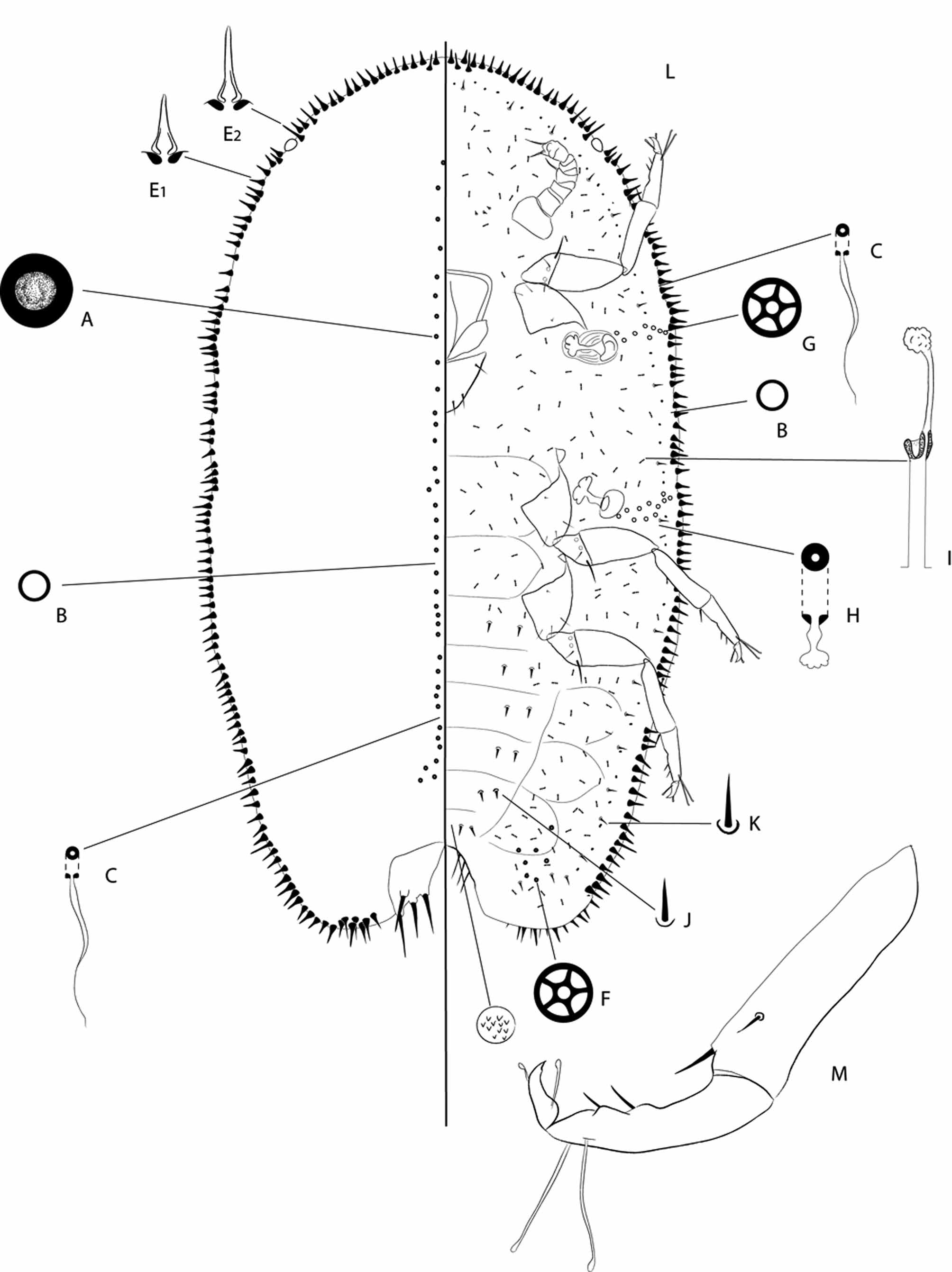
Adult female ( Figs 1 View FIGURE 1 and 2 View FIGURE 2 )
Unmounted material. Not available but the following information given by Goux (1933): adult female “covered by a waxy, glassy test, easily removable from the body, and composed of two symmetrical valves, lamellibranch-like with a hooked edge, each of the two hooks being directed backwards. The line joining the valves forms a deep furrow, which becomes narrower next to the hooks, becoming wider anteriorly and particularly posteriorly. Each valve has small ridges radiating from the top. In general, the test just resembles a small lamellibranch shell. When separated from the rest of the body, it is almost transparent; when in place, it is yellowish-grey and slightly iridescent. Length 3.0 mm, width 2.0 mm and height 2.5 mm. The dorsal surface bears two prominences separated by a longitudinal and median groove that corresponds to the valve sutures. The ventral surface is flat and even concave.”
Slide-mounted material. Fig. 1 View FIGURE 1 (from 14749-1) and Fig. 2 View FIGURE 2 (from 14749-3) (letters in the description refer to those on the figures). Body broadly oval, pear-shaped, with a broader posterior region; anal cleft short with divergent sides, forming an obtuse angle. Dorsum wider than venter. Body length 1.0–4.0 mm (lectotype: 3.15 mm); width 0.5–2.59 mm (lectotype: 2.44 mm).
Dorsum. Derm membranous, apart from an incomplete sclerotised crescent around anal plates on old adult females ( Fig. 1 View FIGURE 1 ). Dorsal setae entirely absent. Preopercular pores (A) round, concave with a granular surface in centre, each 5.–6 μm wide, present in a single medial longitudinal line anterior to anal plates (total number of pores: 42–44). Other dorsal pores of 2 types present, all distributed in same area as preopercular pores: (i) simple pores (B), circular, each 2.5 μm wide, inserted between preopercular pores; and (ii) minute microducts (C), each about 0.5–1.0 μm wide with a long narrow inner ductule, with same distribution as simple pores. Anal plates (D) elongate, each sub-triangular with inner margin diverging posteriorly; dimensions of each plate (μm): 55–75 μm wide, anterior margin 60–85 μm long, posterior margin 78–90 μm long, inner margin 100–130 μm long; each plate with two spinose setae along inner margin, each 48–52 μm long, plus an apical seta 53 μm long, and a discal seta of same shape and size. Ano-genital fold with 3 or 4 pairs of setae on lateral margin. Anal ring with three pairs of long setae.
Margin. Marginal setae (E1 and E2) numerous, short, strongly and sharply spinose, conical, distributed along body margin in a single line, each seta 19–25 μm long and about 4.0 μm wide at base; longest setae mainly on head and on anal lobes. With 18 or 19 setae on each side between stigmatic areas. Setae absent along inner margin of anal cleft. Stigmatic clefts absent; stigmatic setae not differentiated from marginal setae. Two types of pores present among marginal setae, each irregularly distributed: (i) simple pores (B), each 2.5 μm wide; and (ii) minute microducts (C), each 0.5–8.0 μm wide. The structure of these pores and microducts similar to those medially on dorsum. Width of each eyespot lens about 25 μm.
Venter. Derm membranous. Pregenital disc-pores (F) each about 4.0 μm in diameter with 5 loculi, present in a small group of 4–7 pores on either side of vulva. Spiracular disc-pores (G), each about 4 μm in diameter and with 5 loculi, similar to pregenital disc-pores, in an incomplete band extending from each spiracle to margin, most numerous submarginally. Ventral microducts each with a short inner ductule (H) frequent submarginally. Ventral tubular ducts (I) of one type; each with a fairly broad outer ductule 9.0–11 μm long and 2.5 μm wide, and a thin inner ductile, with a well-developed terminal gland; present in a broad submarginal band; similar ducts also present medially on head and thorax. Ventral setae (J) short, each 10–14 μm long, few, with two pairs on each abdominal segments; long pregenital setae absent; other setae (K) each 9.0–10 μm long, in a submarginal row. Three pairs of short interantennal setae present, each 9.0–10 μm long. Antennae (L) 8 segmented, each 120–141 μm long. Labium with 4 pairs of setae. Spiracles small, width of peritremes: anterior 24 μm, posterior 32 μm. Legs (M) well developed, lengths (μm): trochanter + femur 112–120 μm, tibia + tarsus 132–153 μm, without an articulatory sclerosis; tarsal digitules 48–55 μm; claw 19–21 μm, with denticle; claw digitules each 15–17 μm, finely knobbed.
Comment. Four of the six adult specimens were very young teneral adult females (L= 0.96–2.6 mm) ( Fig. 2 View FIGURE 2 ). An important newly discovered feature is the presence of pregenital disc-pores, each with five loculi, on either side of anal cleft. Goux (1933) mentions the possible absence of dorsal setae, and this is confirmed.
First-instar nymph ( Fig. 3 View FIGURE 3 )
Described from 2 specimens on the same slide: MNHN 14749-9 and one specimen on slide MNHN 14749-6.
Mounted material. Body elongate oval, length 0.48–0.56 mm, width 0.23–0.25 mm.
Dorsum. Derm membranous; anal lobes well defined. Dorsal simple pores (A), each about 1 μm wide, present in submarginal and submedial lines (perhaps 1 in each line in each segment but unclear on thorax). Anal plates (B) each sub-triangular, with a divergent inner margin; dimensions of each plate: 18 μm wide, 36 μm long; each with 2 setae along inner margin, 30–50 μm long, plus an apical seta 135 μm long and one seta on outer margin. Anal cleft bearing 1 pair of thin setae, each 15 μm long. Anal ring with 6 setae.
Margin. Marginal setae of two sizes, (i) longest setae bent and bluntly spinose, each 17–33 μm long and about 5.0 μm wide at base, distributed along body margin as follows: 3 pairs between eyespots, plus 1 (generally slightly shorter) in each stigmatic area (C1-C3)), and (ii) shortest conical and spinose, each 5.0–9.0 μm long and 4.0–4.5 μm wide at base; with 2 between eyespot and anterior stigmatic area; 3 between stigmatic areas and 9 between posterior stigmatic area and anal lobe, posteriormost 2 pairs a little longer. Stigmatic clefts absent. Eyes present.
Venter. Membranous, with well-defined segmentation. Dermal spinules (D) fairly well defined on abdomen, most visible on posterior abdominal segments. Preantennal pores: 1 pair present anterior to each scape. With 1 spiracular disc-pore (E), 4 μm wide with 5 loculi, near each anterior and posterior spiracle. Ventral microducts: (F) a single pair present between pro- and mesocoxae. Small ventral setae, each 7–8 μm long, distributed in medial, inner submarginal and outer submarginal longitudinal rows on all abdominal segments; with one pair of interantennal setae, each 15 μm long; with one pair of short setae on head, near margin, each 9.0 μm long. Antennae (G), 6 segmented, 112–120 μm long, third segment longest; setal distribution showing nothing distinctive. Normally developed clypeolabral shield about 55 μm long; labium with 4 pairs of setae. Width of spiracular peritreme about 9 μm. Sclerotised spiracular plates present anterior to each spiracle, best developed near anterior spiracles. Legs (H) well-developed, without a tibio-tarsal articulatory sclerosis; claws about 21 μm long, with a denticle; claw digitules each with a slightly knobbed apex and 19 μm long; tarsal digitules offset, each 40–45 μm long; slightly knobbed at apex, those on protarsi similar to those on other legs; tibia + tarsus 135–150 μm long; trochanter + femur 90–105 μm long.
Comment. The first-instar nymphs of C. conchioides are very similar to species in 3 other genera within the Cardiococcinae, namely Ceroplastodes dugesii ( Pellizzari et al., 2008) , Pseudokermes nitens (Cockerell) and Inglisia patella Maskell ( Hodges & Williams, 2003) . No dorsal microducts or trilocular pores could be detected on the nymphs of C. conchioides (present on the nymphs C. dugesii ) and the protarsal digitules were similar to those on the other legs (currently unknown on other 1st -instar Coccidae nymphs).
Distribution. Corsica ( Goux, 1933) (and doubtfully Italy (Marotta, 1987)).
Biology. According to Goux (1933), the adult females become mature in August and most of them were sheltering completely developed nymphs, suggesting that they are ovoviviparous. The second-instar nymph observed by Goux (1933: 120) is in fact a young teneral adult female (slide MNHN 14749-3). Males not observed.


| MNHN |
Museum National d'Histoire Naturelle |
No known copyright restrictions apply. See Agosti, D., Egloff, W., 2009. Taxonomic information exchange and copyright: the Plazi approach. BMC Research Notes 2009, 2:53 for further explanation.
|
Kingdom |
|
|
Phylum |
|
|
Class |
|
|
Order |
|
|
Family |
Chlamydolecanium Goux, 1933
| Vea, Isabelle M. 2011 |
Chlamydolecanium conchioides
| Foldi 2003: 150 |
| Longo 1995: 122 |
| Hodgson 1994: 173 |
| Ben-Dov 1993: 63 |
| Marotta 1987: 100 |
| Kozar 1985: 76 |
| Morrison 1966: 35 |
| Borchsenius 1957: 138 |
Chlamydolecanium
| Goux 1933: 119 |