
Diplopodomyces liguliphorus Santam., Enghoff & Reboleira, 2018
|
publication ID |
https://doi.org/10.5852/ejt.2018.429 |
|
DOI |
https://doi.org/10.5281/zenodo.6486169 |
|
persistent identifier |
https://treatment.plazi.org/id/03E287DE-8A05-FFC1-72EA-FBEEEFB0FE14 |
|
treatment provided by |
Plazi |
|
scientific name |
Diplopodomyces liguliphorus Santam., Enghoff & Reboleira |
| status |
sp. nov. |
Diplopodomyces liguliphorus Santam., Enghoff & Reboleira View in CoL sp. nov.
MycoBank No: MB824137
Fig. 2 View Fig. 2
Diagnosis
The presence of a ligula-like protuberance on the perithecial apex is the most striking characteristic of this species which does not seem to be closely related to any other species in this genus.
Etymology
The species epithet means “bearing a tongue”, in relation to the perithecial protuberance.
Material examined
Holotype SRI LANKA: s.d., on Spirobolida indet. (family uncertain), slide C-F-92267 .
Isotypes SRI LANKA: Same data as holotype. Slides SS E575a and SS E575c ( BCB-Mycotheca).
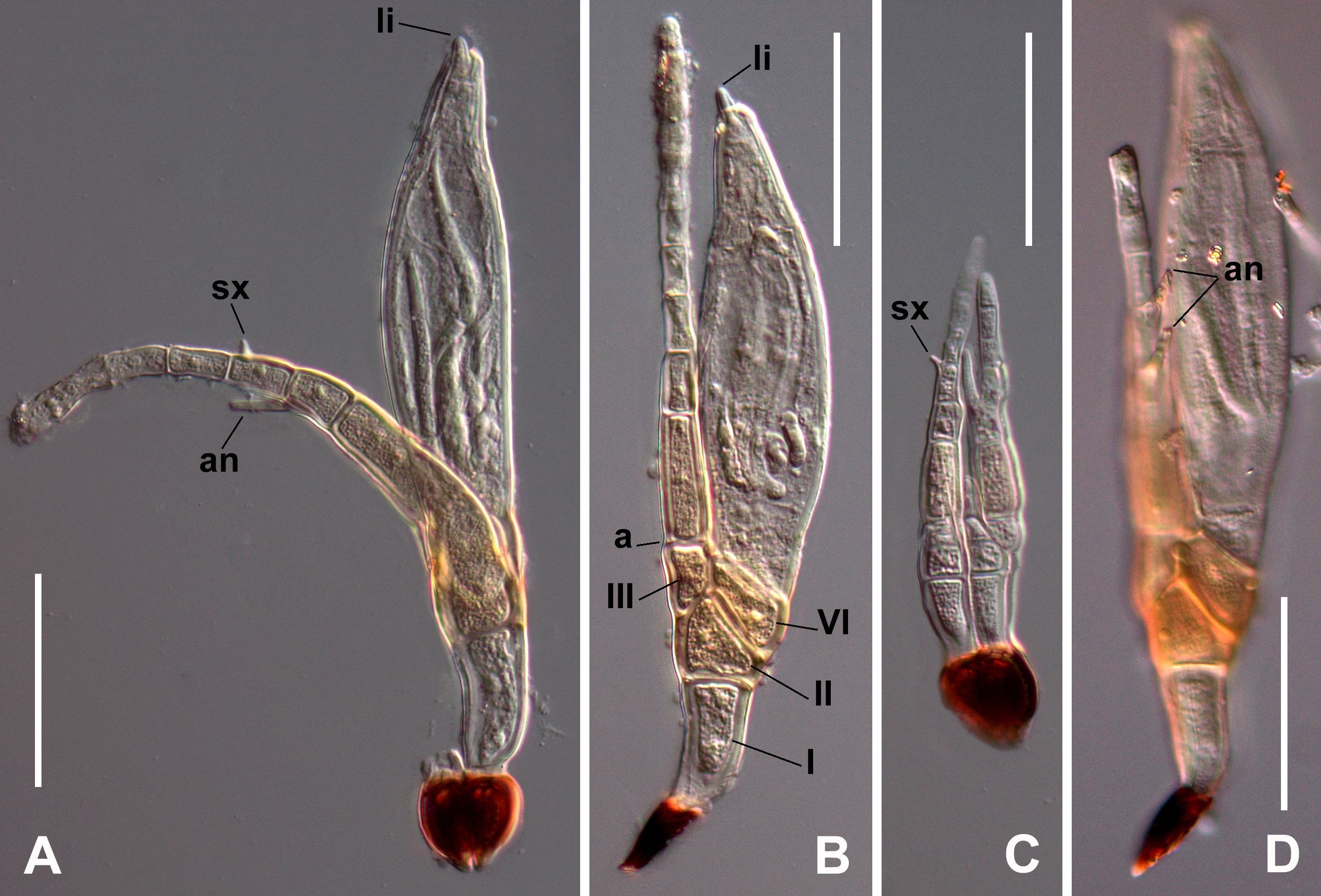
Description
Cells II, III, VI and basal cell of primary appendage bright reddish coloured; the rest of the thallus is hyaline, except for the blackened foot.
Basal cell (I) two times as long as broad, typically obconical, narrow at the base and gradually broadening distally. Suprabasal cell (II) more or less trapezoidal and isodiametric. Cell III smaller than II, slightly longer than broad, laterally adnate and exceeding in length cell VI. Septum I-II horizontal. Septum II-III oblique, almost half in length than I-II. Septum II-VI very oblique, as long as septum I-II.
Primary appendage unbranched, variably bent, consisting of up to 8 cells, gradually tapering distally. Basal cell of primary appendage three times as long as broad, distinctly larger and darker than other cells of appendage. Typically a single flask-shaped antheridium without supporting cell is borne on the second cell of appendage. Spiny remains of spore apex (sx) conspicuous, stout, on the third cell, rarely on the fourth cell of appendage.
Perithecial stalk cell (VI) triangular to rhomboid, obliquely arranged above cell II and under the perithecial body. Secondary stalk cell (VII) and basal cells of perithecium (n, n’, m) hardly distinguishable. Perithecium fusiform, with a blunt apex which bears a small but evident, erect, rounded, ligulate protuberance (li), formed by a w4 perithecial wall cell.
Length from foot to apex of perithecium 119‒192 µm. Perithecium (including basal cells) 68‒121 × 21‒31 µm. Primary appendage (from primary septum to apex, undamaged) 76‒123 µm.
Remarks
Abundant thalli of the new species were scattered on legs and body rings (most on ventral side) of the frontal part of the millipede.
Rarely a second, additional antheridium was seen on the third appendage cell (an, Fig. 2D View Fig. 2 ). Interestingly, the nuclei appear to be conspicuous in all cells of the appendage and also inside the basal cell (I) of the receptacle when seen with DIC optics.
This species is well distinguished by the protuberant lip on the perithecium, the reddish colour in some receptacle cells, and poorly defined walls of the basal cells of the perithecium, a characteristic unknown in the tribe Laboulbenieae but found in other genera of other tribes.
No known copyright restrictions apply. See Agosti, D., Egloff, W., 2009. Taxonomic information exchange and copyright: the Plazi approach. BMC Research Notes 2009, 2:53 for further explanation.
|
Kingdom |
|
|
Phylum |
|
|
Class |
|
|
Order |
|
|
Family |
|
|
Genus |