
Makarovaia, Moraza, María L. & Lindquist, Evert E., 2015
|
publication ID |
https://doi.org/ 10.11646/zootaxa.3931.3.1 |
|
publication LSID |
lsid:zoobank.org:pub:5C5A381C-7EB6-4C1F-BAD2-30B45B019E1F |
|
DOI |
https://doi.org/10.5281/zenodo.6115408 |
|
persistent identifier |
https://treatment.plazi.org/id/03E287ED-1A6C-CE56-EAFA-F980FCE1D448 |
|
treatment provided by |
Plazi |
|
scientific name |
Makarovaia |
| status |
gen. nov. |
Makarovaia new genus
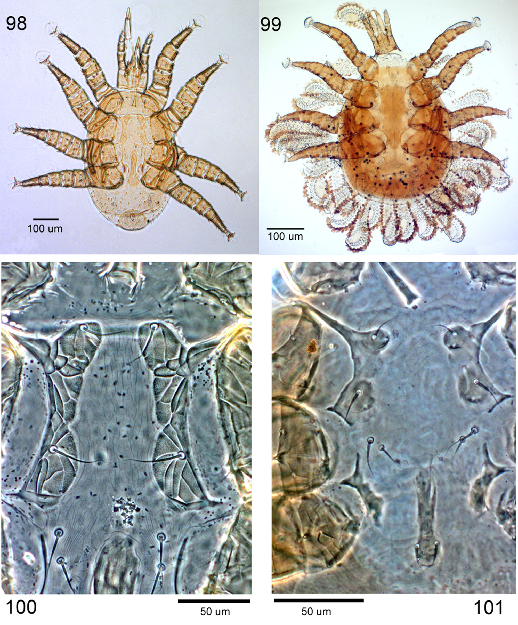
( Figs 1–40 View FIGURES 1 – 4 View FIGURES 5 – 7 View FIGURES 8 – 15 View FIGURES 16 – 20 View FIGURES 21 – 24 View FIGURES 25 – 28 View FIGURES 29 – 33 View FIGURES 34 – 40 , 98–100 View FIGURES 98 – 101 )
Type species. Makarovaia ornata new species. Genus based on adult female, male, nymphal and larval material representing one newly described species.
Diagnosis. Adults of Makarovaia are distinguished from those of Hispiniphis in having the dorsal shield more extensive anterolaterally, so as to bear setae z2, s1, s4, r2, r3, and in having more strongly sclerotised and ornamented shields on the venter, including a moderately large, obovate anal shield slightly wider than long. Male sternigenital shield fully formed, with endopodal extensions between leg bases, a convex posterior margin between bases of legs IV, and bearing all five pairs of sternal setae. Hypostome of nymphs and adults elongate, resulting in corniculi inserted relatively posteriorly, at level about halfway between insertions of hypostomatic setae hp1 and hp3. Movable cheliceral digit of female and female nymphs multidenticulate (≥four teeth), and with bluntly pointed process distinct on midventral face. Male chelicera with ciliated, flexible, digitiform process paraxially at level where movable digit hinged to fixed digit, and with spermatodactyl abruptly bent downward near it base, directing the freely extending part posteroventrally. In addition to leg chaetotactic reductions noted for the genusgroup description, genua II and III lacking ventral setae (av absent); femora I–II and tibia I retaining seta ad -3, genu II retaining al -2. Tarsus I without ventrodistal acuminate process extending under pretarsus; tarsi II–IV with ventral projection under pretarsus acuminate in immature instars but truncate in adult. Nymphs with opisthosoma normal in fully-developed form, size, and not closely flanked by legs IV, which directed posterolaterally.
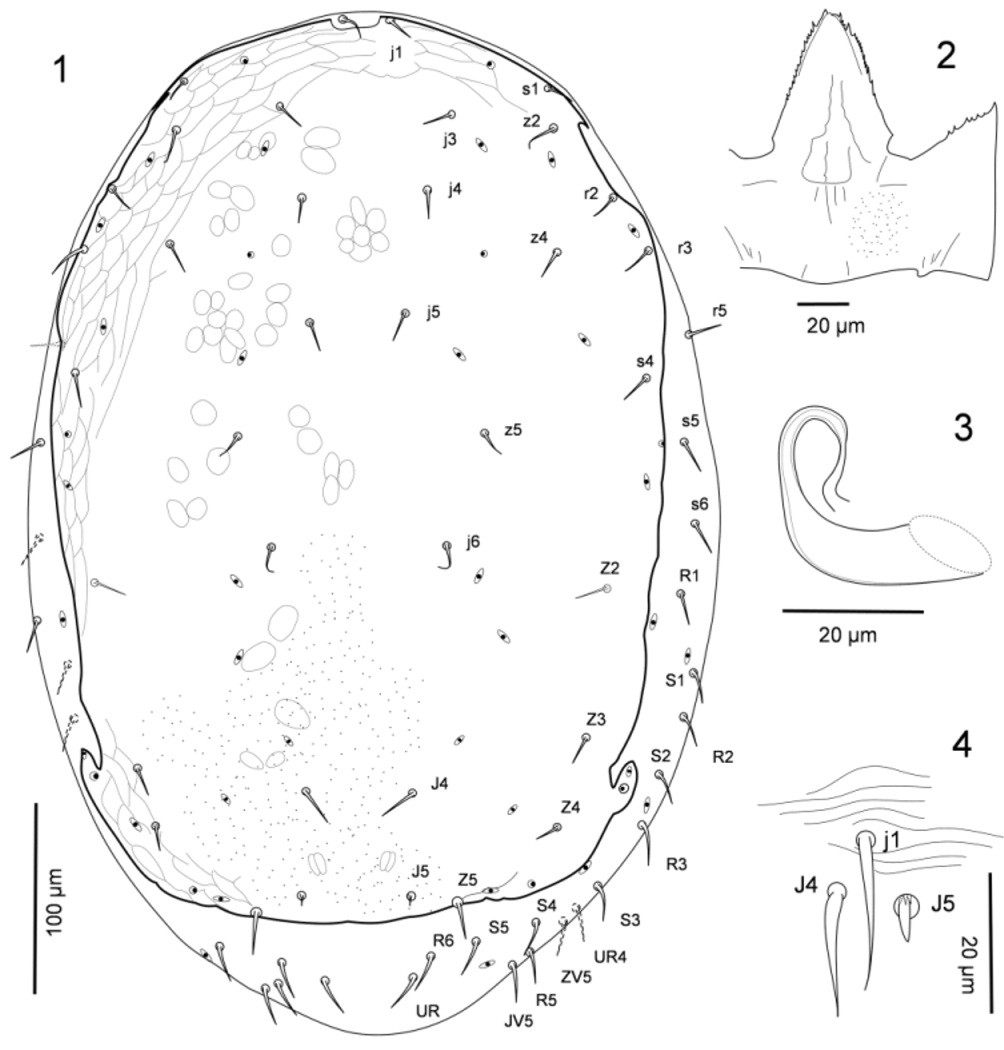
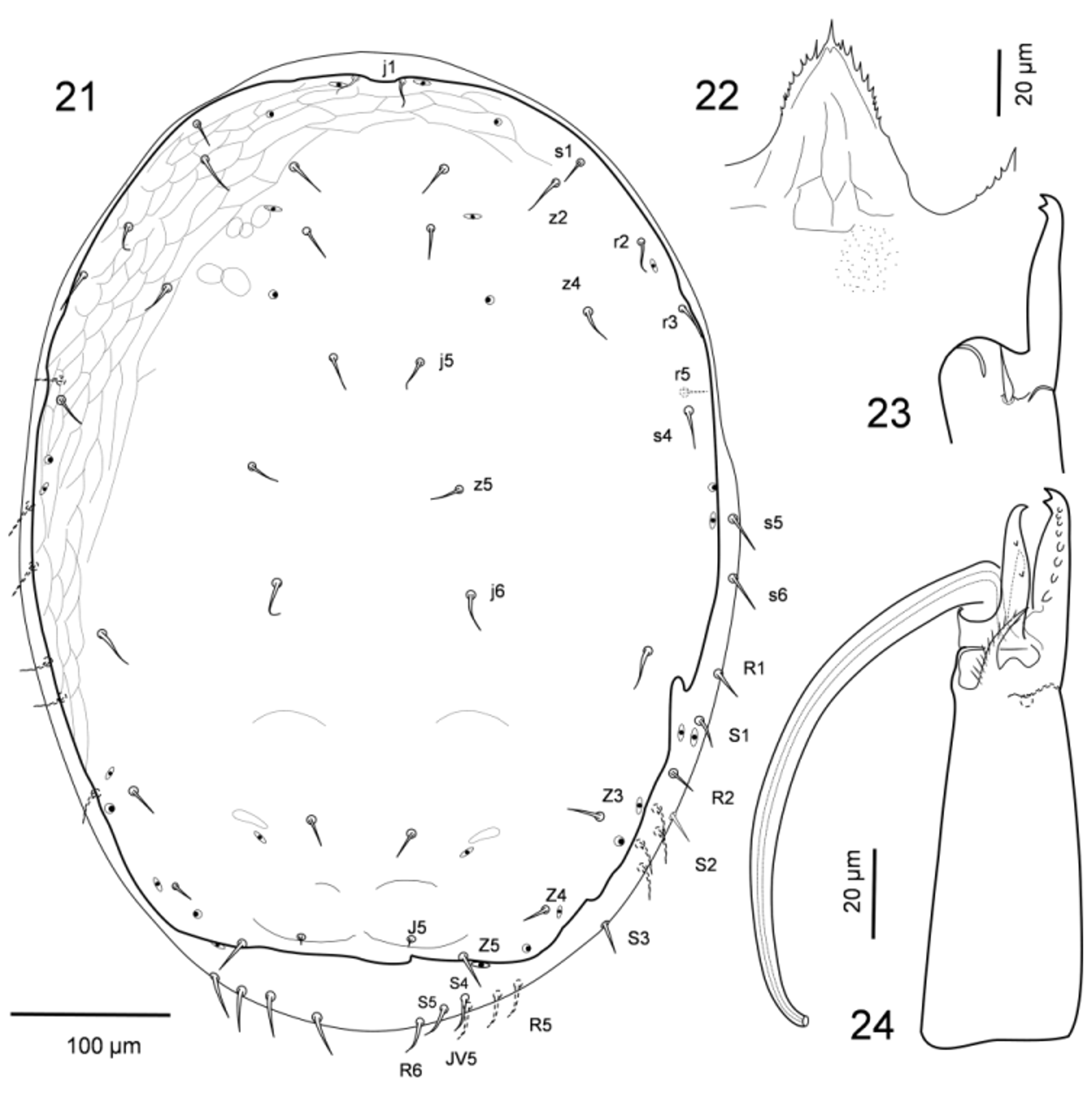
Description. Idiosomatic dorsum. Adult female ( Fig. 1 View FIGURES 1 – 4 ). Dorsal shield well sclerotised, sufficiently expansive anterolaterally to bear setae s1, z2, r2, r3; surrounding soft cuticle thickly formed and ornamented, in distinction to simply striated ventral cuticle, as noted for family. Dorsal shield with maximum complement of 18 pairs of setae, including 12 podonotal (j1, j3–j6, z2, z4, z5, s1, s4, r2, r3) and six opisthonotal pairs (J4, J5, Z2–Z5); dorsal setae similar in form and general size except J5 always minute. Dorsal shield with complement of 17 pairs of discernible pore-like structures (nine podonotal, eight opisthonotal), of which five pairs (three podonotal, two opisthonotal) superficially appear secretory (gland pores) and 12 pairs (six podonotal, six opisthonotal) non-secretory (poroids). Soft lateral cuticle with ca. 20–25 pairs of lateral and marginal setae (s5, s 6, r5, S1–S5, R1–R6, and six or more UR ’s), and at least six pairs of marginal poroids, including idRp.
Adult male ( Fig. 21 View FIGURES 21 – 24 ). Dorsal shield similar in size, ornamentation and setation to that of female; form and relative lengths of setae on dorsal shield and dissimilar structure of surrounding soft cuticle as in female.
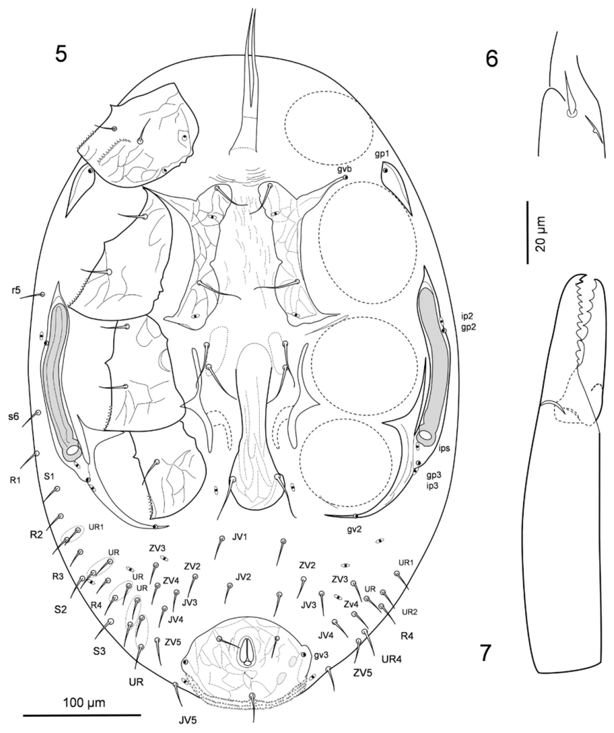
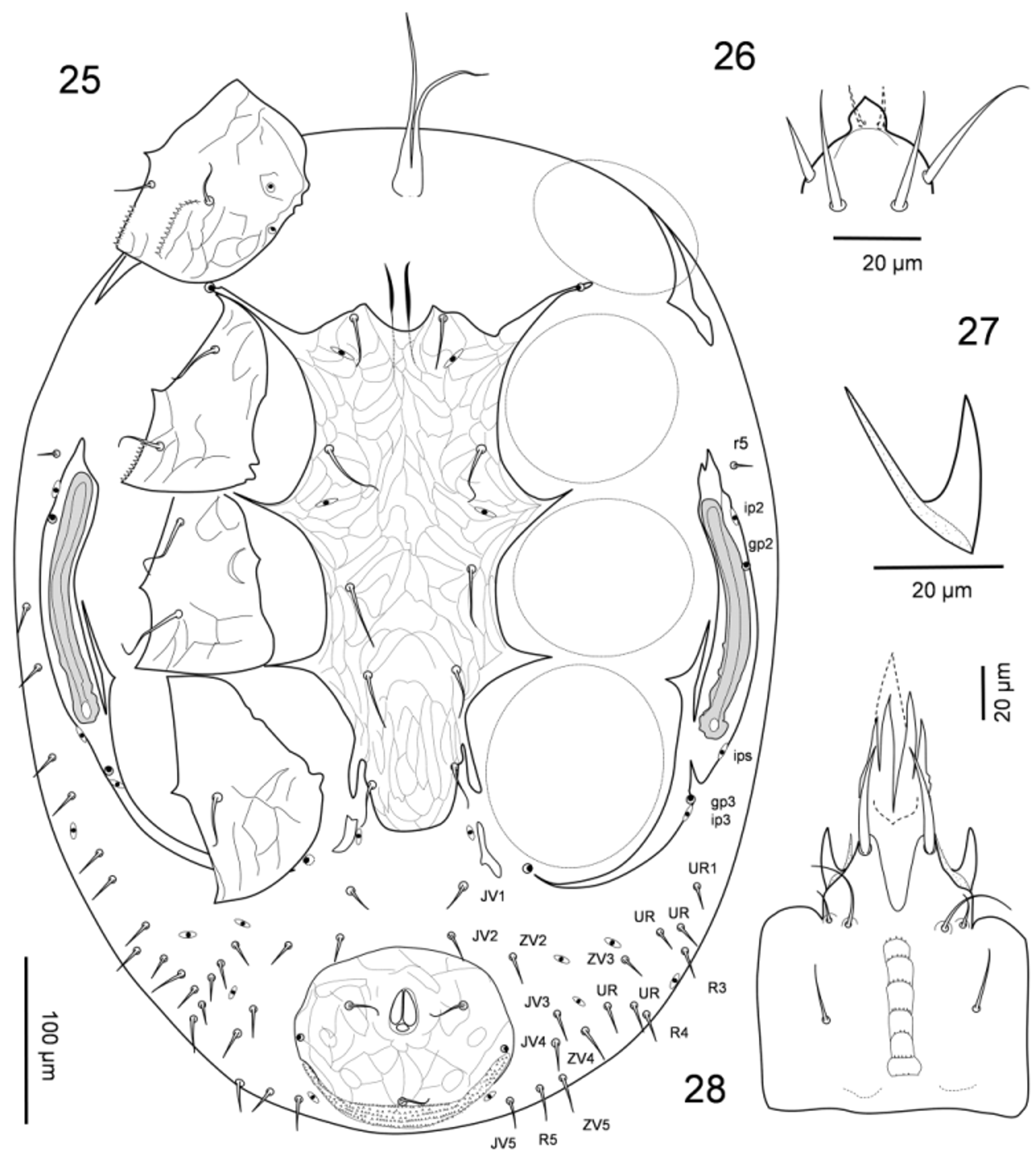
Idiosomatic venter. Adult female ( Figs 5–11 View FIGURES 5 – 7 View FIGURES 8 – 15 ). Tritosternum with laciniae free for most of length, without basal elaborations; laciniae nearly devoid of pilosity ( Fig. 11 View FIGURES 8 – 15 ). Ventral shields well sclerotised and ornamented. Presternal region faintly lineated, without platelets ( Fig. 5 View FIGURES 5 – 7 ). Sternal shield fragmented, form and setation as noted for genus-group description; sternal shield pieces reticulated, and unsclerotised medial region faintly longitudinally lineate or reticulate; setae st3 and st4 on smooth cuticular area on either side (perhaps unsclerotised sternal remnants). Epigynal shield well sclerotised and ornamented, narrow, with hyaline anterior margin not widened between legs III, but with posterior margin widened, strongly convex. Opisthosomatic venter lacking metapodal platelets, with soft cuticle normally striated but not further ornamented, and with ca. nine pairs of opisthogastric setae (JV1–JV5, ZV2–ZV5) and four pairs of poroids flanked by several pairs of R - or UR -setae. Anal shield moderately large, well sclerotised and ornamented, ovoid, wider than long; para-anal setae inserted near anterior level of anus, similar in length to postanal seta; shield with pair of gland pores gv3 slightly within lateral margins posterior to level of insertions of para-anal setae, and with extensive cribrum along posterior margin. Peritrematal shield poorly developed, interrupted at level of coxae II and with a separate platelet bearing gland pore gp1 at level between coxae I and II ( Fig. 5 View FIGURES 5 – 7 ); exopodal strip continuous alongside peritrematal shield only to level of coxae III, extensions between bases of coxae I–II, II–III absent. Peritremes wide, shortened, reaching at most to mid-level of coxa II. Spermathecal apparatus with small section of tubular piece discernibly sclerotised, its distal portion widened ( Fig. 3 View FIGURES 1 – 4 ).
Adult male ( Fig. 25 View FIGURES 25 – 28 ). Form of presternal area as in female, tritosternum with laciniae similar in length to those in female. Sternitigenital shield anterior margin strongly defined at level of genital opening; shield well sclerotised, united with endopodal strips between coxae I–II, II–III, III–IV, with five pairs of setae and two pairs of poroids. Form and extent of exopodal and peritrematal structures much as in female, except fragment absent between coxae I–II, and peritremes slightly shorter. Opisthogastric region much as in female, with same pairs of opisthogastric setae and poroids, flanked by several (fewer) pairs of R - or UR -setae. All ventral idiosomatic setae smooth, similar in shape and length as in female. Anal shield much as in female, but with para-anal setae inserted at mid-level of anus.
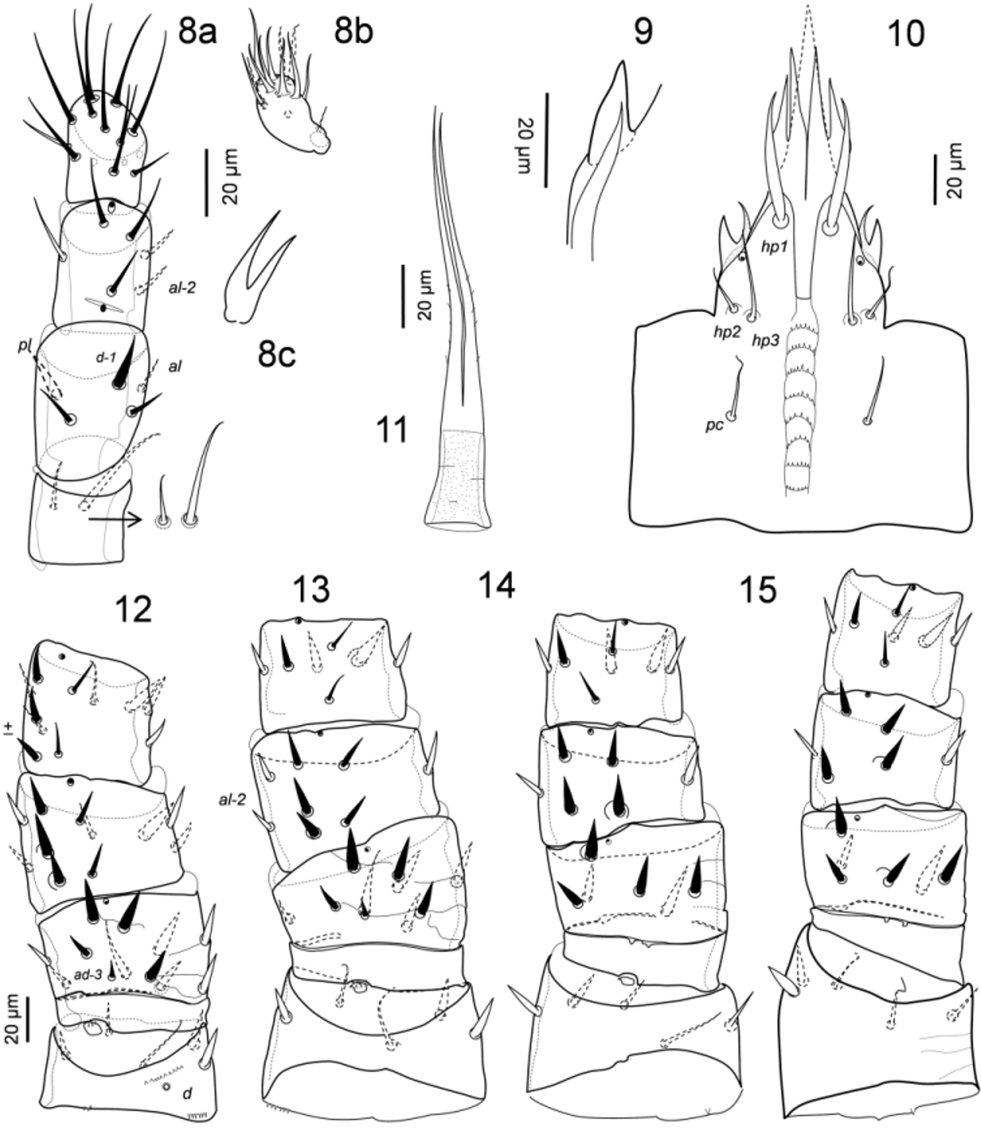
Gnathosoma . Female and male. Gnathotectum with anterior margin convex to nearly angular, denticulate ( Figs 2 View FIGURES 1 – 4 , 22 View FIGURES 21 – 24 ). Chelicerae generally as described for the genus-group description ( Figs 6, 7 View FIGURES 5 – 7 , 23, 24 View FIGURES 21 – 24 ). Fixed cheliceral digit multidentate, with pilus dentilis reduced to an alveolar remnant; movable digit of female multidentate ( Fig. 7 View FIGURES 5 – 7 ), with a small blunt process on mid-ventral face. Movable cheliceral digit of male bidentate, lacking pointed process on ventral face; spermatodactyl digit-like, abruptly curved ventrally near base and directed posteroventrally ( Fig. 24 View FIGURES 21 – 24 ); male chela with conspicuous ciliated arthrodial process on paraxial surface at base of movable digit ( Fig. 24 View FIGURES 21 – 24 ). Corniculi of female and male similar in form, short, stout, inserted at level midway between insertions of subcapitular setae hp1 and hp3, and with tip of paraxial acuminate process reaching slightly beyond that of main corniculus tip ( Figs 10 View FIGURES 8 – 15 , 27, 28 View FIGURES 25 – 28 ); bifid internal malae elongate, extending well beyond apex of palpgenu ( Figs 10 View FIGURES 8 – 15 , 28 View FIGURES 25 – 28 ); apex of labrum extending to mid-level or apex of palptibia. Subcapitulum with a pair of pore-like structures at level of insertion of corniculi ( Fig. 10 View FIGURES 8 – 15 ). Subcapitular setae hp1 much longer and thicker than other pairs. Deutosternum with seven or eight moderately narrow rows of denticles, basal rows slightly narrower; rows similarly multidenticulate, connected by lateral margins. Palpal setation and form of setae as described for genusgroup description ( Figs 8 View FIGURES 8 – 15 a, b, c); palptrochanter with inner seta twice as long as external seta ( Fig. 8 View FIGURES 8 – 15 a).
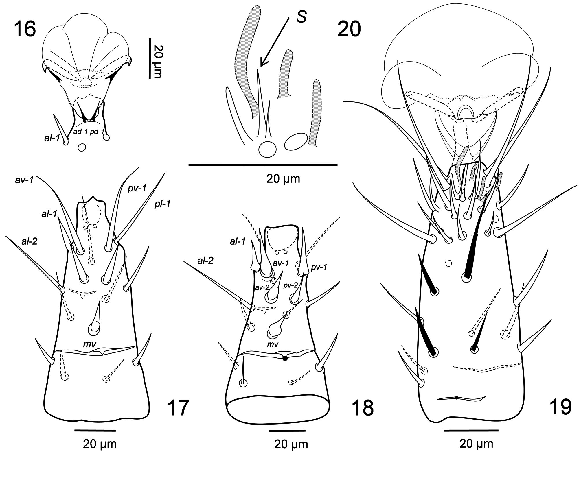
Legs. Female and male. Legs relatively short, clearly shorter than dorsal shield. Legs similar in size, II and III slightly thicker than legs I and IV ( Figs 12–15 View FIGURES 8 – 15 ). Distal posterior ventral and dorsal margins of coxae I–IV serrated, and coxa I with a serrated ridge on ventral face. Pretarsi of legs I to IV with slender claws and large, rounded pulvillus; those of legs II–IV with short, inconspicuous paradactyli ( Fig. 16 View FIGURES 16 – 20 ). Tarsus I with sensilla s inconspicuous, short and acute ( Fig. 20 View FIGURES 16 – 20 ). Legs II to IV with tarsus (excluding pretarsus) about twice as long as tibia. Tarsi II–IV with apical setal processes ad-1, pd-1 reduced, shorter than short pretarsi ( Fig. 16 View FIGURES 16 – 20 ), and with apical ventral process short, truncated, unhinged basally ( Figs 17, 18 View FIGURES 16 – 20 ). Setation and its ontogeny on segments of legs I to IV deficient from full complement of Melicharidae (as presented by Lindquist & Evans 1965 for Melicharini): coxae, 2-2-2-1; trochanters, 5-5-5-5; femora, 12 (2 3/1 2/2 2) – 10 (1 3/1 2/2 1) – 6 (1 2/1 1/0 1) – 6 (1 2/1 1/0 1); genua, 11 (2 3/1 2/1 2) – 8 (2 3/0 2/0 1) – 6 (1 2/0 2/0 1) – 6 (1 2/0 2/0 1); tibiae, 11 (2 3/1 2/1 2) – 7 (1 1/1 2/1 1) – 7 (1 1/1 2/1 1) – 7 (1 1/1 2/1 1); trochanter I seta ad vestigial, only alveolus discernible; femora III–IV with seta pd -2 inserted relatively laterally, in pl position. In addition to setal absences noted for family: genua II–III lacking av, and genu IV lacking pd -3; however, femora I–II and tibia I retaining seta ad -3. Several setae moderately thickened, spinelike: pl on trochanters I and II, and al on trochanter IV; ad -1, pd -1, pd -2 on femora I–II and ad1, ad -2, pd, pl on femora III–IV; some ventral setae on femora I–IV (on female and male); pv on genu and tibia I, and pv and to less extent av on tibiae II–IV. Seta pv -2 on trochanters I–IV attenuate, thinner and longer than other setae. Male leg setal dimorphism limited to form of some ventral and lateral setae on tarsi II to IV. Tarsi II–IV in both sexes with setae al -2, pl -1, slightly longer, more attenuated than other tarsal setae; on female, ventral setae av -1, pv -1 similar to pl -1, while al -1, av -2, pv -2 and moreso mv moderately spine-like ( Fig. 17 View FIGURES 16 – 20 ); on male, al -1, av -1, pv - 1, av -2, pv -2 and mv thickened, similarly spine-like with swollen, bulbous base ( Fig. 18 View FIGURES 16 – 20 ); apical ventral tarsal process truncate-acuminate on female ( Fig. 17 View FIGURES 16 – 20 ) but truncate-straight on male ( Fig. 18 View FIGURES 16 – 20 ). Other leg setae simple, not strongly differentiated on either sex.
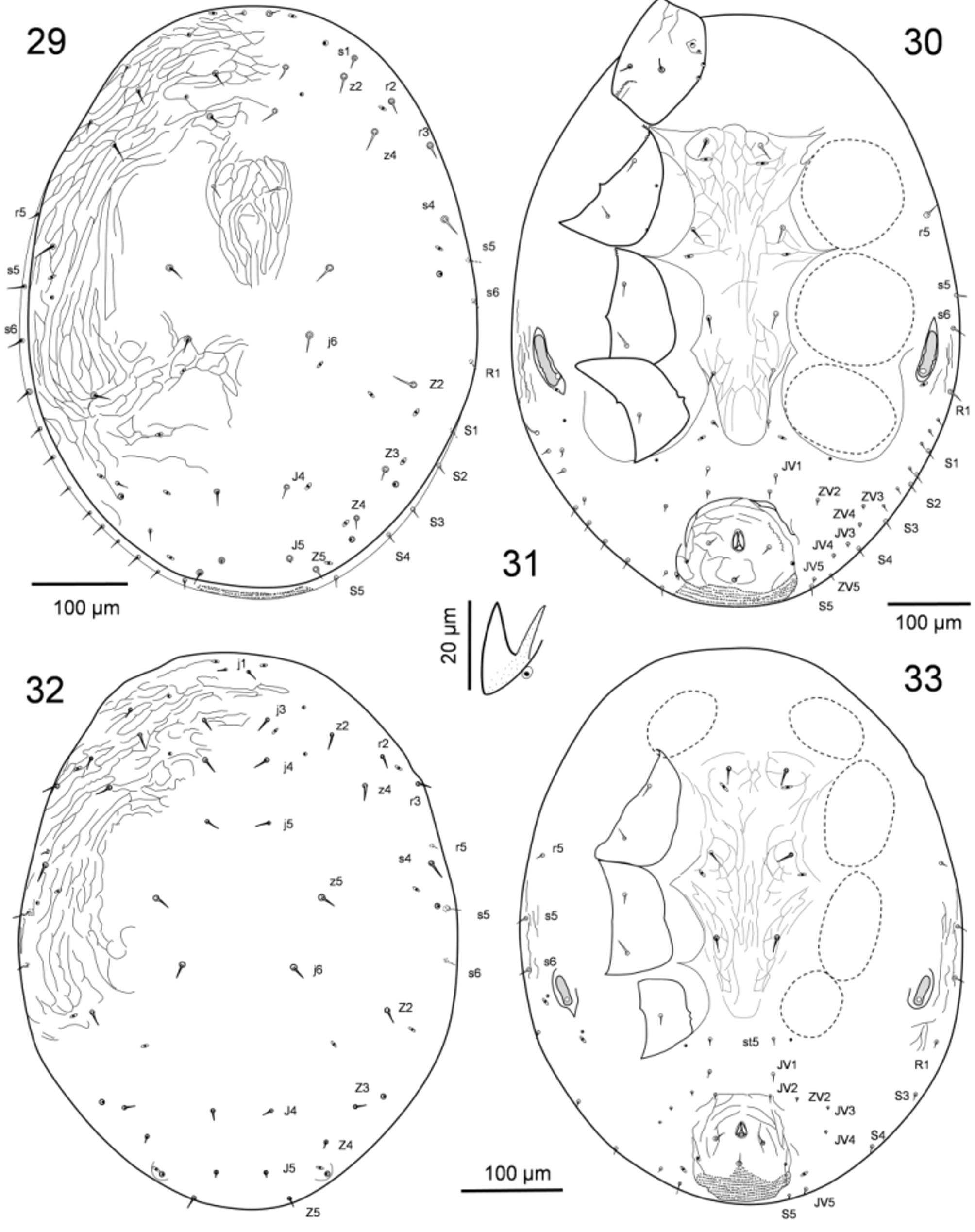
Deutonymph. Idiosomatic dorsum. Dorsal shield extensive, poorly sclerotised, without lateral incisions, lightly lineated, with 18 pairs of similarly smooth, short setae except J5 minute: 12 pairs (j1, j3–j6, z2, z4, z5, s1, s4, r2, r3) on podonotal region, and six pairs (J4-J5, Z2–Z5) on opisthonotal region ( Figs 29, 30 View FIGURES 29 – 33 ). Lateral soft cuticle with s5, s6, r5 on podonotal region and S1-S5, R1-R6 and variably few UR -setae on opisthonotal region. Dorsal idiosomatic complement of poroids and gland-pores similar to that in female.
Idiosomatic venter. Sternal shield lightly sclerotised and ornamented, with endopodal extensions between coxae I–II, II–III and III–IV, with four pairs of setae st1-st4 and poroids iv1 and iv2; poroids iv3 absent; setae st5 on soft cuticle together with poroids iv5 between coxae IV ( Fig. 30 View FIGURES 29 – 33 ). Anal shield lightly sclerotised and ornamented, with circumanal setae short and similar in length, adanal gland pores (gv3) at level between para-anal and postanal setae; cribrum large as in adult. Opisthogaster with nine pairs of ventral setae in deutonymph (the four larval JV1, JV2, JV5, ZV2 plus five minute deutonymphal JV3, JV4, ZV3–ZV5, flanked by a few either S - or R - or UR - setae) ( Fig. 30 View FIGURES 29 – 33 ). Peritrematal shields and peritremes shortened, paedomorphic, as in protonymphal instar, developed only between midlevels of coxae III-IV ( Fig. 30 View FIGURES 29 – 33 ). Peritrematal gland pore gp2 and poroid ip2 indistinct on soft cuticle; poststigmatic poroid ips adherent to stigma on posterior edge of peritrematal shield, but poststigmatic gland pore gp3 and poroid ip3 indistinct on soft cuticle in that area. Rim of exopodal plate behind coxa IV inconspicuous, with gland pore gv2.
Gnathosoma . Gnathotectum, chelicerae and other mouthpart structures, corniculi and adjacent structures as in adult female ( Fig. 31 View FIGURES 29 – 33 ), but fixed cheliceral digit less hooked apically; deutosterum with seven rows of denticles (two basal rows narrower and less arched than anterior ones), with structures otherwise as in adult; palpi similar to those in adult female, including similar form of al setae on palpfemur and palpgenu. Chelicerae of male deutonymph with movable digit thick, with apical hook and one distal minute tooth and formation of primordial sperm duct present ( Fig. 40 View FIGURES 34 – 40 ).
Legs. Pretarsal structures and chaetotaxy of legs I–IV as in adult, including these additions to protonymphal setal complement: av -2 on trochanters I-IV; ad -3 on femur I, pd -2 on femora I, III, IV, pv -2 on femora I, II, al -2 and pl -2 on genu I, ad -3 on genua I–II, pl -1 on genu IV, al -2 and pl -2 on tibia I. Size and shape of leg setae similar to those on adult female. Trochanter I with postero-dorsal serrated ridge. Tarsi II–IV with form of setae and acuminate form of apical ventral process as in protonymph ( Fig. 26 View FIGURES 25 – 28 ).
Protonymph. Idiosomatic dorsum. Body with dorsal shielding not clearly delimited (outlines of podonotal shield, mesonotal scutellae, pygidial shield not evident), but with faint lateral lineation, less than in deutonymph; dorsum maximally with 24 pairs of similarly smooth, short setae, except J5 minute (protonymphal setae r2, r3, r5, R1, Z2 added to larval complement; j2, J1, Z1 absent, as on deutonymph and adult, but S2 not discerned). Body dorsum with complement of 12 pairs of discernible pore-like structures, of which seven non-secretory (poroids) and five superficially appear to be secretory (gland pores); paravertical poroids idj1 added to larval complement.
Idiosomatic venter. Sternal shield weakly sclerotised and ornamented, with faint endopodal extensions between coxae I–II, and II–III, with three pairs of setae st1-st3 and two pairs of poroids (iv2 added to larval complement); setae st5 on soft cuticle at level of posterior border of coxae IV ( Fig. 33 View FIGURES 29 – 33 ); paragenital poroids absent. Anal shield faintly sclerotised and ornamented, roughly as wide as long, with circumanal setae similarly short; para-anal setae inserted at level slightly posterior to anal opening; adanal gland pores (gv3) at level of postanal seta; cribrum large, as in deutonymph. Opisthogaster with four to six pairs of ventral setae; larval complement sometimes augmented by normally deutonymphal setae JV3, JV4 ( Fig. 33 View FIGURES 29 – 33 ). Peritrematal region on each side with poroid and gland pore on soft cuticle alongside weakly formed peritreme between coxae III and IV, and with poststigmatic poroid and gland pore on soft cuticle behind peritreme ( Fig. 33 View FIGURES 29 – 33 ). Exopodal rim behind coxa IV not discernible, but gland pore gv2 present on inguinal area.
Gnathosoma . Form of gnathotectum, corniculus and its paraxial acuminate process, elongated internal malae, and other gnathosomatic structures similar to those in deutonymph, except palpi with normal protonymphal complement of setae (see Evans 1964), including only one trochanter seta; corniculi inserted clearly posterior to level of insertions of setae hp1, and with tip of inner acuminate process reaching beyond that of main corniculus tip ( Fig. 37 View FIGURES 34 – 40 b). Cheliceral movable digit with three or usually four teeth and a small mid-ventral projection; fixed digit multidentate, with offset subapical tooth and sparse (five to seven), well-formed teeth along masticatory surface ( Fig. 39 View FIGURES 34 – 40 ).
Legs. Legs I to IV with pretarsi endowed by well-developed claws and normal protonymphal complement of setae as described for Ascidae by Lindquist & Evans (1965): coxae, 2-2-2-1; trochanters 4-4-4-4 (1 0/2 1 on I, II, 1 1/2 0 on III, IV); femora 10 (2 2/1 2/1 2) – 8 (1 2/1 2/1 1) – 5 (1 2/1 1/0 0) – 4 (1 2/0 1/0 0); genua 8 (1 2/1 2/1 1) – 6 (1 2/0 2/0 1) – 6(1 2/0 2/0 1) – 6 (1 2/0 2/0 1); tibiae 8 (1 2/1 2/1 1) – 7 (1 1/1 2/1 1) – 7 (1 1/1 2/1 1) – 7 (1 1/1 2/1 1); seta pl -1 accelerated in first presence in protonymph, rather than deutonymph, on genu IV. Coxae I–II distal rims with serrated posterodorsal strips. Leg setae generally simple, not markedly differentiated. Tarsi II–IV with setae ad -1, pd -1 small but as long as short pretarsus; al -2, a v -1, pv -1, pl -1 and md longer, thinner than other tarsal setae; apical ventral tarsal process present, bluntly triangular.
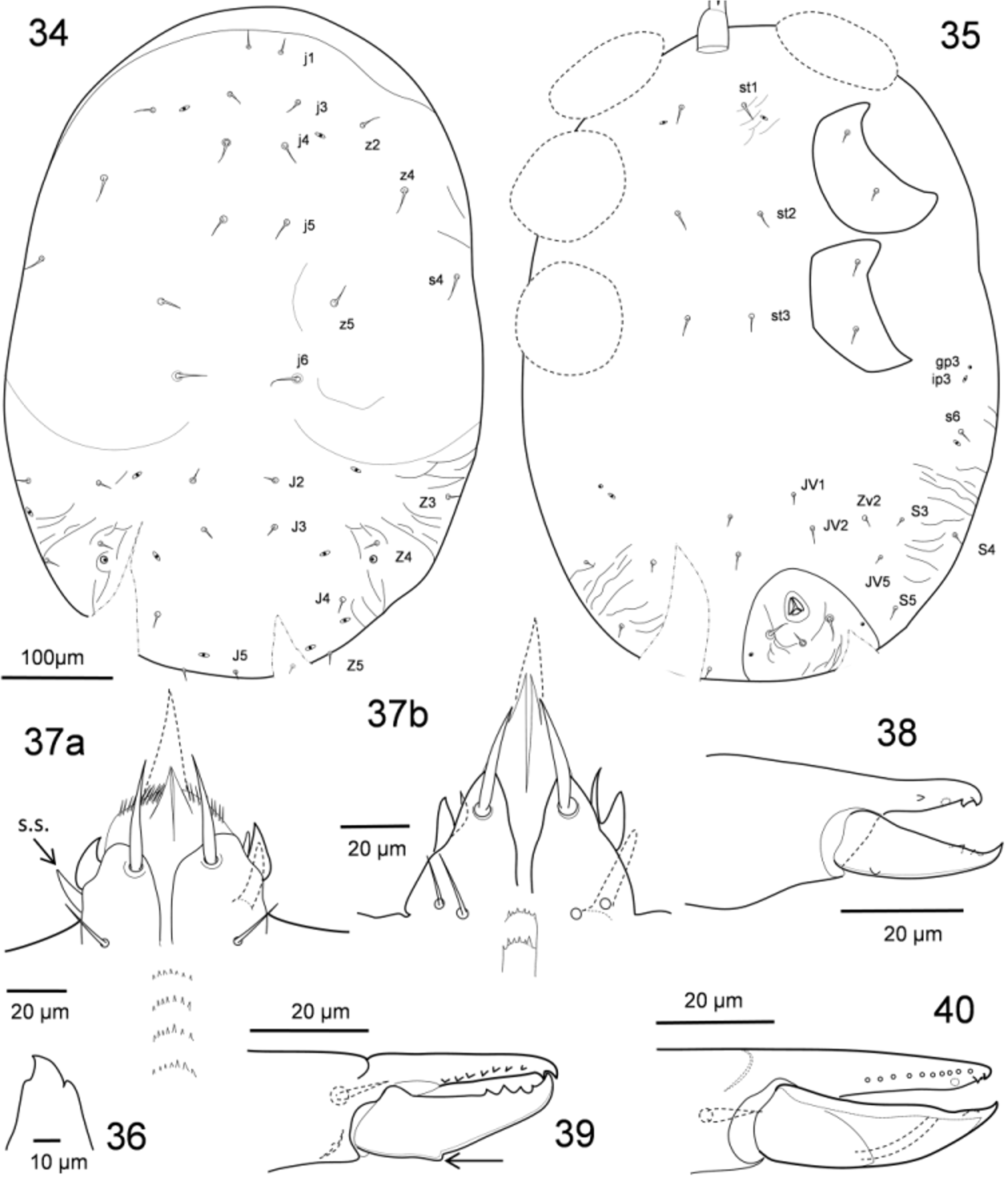
Larva. Idiosomatic dorsum. Body with dorsal shielding not clearly delimited, and surface not discernibly ornamented as on nymphal instars; mesonotal scutellae indiscernible ( Fig. 34 View FIGURES 34 – 40 ). Body dorsum with 20 pairs of smooth setae: ten podonotal pairs, and ten opisthonotal pairs (J1 absent, J5 miniscule, S3–S5 inserted ventrolaterally); discernible pore-like structures include five (one podonotal, four opisthonotal) poroids, and one (opisthonotal) gland pore.
Idiosomatic venter. Tritosternum normally developed, similar in form as in subsequent instars. Sternal shield not distinguishable, intercoxal region with setae st1–st3 and poroids iv1; pair of subcutaneous structures (prevalent among larvae of Gamasina ) indiscernible at level of posterior margins of coxae III. Anal shield faintly sclerotised, roughly as wide as long; para-anal setae inserted at level slightly posterior to anal opening, and longer than postanal seta; adanal gland pores (gv3) at level of postanal seta; cribrum absent. Venter with four pairs of opisthogastric setae on soft cuticle anterolateral to anal shield, flanked by opisthonotals (S3–S5). Poststigmatic poroid ip3 and gland pore gp3 on soft cuticle near level of posterior margin of coxae III ( Fig. 35 View FIGURES 34 – 40 ).
Gnathosoma . Gnathotectum with convex anterior margin less denticulate than in subsequent instars ( Fig. 36 View FIGURES 34 – 40 ). Cheliceral movable digit with two teeth and a small mid-ventral projection; dentition of fixed digit weakly developed, with offset subapical tooth and masticatory ridge with a weak tooth proximally ( Fig. 38 View FIGURES 34 – 40 ). Form of corniculus and its inner acuminate process much as in subsequent instars, but inserted at same transverse level as alveoli of setae hp1, and with inner process shorter relative to subsequent instars, its tip not reaching that of main corniculus tip; internal malae widened and shorter than in subsequent instars, fringed, not bifid ( Fig. 37 View FIGURES 34 – 40 a). Deutosternum with rows of denticles similar to those in nymphs. Palpus with normal larval complement of setae (see Evans 1964); palp-trochanter nude.
Legs. Legs I to III with pretarsi having well-developed claws, and with normal larval complement of setae as described for Ascidae by Lindquist & Evans (1965): coxae, 2-2-2; trochanters 4-4-4; femora 10 (2 2/1 2/1 2) – 7 (1 2/1 2/0 1) – 5 (1 2/1 1/0 0); genua 8 (1 2/1 2/1 1) – 6 (1 2/0 2/0 1) – 6(1 2/0 2/0 1); tibiae 8 (1 2/1 2/1 1) – 7 (1 1/1 2/1 1) – 7 (1 1/1 2/1 1). Tarsus I with 34 setae. Coxa I with distal rim serrated posterodorsally; distal rims of coxae otherwise smooth. Leg setae generally simple, not markedly differentiated. Tarsi II and III with setae ad -1, pd -1 as long as short pretarsus; apical ventral tarsal process triangular.
Etymology. The generic name is in honor of our esteemed Russian colleague, Olga Makarova, whose many contributions to the systematic and ecological knowledge of gamasine mites are exemplary.
Distribution and habitats. The new genus is currently based on one newly described species. All instars of mites of the genus Makarovaia are known only as subelytral symbionts of adult hispine beetles of the genus Chelobasis , tribe Arescini. Only four species of the neotropical genus Chelobasis are recognised; their larvae are all restricted to unfurled leaves of Heliconia ( Strong 1977; Staines 2009).
Remarks. The larval and protonymphal complement of four dorsal setae on femur I differs from the general pattern given by Evans (1963) in having two anterodorsals and two posterodorsals, instead of three and one, respectively. This is probably a posterolateral shift in position of one of the more basal dorsal setae, rather than an unlikely ontogenetic retardation of one anterodorsal seta and acceleration of one posterodorsal seta.
Adult males of the one known species of Makarovaia have a peculiarly formed structure at the base of the movable cheliceral digit. Quite slender, finely ciliated, and apparently flexible, it seems to be an outgrowth from the paraxial surface of the arthrodial envelope ( Fig. 24 View FIGURES 21 – 24 ). A somewhat similar but more strongly elaborated excrescence, restricted to males, has been illustrated for many free-living parasitid mites ( Micherdzinski 1969); otherwise, such a structure has not been noted or illustrated among males of other families of Gamasina , although it may be readily overlooked. Similar or more elaborate arthrodial brushes are evident among members of the families of Eviphidoidea, but they are featured on immatures and adults of both sexes. The structure is not formed in the immatures or adult female of Makarovaia ornata , and is not evident among members of the species of the sister genus Hispiniphis at hand.
Adult males of the one known species of Makarovaia are frequently infected with an undescribed form of the laboulbeniaceous genus Rickia (see discussion). Adults of species of Hispiniphis have rarely been found infected with this fungus, despite the coexistence of the beetle hosts.
No known copyright restrictions apply. See Agosti, D., Egloff, W., 2009. Taxonomic information exchange and copyright: the Plazi approach. BMC Research Notes 2009, 2:53 for further explanation.