
Stenotarsus
|
publication ID |
https://doi.org/ 10.11646/zootaxa.3645.1.1 |
|
publication LSID |
lsid:zoobank.org:pub:9DC9FDE7-C9BB-4748-B23C-9DE780A1D375 |
|
DOI |
https://doi.org/10.5281/zenodo.6164176 |
|
persistent identifier |
https://treatment.plazi.org/id/03E287F6-305E-FFAF-0B83-FE85FA53FDA6 |
|
treatment provided by |
Plazi |
|
scientific name |
Stenotarsus |
| status |
|
Key to the species of Stenotarsus View in CoL from México, Guatemala and Belize
[Note: Stenotarsus orbicularis Gerstaecker is not included in the key since neither the type nor any other unambiguously identified specimens were available for study]
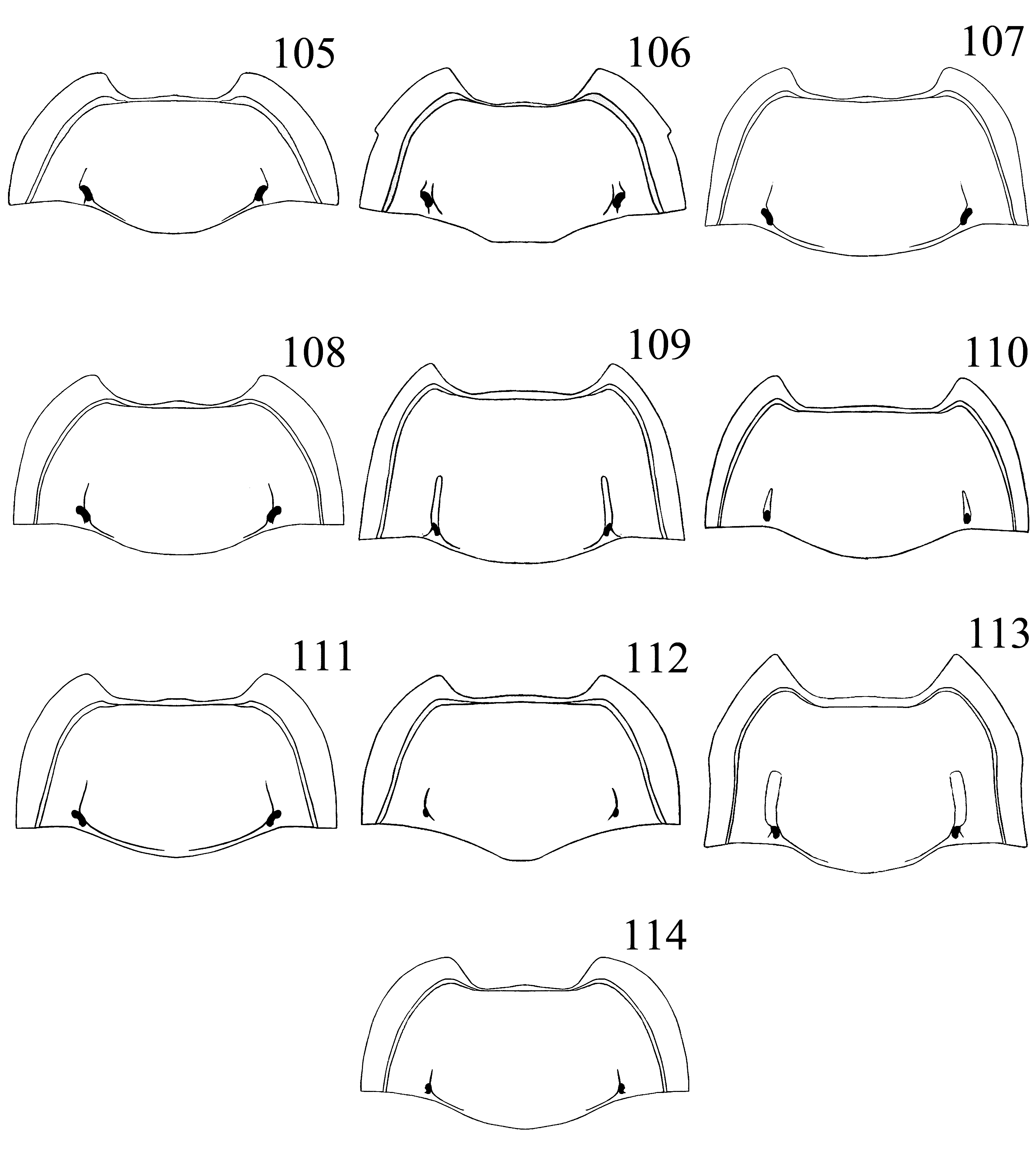
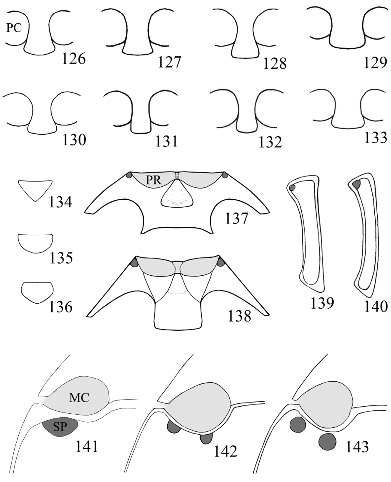
1. Elytra with foveolate punctures arranged in longitudinal striae, lacking in apical third ( Figs. 123, 125 View FIGURES 115 – 125 ); pronotum with longitudinal sulci rather deeply and widely excavated, long, reaching almost middle of pronotum ( Figs. 109, 113 View FIGURES 105 – 114 ); mesoventrite with carinae defining three subtriangular areas ( Fig. 138 View FIGURES 126 – 143 ); median lobe curved and twisted ( Figs. 234–235, 240–241 View FIGURES 224 – 243 ).......... 2
– Elytra with foveolate punctures sparse, not forming longitudinal striae ( Figs. 115–122, 124 View FIGURES 115 – 125 ); pronotum with longitudinal sulci moderately impressed, narrow, short, hardly reaching middle of pronotum, sometimes greatly reduced or almost absent ( Figs. 90–108, 110–112, 114 View FIGURES 90 – 104 View FIGURES 105 – 114 ); mesoventrite without carinae ( Fig. 137 View FIGURES 126 – 143 ); median lobe curved in ventral view, linear or weakly sinuate in lateral view ( Figs. 200–233, 236–239, 242–243 View FIGURES 200 – 223 View FIGURES 224 – 243 )........................................................... 3
2. Foveolate punctures of elytra small, diameter 2X the diameter of setiferous punctures ( Fig. 123 View FIGURES 115 – 125 ); antennae black, with scape light brown; pronotum ( Fig. 109 View FIGURES 105 – 114 ) with lateral margins moderately wide (width slightly less than 1/4 of the distance between midpoint of basal pore and hind angle), front angles moderately produced; longitudinal sulci moderately wide............................................................................................ Stenotarsus rulfoi sp. nov.
– Foveolate punctures of elytra large, diameter 4X the diameter of setiferous punctures ( Fig. 125 View FIGURES 115 – 125 ); antennae black, with first three or four antennomeres light brown; pronotum ( Fig. 113 View FIGURES 105 – 114 ) with lateral margins wide (width slightly less than 1/3 of the distance between midpoint of basal pore and hind angle), front angles markedly produced; longitudinal sulci distinctly wide....................................................................................... S. spiropenis sp. nov.
3. Basal pores of pronotum large, oblique ( Figs. 79 View FIGURES 79 – 89. 79 – 83 , 98 View FIGURES 90 – 104 ); metaventrite with one large setose pore posterior to each mesocoxa, its width almost half width of mesocoxa ( Fig. 141 View FIGURES 126 – 143 )............................................ S. marginalis Arrow
– Basal pores of pronotum small to moderately large, variable in shape ( Figs. 80–83 View FIGURES 79 – 89. 79 – 83 , 91–97, 100–105, 107–108, 110–112, 114 View FIGURES 90 – 104 View FIGURES 105 – 114 ); metaventrite with pair of small setose pores posterior to each mesocoxa, width of each one approximately a third of width of mesocoxa ( Fig. 142 View FIGURES 126 – 143 ).................................................................................... 4
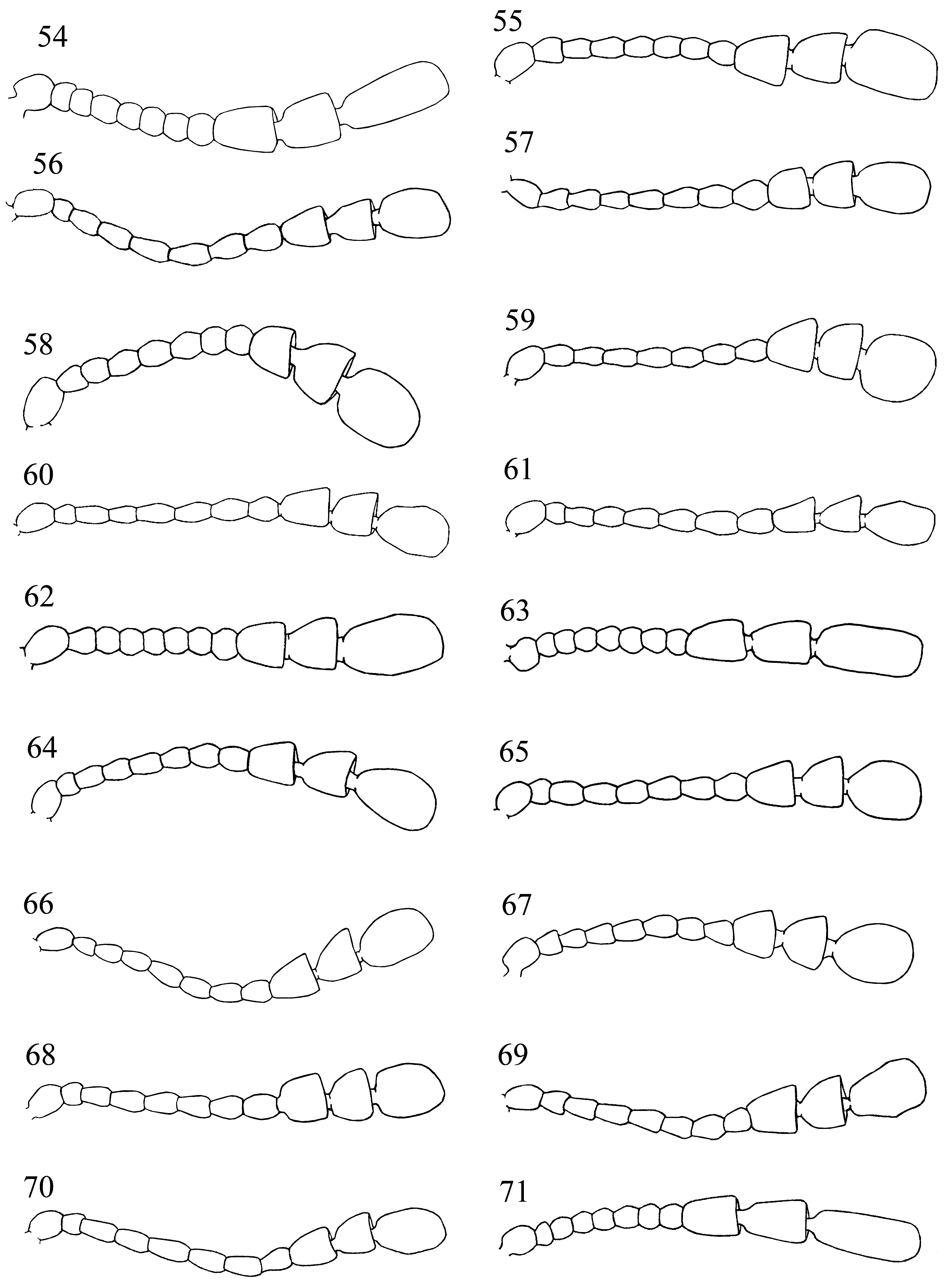
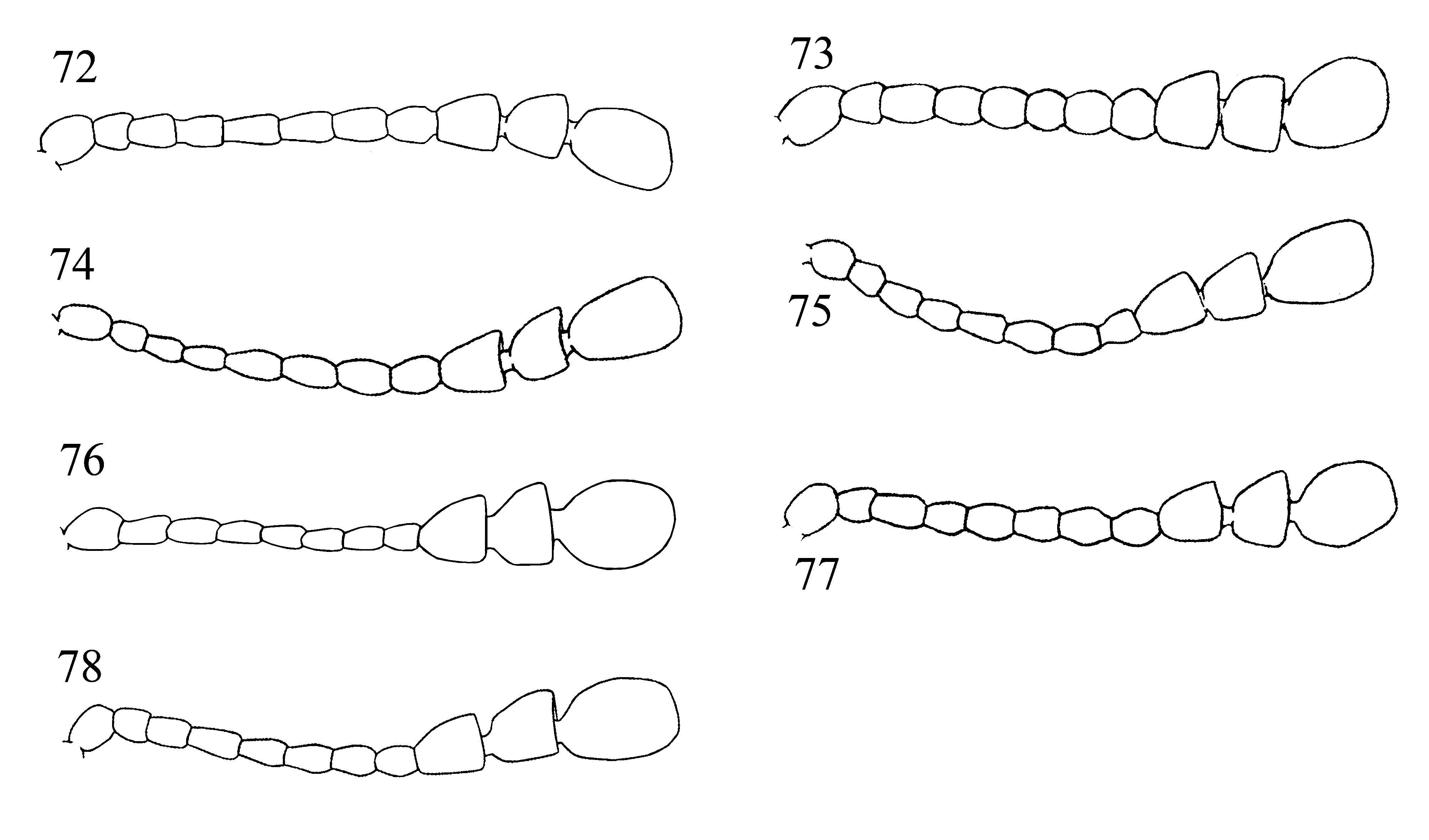
4. Antennomeres 2–8 moniliform, as long as wide; club slightly longer than rest of antenna, its articles elongate, almost parallel sided ( Figs. 54, 63 View FIGURES 54 – 71 ; 71)................................................................................. 5
– Antennomeres 2–8 elongate, distinctly longer than wide ( Figs. 55–62, 64–70 View FIGURES 54 – 71 ; 72–78); club shorter than rest of antenna, its articles widened apically.................................................................................. 7
5. Pronotum ( Fig. 90 View FIGURES 90 – 104 ) with lateral margins lacking tooth at midlength, with longitudinal sulci comparatively long and deeply marked; median lobe distinctly broadened apically in ventral view ( Fig. 201 View FIGURES 200 – 223 )........................ S. cortesi sp. nov.
– Pronotum ( Figs. 99 View FIGURES 90 – 104 , 106 View FIGURES 105 – 114 ) with lateral margins bearing small tooth at midlength, with longitudinal sulci comparatively short and feeble; median lobe weakly widened apically in ventral view ( Figs. 219 View FIGURES 200 – 223 , 233 View FIGURES 224 – 243 ).................................. 6
6. Pronotum and elytra red, each with a large, central, black macula (Fig. 24); median lobe strongly widened apically, in lateral view ( Fig. 218 View FIGURES 200 – 223 )................................................................ S. mesoamericanus sp. nov.
– Pronotum and elytra entirely reddish brown ( Fig. 34 View FIGURES 26 – 37 ); median lobe weakly widened apically, in lateral view ( Fig. 232 View FIGURES 224 – 243 )..................................................................................... S. parallelicornis sp. nov.
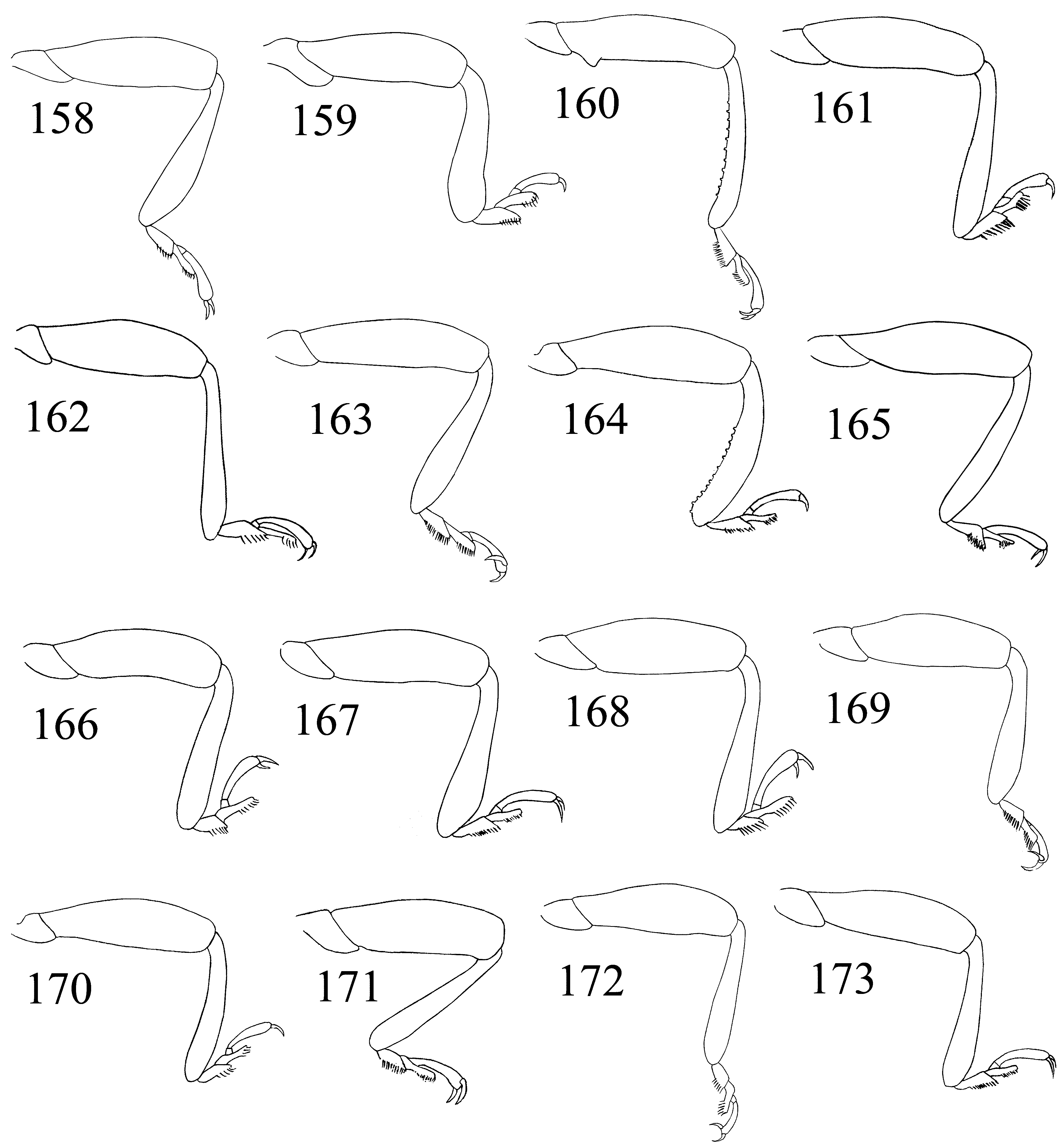
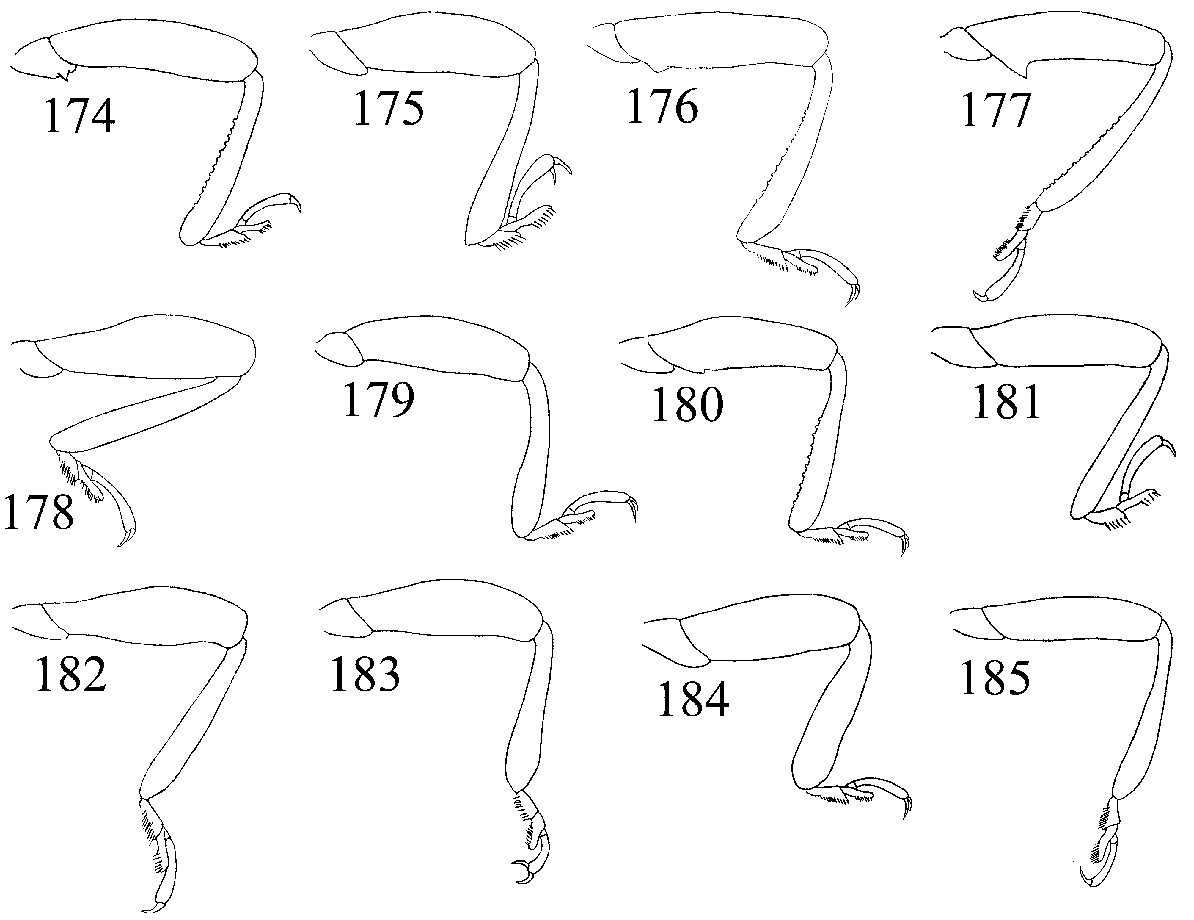
7. Metatrochanter and metafemur with unusually long and sparse setae, their length 0.3–0.5X length of the metafemur ( Fig. 144 View FIGURES 144 – 157 ); metatibia stout and slightly sinuate, more conspicuously in males ( Figs. 144 View FIGURES 144 – 157 , 159 View FIGURES 158 – 173 , 184 View FIGURES 174 – 185. 174 – 183 ); body size 3.4–3.9 mm, completely dark brown except black antennal club (Fig. 15)............................................. S. exiguus Gorham
– Metatrochanter and metafemur densely covered with normal, comparatively short, decumbent setae, their length much smaller than 0.3X length of metafemur; metatibia linear or rarely slightly curved, bent medially and rather narrow (e.g. Figs. 145–149 View FIGURES 144 – 157 ); body size and color variable............................................................................. 8
8. Scutellum semicircular ( Fig. 135 View FIGURES 126 – 143 ); elytral epipleuron distinctly wide at base, 1.2X wider than anterior border of intercoxal process of metaventrite; male metatrochanter with a small sharp spine ( Figs. 148 View FIGURES 144 – 157 , 174 View FIGURES 174 – 185. 174 – 183 ); median lobe with an accessory process ( Figs. 231 View FIGURES 224 – 243 ).............................................................................. S. ovali s Arrow
– Scutellum triangular ( Figs. 134 View FIGURES 126 – 143 ); elytral epipleuron narrow to moderately wide at base, as wide or narrower than anterior bor- der of intercoxal process of metaventrite; metatrochanters unarmed in both sexes; median lobe without accessory process... 9
9. Lateral margins of pronotum scarcely raised, very narrow, about as wide as a sixth of distance between basal pore and hind angle, ( Fig. 96 View FIGURES 90 – 104 ); meso- and metatibiae wide, flattened, strongly curved in males ( Figs. 146 View FIGURES 144 – 157 , 166, 170 View FIGURES 158 – 173 ); body brown with antennae black except scape, or scape and pedicel wich are brown, legs uniformly brown or femur brown and tibiae and tarsi black (Figs. 21, 28)........................................................................................ 10
– Lateral margins of pronotum raised, comparatively wide, as wide or wider than a fifth of distance between basal pore and hind angle ( Figs. 92–95, 97, 100–104 View FIGURES 90 – 104 , 107–108, 110–112 View FIGURES 105 – 114 ); mesotibia always linear, metatibia most often linear ( Figs. 161–163, 168–169, 171–173 View FIGURES 158 – 173 , 178, 181–183 View FIGURES 174 – 185. 174 – 183 ), if weakly bent or curved in males of some species then distinctly narrow ( Figs. 160, 165 View FIGURES 158 – 173 ); body coloration variable; if body completely brown, then antennae have at least first four articles brown and others are black.................................................................................................... 11
10. Tibiae and tarsi reddish brown (Fig. 21); metatibia broad, markedly curved ( Fig. 146 View FIGURES 144 – 157 ), with medial row of tubercles in male
( Fig. 164 View FIGURES 158 – 173 ); median lobe widened at basal third in ventral view ( Figs. 213 View FIGURES 200 – 223 )............................ S. latipes Arrow – Tibiae and tarsi dark fuscous or black ( Fig. 28 View FIGURES 26 – 37 ); metatibia moderately wide and curved, without tubercles on medial face ( Fig. 170 View FIGURES 158 – 173 ); median lobe ( Figs. 223 View FIGURES 200 – 223 ) narrow at basal third in ventral view.............................. S. molgorae sp. nov.
11. Pronotum with basal pores moderately large, reniform, markedly oblique; basal sulcus present, complete or occasionally lacking medially ( Figs. 92–95, 100–101 View FIGURES 90 – 104 , 105, 107–108, 111, 114 View FIGURES 105 – 114 ).................................................. 12
– Pronotum with basal pores comparatively small, rounded or elongate, rarely oblique; basal sulcus indistinct or absent ( Figs. 93–94, 97, 102, 104 View FIGURES 90 – 104 , 110, 112 View FIGURES 105 – 114 ).......................................................................... 20
12. Antennomeres 4–7 thin, about 2X as long as wide; antennomere 9 2.2X wider than 8 ( Fig. 59 View FIGURES 54 – 71 ); pronotum with lateral margins comparatively narrow ( Fig. 95 View FIGURES 90 – 104 ); elytra with setae relatively short and decumbent; head black............ S. kafkai sp. nov.
– Antennomeres 4–7 comparatively stout, 1.7X as long as wide or shorter; antennomere 9 about 1.8X wider than 8 (56, 64–65, 68, 72, 75, 78); pronotum with lateral margins comparatively wide ( Figs. 92, 100–101, 103 View FIGURES 90 – 104 , 107–108, 111, 114 View FIGURES 105 – 114 ); elytra with setae comparatively long and suberect; head red or brown..................................................... 13
13. Ventrite V of females markedly emarginate medially ( Fig. 189 View FIGURES 186 – 195 ); pronotum with basal sulcus complete ( Fig. 101 View FIGURES 90 – 104 ); antenna with terminal antennomere short, subovate ( Fig. 65 View FIGURES 54 – 71 ), 1.4X as long as wide, 3.5X as long as pedicel; median lobe constricted apically, in lateral view ( Fig. 220 View FIGURES 200 – 223 )....................................................... S. militaris Gerstaecker
– Ventrite V of females truncate ( Figs. 187, 191 View FIGURES 186 – 195 ); pronotum with basal sulcus complete, or vanishing medially ( Figs. 92, 100, 103 View FIGURES 90 – 104 , 107–108, 111, 114 View FIGURES 105 – 114 ); antenna with terminal antennomere elongate, oval or subrectangular ( Figs. 56, 64, 68 View FIGURES 54 – 71 , 72, 75, 78 View FIGURES 72 – 78 ), about or longer than 1.4X as long as wide and 3.5X as long as pedicel; median lobe not distinctly constricted apically, in lateral view............................................................................................... 14
14. Legs long and slender, ( Figs. 145 View FIGURES 144 – 157 , 160 View FIGURES 158 – 173 ; 176–177) slightly less pronounced in females ( Fig. 185 View FIGURES 174 – 185. 174 – 183 ); male metafemur with a tooth basally ( Figs. 150–153 View FIGURES 144 – 157 ), metatibia slightly bent medially at apical third, with a row of small tubercles; metaventrite of males with a transverse, densely punctate concavity, at anterior margin between mesocoxae ( Figs. 88–89 View FIGURES 79 – 89. 79 – 83 ); median lobe gradually widening apically, in lateral view ( Fig. 204 View FIGURES 200 – 223 ), gradually narrowing apically, in ventral view, slightly flexed laterally with a small apical crest ( Fig. 205 View FIGURES 200 – 223 )................................................................................. 15
– Legs moderately long and slender, unarmed ( Figs. 168, 172 View FIGURES 158 – 173 , 183 View FIGURES 174 – 185. 174 – 183 ); metatibia linear; metaventrite without cavities or other modifications near anterior margin in both sexes; median lobe not as above, wider apically or slightly sinuate, in lateral view (210– 211, 226–227, 242–243)............................................................................... 18
15. Prosternal process narrow at base, scarcely widened apically ( Fig. 131 View FIGURES 126 – 143 ); body size 5.1–5.4 mm ( Fig. 39 View FIGURES 38 – 42 ); tooth of male metafemur scarcely developed ( Fig. 153 View FIGURES 144 – 157 )........................................................ S. shockleyi sp. nov.
– Prosternal process moderately narrow at base, strongly widened apically ( Fig. 127 View FIGURES 126 – 143 ); body size 5.4–7.9 mm; tooth on male metafemur moderately to well developed ( Fig. 150–152 View FIGURES 144 – 157 )..................................................... 16
16. Tooth on male metafemur widely truncate ( Fig. 151 View FIGURES 144 – 157 ); foveolate punctures on elytra comparatively large and deep ( Fig. 122 View FIGURES 115 – 125 ); body size 6.9–7.9 mm, 1.80–1.83X as long as wide; elytra completely reddish brown ( Figs. 35 View FIGURES 26 – 37 )........ S. raramuri p. nov.
– Tooth on male metafemur with apex narrowly blunt or acute ( Fig. 150, 152 View FIGURES 144 – 157 ); foveolate punctures on elytra rather small and shallow ( Fig. 117 View FIGURES 115 – 125 ); body size 5.4–7.4 mm, 1.56–1.75X as long as wide; elytron red with large black macula, or completely brown (Figs. 16–17, 36)................................................................................ 17
17. Tooth on male metafemur large, pointed ( Fig. 152 View FIGURES 144 – 157 ); body size 5.4–6.6 mm ( Fig. 36 View FIGURES 26 – 37 ); antenna red with antennomeres 7–11 black; legs red; pronotum red with large triangular macula not surpassing pores at base; elytron red with large, central, black macula, macula always reaching apical third.......................................... S. rubrocinctus Gerstaecker
– Tooth on male metafemur moderately large, blunt or rounded apically ( Fig. 150 View FIGURES 144 – 157 ); body size 6.4–7.4 mm (Fig. 16–17); antenna black or with antennomeres 1–6 red, brown or infuscate; legs black, brown or red; body completely brown or contrastly colored: pronotum uniformly black or with angles red, black part sometimes subtriangular, surpassing basal pores at base; elytron uniformly reddish-brown or red with large, central, black macula, macula sometimes hardly reaching apical third..................................................................................... S. globosus Guérin-Méneville
18. Terminal antennomere elongate, 1.7X as long as wide, 4.4X longer than pedicel ( Fig. 64 View FIGURES 54 – 71 ); median lobe distinctly thin ( Figs. 210–211 View FIGURES 200 – 223 ).......................................................................... S. mexicanus sp. nov.
– Terminal antennomere moderately elongate, 1.4–1.5X as long as wide, 3.5–3.7X longer than pedicel ( Figs. 68, 69 View FIGURES 54 – 71 ); median lobe moderately broad ( Figs. 226–227, 242–243 View FIGURES 224 – 243 )........................................................... 19
19. Pronotum with basal pores small, basal sulcus incomplete, lateral margins wide, width 1/3 of distance between midpoint of basal pore and hind angle ( Fig. 114 View FIGURES 105 – 114 ); median lobe rather linear, narrowing apically, in lateral view ( Fig. 242 View FIGURES 224 – 243 ); elytron orangered with large, black macula ( Fig. 42 View FIGURES 38 – 42 ).................................................... S. thoracicus Gorham
– Pronotum with basal sulcus complete, basal pores moderately large, lateral margins comparatively narrow, width 1/4 of distance between midpoint of basal pore and hind angle ( Fig. 103 View FIGURES 90 – 104 ); median lobe sinuate, same width throughout ( Fig. 226 View FIGURES 224 – 243 ); elytron uniformly brown ( Fig. 30 View FIGURES 26 – 37 )....................................................... S. nigricans Gorham
20. Tarsomere 2 of all legs narrow, not widened distally, at most 1.3X wider than tarsomere 4 at midlength ( Fig. 155 View FIGURES 144 – 157 )........ 21
– Tarsomere 2 of all legs widened distally, at least 2.0X wider apically than tarsomere 4 at midlength ( Figs. 157 View FIGURES 144 – 157 ).......... 23
21. Base of pronotum clearly emarginate near scutellum ( Fig. 97 View FIGURES 90 – 104 ); antenna distinctly long, 0.40–0.45X as long as body in females, 0.45–0.50X in males, terminal antennomere elongate (Fig. H8); median lobe strongly widened apically, in lateral view ( Fig. 214 View FIGURES 200 – 223 )............................................................................. S. lemniscatus Gorham
– Base of pronotum rounded or truncate near scutellum ( Figs. 94, 103 View FIGURES 90 – 104 ); antenna short, 0.33–0.38X as long as body in both sexes, terminal antennomere short to moderately long oval ( Figs. 58, 67 View FIGURES 54 – 71 ); median lobe of same width throughout, in lateral view ( Fig. 224 View FIGURES 224 – 243 ) or slightly narrowing apically ( Fig. 208 View FIGURES 200 – 223 ).............................................................. 22
22. Base of pronotum rounded ( Fig. 94 View FIGURES 90 – 104 ); terminal antennomere parallel sided ( Fig. 58 View FIGURES 54 – 71 ); pronotum and elytra contrastly colored (Fig. 19); metaventrite of males with a deep longitudinal pit or incision near anterior margin ( Fig. 84 View FIGURES 79 – 89. 79 – 83 ); median lobe moderately
broad, in ventral view, somewhat constricted after midlength ( Fig. 209 View FIGURES 200 – 223 )............................. S. incisus sp. nov. – Base of pronotum truncate near scutellum ( Fig. 102 View FIGURES 90 – 104 ); terminal antennomere oval ( Fig. 67 View FIGURES 54 – 71 ); pronotum and elytra uniformly colored ( Fig. 29 View FIGURES 26 – 37 ); metaventrite of males without an excision near anterior margin ( Fig. 85 View FIGURES 79 – 89. 79 – 83 ); median lobe very narrow throughout its length, in ventral view............................................................ S. monterrosoi sp. nov.
23. Body small (3.0 mm), short oval ( Fig. 40 View FIGURES 38 – 42 ), 1.7X longer than wide; elytra ovoid; lateral margins of pronotum wide ( Fig. 112 View FIGURES 105 – 114 ); median lobe of same width throughout, in lateral view ( Fig. 238 View FIGURES 224 – 243 )................................. S. smithi Gorham
– Body moderately large (3.60–5.05 mm), long oval (Figs. 18, 31–32, 38); lateral margins of pronotum narrow ( Figs. 93, 104 View FIGURES 90 – 104 , 109 View FIGURES 105 – 114 ); median lobe of different width throughout its length, in lateral view ( Figs. 206 View FIGURES 200 – 223 , 228, 236 View FIGURES 224 – 243 )....................... 24
24. Terminal antennomere with an angle on lateral margin near midlength ( Fig. 69 View FIGURES 54 – 71 ); pronotum and elytra uniformly brown ( Fig. 32 View FIGURES 26 – 37 ) or red-orange, each with large, black macula ( Fig. 31 View FIGURES 26 – 37 ); median lobe moderately wide, in ventral view ( Fig. 229 View FIGURES 224 – 243 )......................................................................................... S. oblongulus Gorham
– Terminal antennomere without protuberance on lateral margin ( Figs. 57 View FIGURES 54 – 71 , 74 View FIGURES 72 – 78 ); pronotum and elytra always uniformly brown (Figs. 18, 38); median lobe narrow, in ventral view ( Figs. 207 View FIGURES 200 – 223 , 237 View FIGURES 224 – 243 )............................................. 25
25. Metatibia slightly curved, almost parallel in basal half, then widened at 3/4 ( Fig. 149 View FIGURES 144 – 157 ); ventrite I of male abdomen with small, conical protuberance ( Fig. 186 View FIGURES 186 – 195 )........................................................... S. sallaei Gorham
– Metatibia gradually widened apically ( Fig. 161 View FIGURES 158 – 173 ); ventrite I of male abdomen without protuberance.... S. guatemalae Arrow


No known copyright restrictions apply. See Agosti, D., Egloff, W., 2009. Taxonomic information exchange and copyright: the Plazi approach. BMC Research Notes 2009, 2:53 for further explanation.