
Stactobia McLachlan, 1880
|
publication ID |
https://doi.org/ 10.11646/zootaxa.4350.2.1 |
|
publication LSID |
lsid:zoobank.org:pub:2692F486-4EEA-4B62-BC88-8EEF71AB9C0F |
|
DOI |
https://doi.org/10.5281/zenodo.6034135 |
|
persistent identifier |
https://treatment.plazi.org/id/03E31C49-FFD5-3239-FF37-FE68FD120695 |
|
treatment provided by |
Plazi |
|
scientific name |
Stactobia McLachlan |
| status |
|
Genus Stactobia McLachlan View in CoL
Stactobia McLachlan 1880 View in CoL , 505. Type species: Hydroptila fuscicornis Schneider , by subsequent designation ( Mosely 1933).
Diagnosis. Nine hydroptilid genera, Hydroptila Dalman , Microptila Ris , Orthotrichia Eaton , Oxyethira Eaton , Plethus Hagen , Pseudoxyethira Schmid , Stactobiella Martynov , Ugandatrichia Mosely , and Stactobia McLachlan , have been found in Japan (Ito 2017). Among these genera, the males of Stactobia are distinguished from those of other genera in Japan by a combination of the following character states: Head with 3 ocelli and large ellipsoidal posterolateral warts, lozenge-shaped mesoscutellum with transverse suture, meso- and metanota without any setal warts, tibial spur formula 1, 2, 4 or 0, 2, 4, and ventral process of abdominal segment VII long and thickened apically.
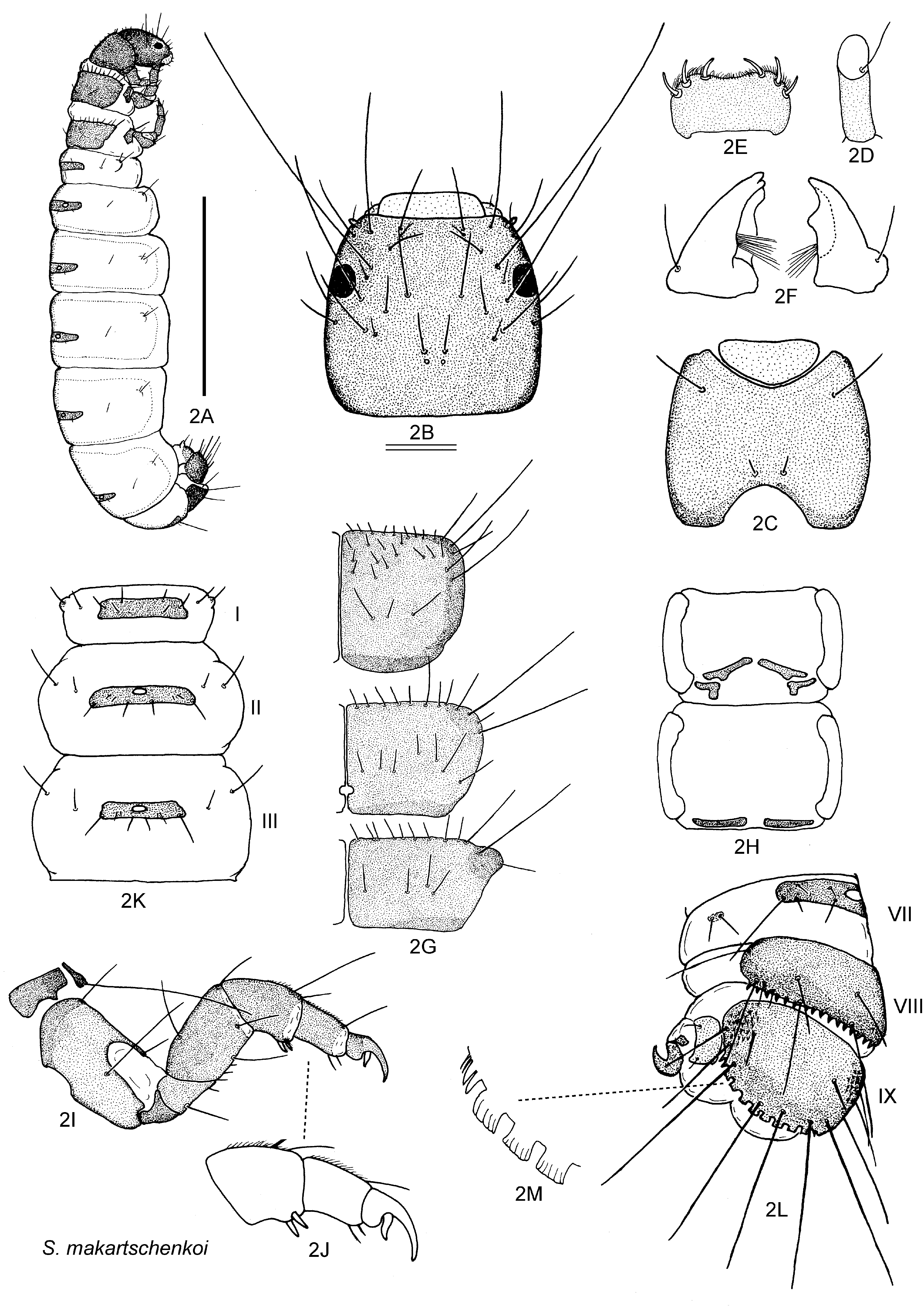
The final instar larva of this genus is clearly discriminated from those of other hydroptilid genera by large semicircular sclerites of tergum IX, each bearing posterior edge with striped ‘crenellations’ or ‘crenels’ at middle and spines laterally.
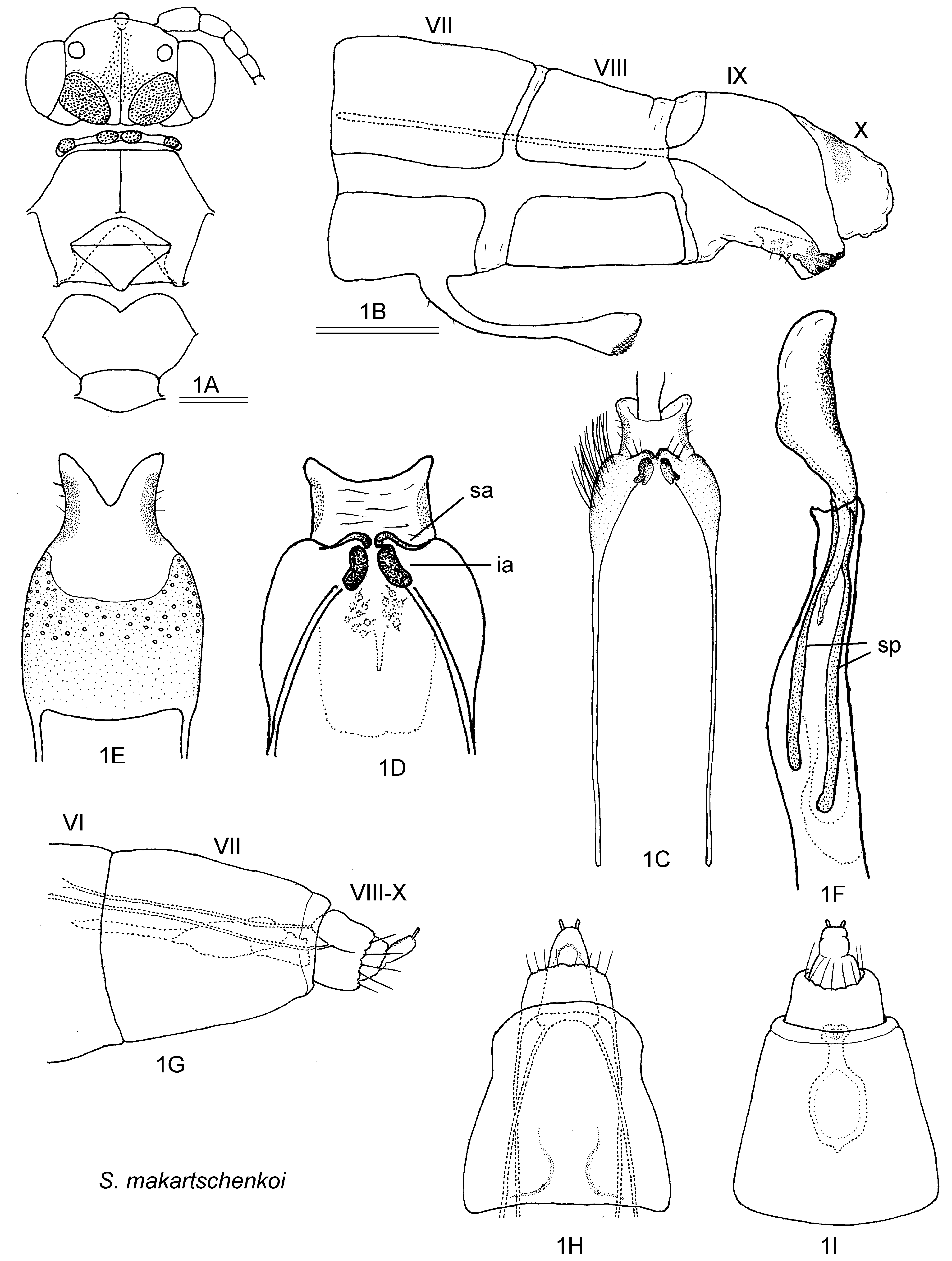
Description. Adult (modified after Marshall 1979). Head with 3 ocelli, posterodorsal warts (postoccipital lobes) large, oval ( Fig. 1A View FIGURE 1 ). Antennae 18–25 segmented. Maxillary palpi each 5-segmented, 2 basal segments short, other segments cylindrical. Labial palpi each 3-segmented, basal segment short, other segments cylindrical. Pronotum with pairs of round mesal and round lateral warts; mesoscutellum lozenge shaped, with horizontal suture; metascutellum trapezoidal ( Fig. 1A View FIGURE 1 ). Wings black, acuminate, densely covered with long black hairs; length of each forewing 1.4–4.0 mm. Tibial spur formula 1, 2, 4 or 0, 2, 4.
Male (e.g., Figs 1B–1F View FIGURE 1 ): Sternite VIII displaced posteriorly. Segment IX reduced ventrally, often produced anteriorly as pair of internal lateral apodemes. Tergite X semimembranous. Subgenital appendages variable in development. Inferior appendages small, rarely elongate. Phallus long, almost straight, often considerably developed but sometimes slender and simple, usually with dilated, heavily sclerotized spines, membranous apex. Ventral process of sternite VII long, sinuous and thickened apically.
Female (e.g., Figs 1G– 1I View FIGURE 1 ): Ventral process of abdominal sternum VI very small, often indistinct due to dense setae. Tergite and sternite of segment VII fused laterally. Segment VIII short, semimembranous with long pair of internal apodemes extending anteriorly and marginal setae posteriorly. Segments IX and X very small, membranous, terminated by pair of cerci. Morphological diagnosis of species generally not possible for females currently.
Larva. Final instar larva (e.g., Figs 2A–2M View FIGURE 2 ): Known for several species ( Vaillant 1951; Botosaneanu & Levanidova 1988; Waringer & Graf 2011). Case-bearing, cases usually fixed on substrate ( Figs 3F, 3G View FIGURE 3 ). Sclerotized parts of body brown to dark brown in most species. Thorax and abdomen dorsoventrally flattened (depressed), massive, well sclerotized. Head subquadrate, suture indistinct, antennae near anterolateral corners, seta 9 (sensu Wiggins 1996) longest, about 1.5 times head width; labrum symmetrical; mandibles robust, slightly asymmetrical. Thorax covered with large subquadrate dorsal plates, with several long setae on anterodorsal corners and many short setae on anterior margins and middle area; in some species sternites with very short, wide sclerites. Thoracic legs robust, all legs similar in form and length, tibiae with 2 short spines on subapical anteroventral corner, basal seta of tarsal claws thick, slightly sinuate. Abdominal segments more or less flattened dorsoventrally (depressed), each with dorsal tergite; tergite on segment I wide short rectangular; tergites on segments II–VII broadly rectangular, each with median small round area of chloride epithelia (dorsal ring of Marshall 1979; chloride epithelia? of Botosaneanu & Levanidova 1988); tergite VIII large subquadrate with spinose posterior margin; tergite IX large semicircular, characteristic, posterior edge with striped ‘crenellations’ (modified flattened setae of Marshall 1979) or ‘crenels’ ( Botosaneanu & Levanidova 1988) at middle edges and spinose at lateral edges. On segment X, lateral sclerites rectangular; paired anal legs strongly curved anterolaterad, anal claws without any accessory hooks.
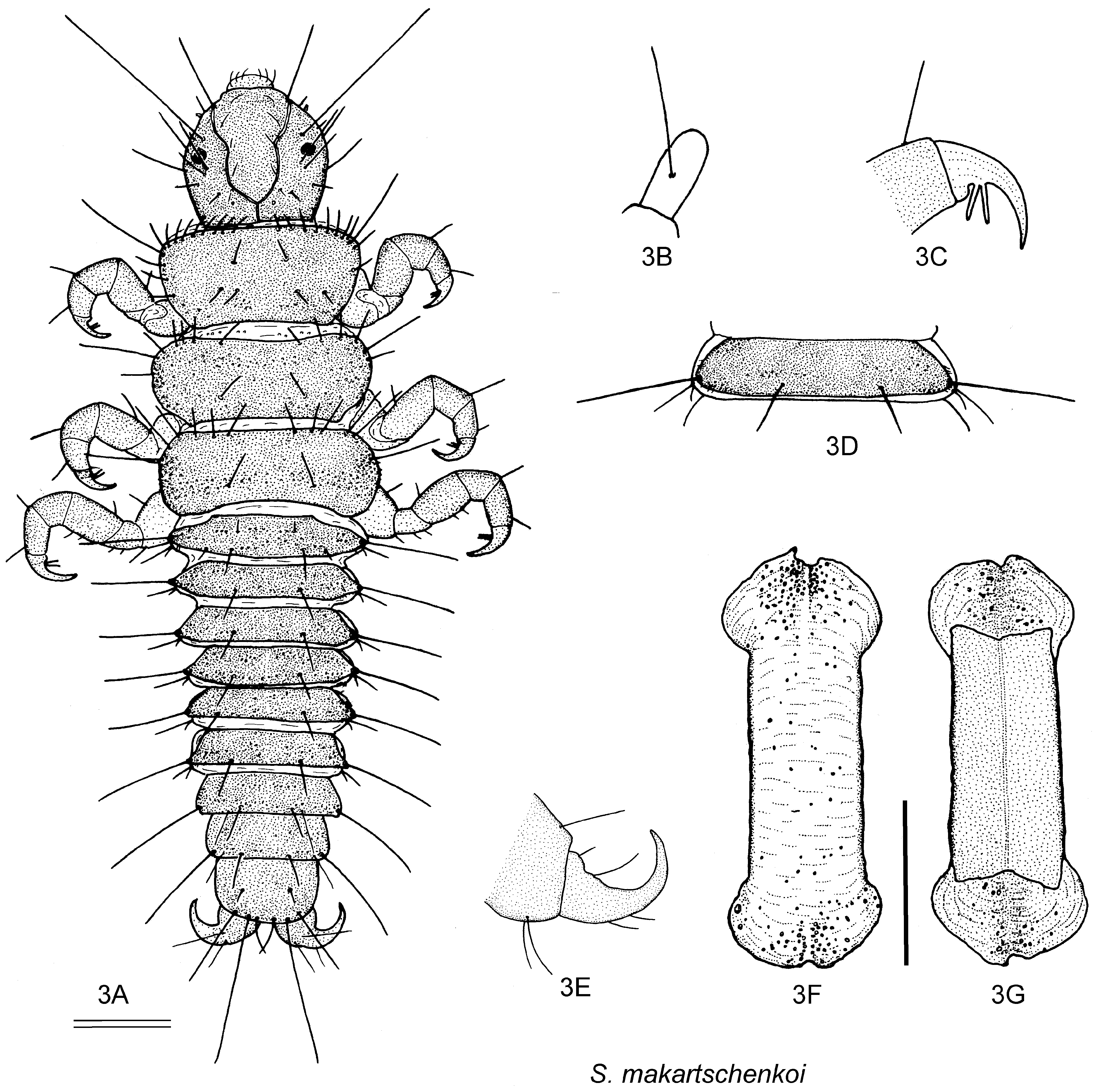
Early instar larva (from Marshall 1979, e.g., Figs 3A–3E View FIGURE 3 ). Free-living, caseless, dorsoventrally flattened (depressed), with dorsal sclerites on all abdominal segments, paired anal claws gently curved posterolaterad. Case (e.g., Figs 3F, 3G View FIGURE 3 ). For most species, cases of final instar larvae made of silk, sometimes together with few mineral particles, composed of ventral and dorsal valves; each with ventral valve forming flat sheet; dorsal valve forming convex dome carried tortoise-like, larger than ventral valve, ends interchangeable, with anterior and posterior hoods concealing larva completely.
Habitat. Adults of most species found near hygropetric zones: Falls and fast flowing streams with boulders. Japanese name. Kaku-himetobikera-zoku.
No known copyright restrictions apply. See Agosti, D., Egloff, W., 2009. Taxonomic information exchange and copyright: the Plazi approach. BMC Research Notes 2009, 2:53 for further explanation.
|
Kingdom |
|
|
Phylum |
|
|
Class |
|
|
Order |
|
|
Family |
Stactobia McLachlan
| Ito, Tomiko 2017 |
Stactobia
| McLachlan 1880 |
Hydroptila fuscicornis
| Schneider 1845 |