
Macrothrix boergeni Studer, 1878
|
publication ID |
https://doi.org/10.1080/00222930701689937 |
|
persistent identifier |
https://treatment.plazi.org/id/03E32C46-B14F-FF9F-FE77-FDB0D99713A0 |
|
treatment provided by |
Felipe |
|
scientific name |
Macrothrix boergeni Studer, 1878 |
| status |
|
Macrothrix boergeni Studer, 1878 View in CoL
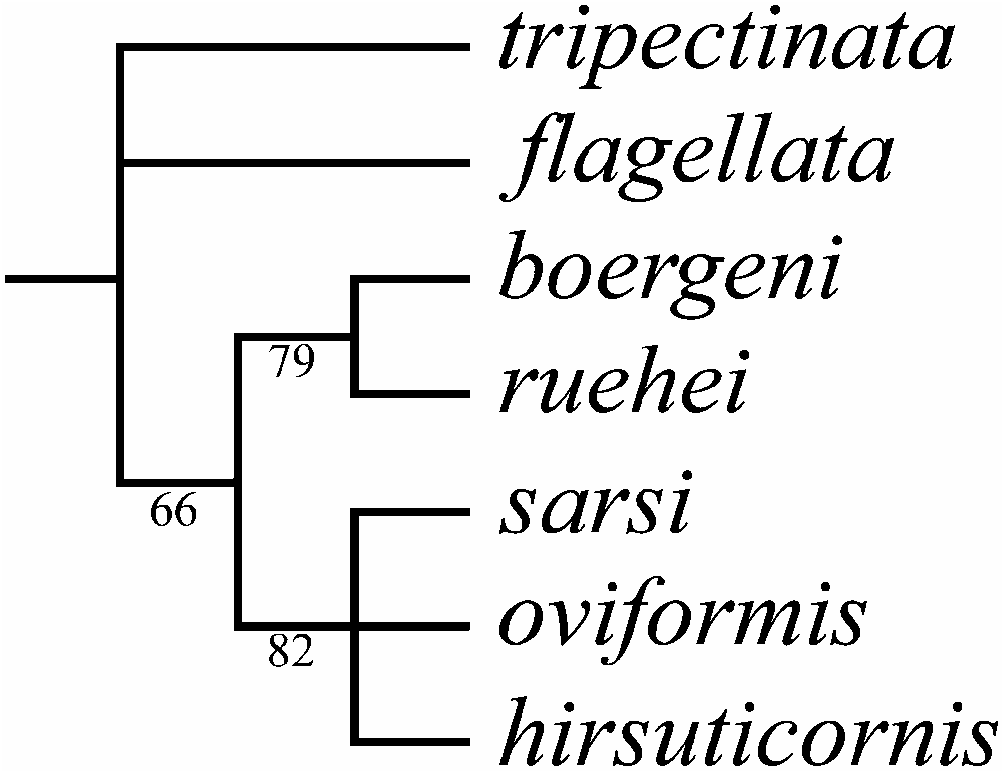
( Figures 1–5 View Figure 1 View Figure 2 View Figure 3 View Figure 4 View Figure 5 , 6A–C View Figure 6 )
Macrothrix Börgeni Studer 1878, p 108 View in CoL , Plate 3, Figure 2 View Figure 2 .
Macrothrix hirsuticornis Norman and Brady View in CoL in Rühe 1914, p 55 –56, Figures 6b View Figure 6 , 19 View Figure 19 (only specimens from Kerguelen!); Brehm 1954, p 41; Gay 1981, p 51, Figures 21–24 View Figure 21 View Figure 22 View Figure 23 View Figure 24 . Macrothrix cf. hirsuticornis in De Smet 2001, p 263 View in CoL .
All records of Macrothrix View in CoL from Kerguelen listed by Pugh et al. (2002) probably present M. boergeni View in CoL .
Type locality (according to neotype selected here)
An un-named pond near Port-aux-Français, Îles Kerguelen, French Subantarctic Territories (approximately 49 ° 219S, 70 ° 139E). The sample was collected 5 February 1988 by W. H. De Smet .
Type material
Neotype (selected here): a parthenogenetic female in 90% alcohol, NHM 2004.2092. Author’s type material is apparently lost.
Label of the neotype: ‘‘ Macrothrix boergeni Studer, 1878 ; 1 parth. ♀, A pond near Portaux-Français , Iles Kerguelen, collected 5 February 1988 by W. H. De Smet, NEOTYPE’ ’.
Other material examined
Kerguelen Archipelago : many parthenogenetic (parth.) ♀♀ taken from the sample, from which the neotype was selected, AAK 2002-027 (tube); 35 parth. ♀♀ taken from the same sample, NHM 2004.2093–2102 (tube) .
Amended diagnosis
Parthenogenetic female. In lateral view body subovoid, cervical depression absent, dorsal margin breached by a ‘‘step’’ in posterior boundary of head, dorsal margin of valves without any serration. Postero-dorsal angle as rounded triangle, lies in level of middle of body height. No dome above eye. Ocellus large. Dorsal organ ovoid, small. Labrum with moderately projected apex bearing several small tubercles.
Postabdomen subovoid, with rounded distal extremity, without a ‘‘heel’’ basally, and without a reticulation on sides. Ventral margin straight, with a few series of small, robust denticles. Dorsal margin distinctly bilobed; preanal margin with transversal series of minute setules, anal margin with groups of thicker setules. Postabdominal seta with short distal segment, densely armed with relatively short setules; basal segment with very few (two to three) short, sparsely located setules. On external side of postabdominal claw, a series of three to five robust denticles; medial row (on ventral margin) of about seven to nine denticles; inner row with numerous denticles, organized in three successive series.
Antenna I widened distally, straight, without a subapical external angulation; sensory seta at distance of about two antennular diameters (at base) from antenna I joint; on anterior face about six to eight transverse rows of spinules, associated with distinct reticulation. Nine short aesthetascs, three of them slightly larger than the rest. Antenna II with distal burrowing spine on basal segment somewhat shorter than proximal segment of exopod. Length of all apical swimming setae subequal. Lateral seta on proximal endopod segment larger than other setae, lacking robust denticles in middle. A spine on second segment of exopod half as long as the next (second) segment. On posterior side of segments 1–3 of exopod there are a series of small additional denticles.
Limb I with outer distal lobe supplied with longest apical seta having distal segment unilaterally armed with relatively robust setules, located more rarely on proximal portion of seta distal segment; inner-distal lobe with three bisegmented setae of different size, unilaterally setulated in distal part, robustness of armature different in different setae, smallest one with numerous fine setules; two ejector hooks of different size, sometimes one of them rudimentary; a remainder of gnathobase I with single fully setulated setae. On limb II, scrapers 1–2 with delicate feathering, but scrapers 3–7 with denticles specially massive; a solitary posterior seta near gnathobase present; filter plate II with four setae, without a rudiment of fifth seta. On limb III epipodite with five setae; a distal group of three long setae, seta 1 shortest, armed with robust denticles; setulated projections proximally to seta 3 and between setae 2 and 3; on inner-distal portion, seta 1 with specially short and robust denticles; seta a with fine setules basally and robust spinules distally, seta b characteristically long; basal endite posteriorly with four soft setae. Limb IV with exopodite small, bearing only a distal group of three bilaterally feathered setae of subequal size; on inner-distal portion of this limb seta 1 with strong setules basally and two to three robust denticles distally; posteriorly a row of five long setae. On limb V there are three setae at inner margin, sometimes seta 3 reduced.
Ephippial female, male. Not adequately described.
Size. Up to 1.06 mm.
Full redescription
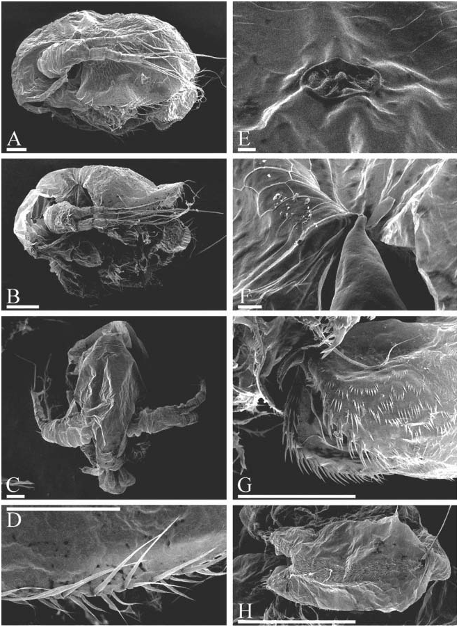
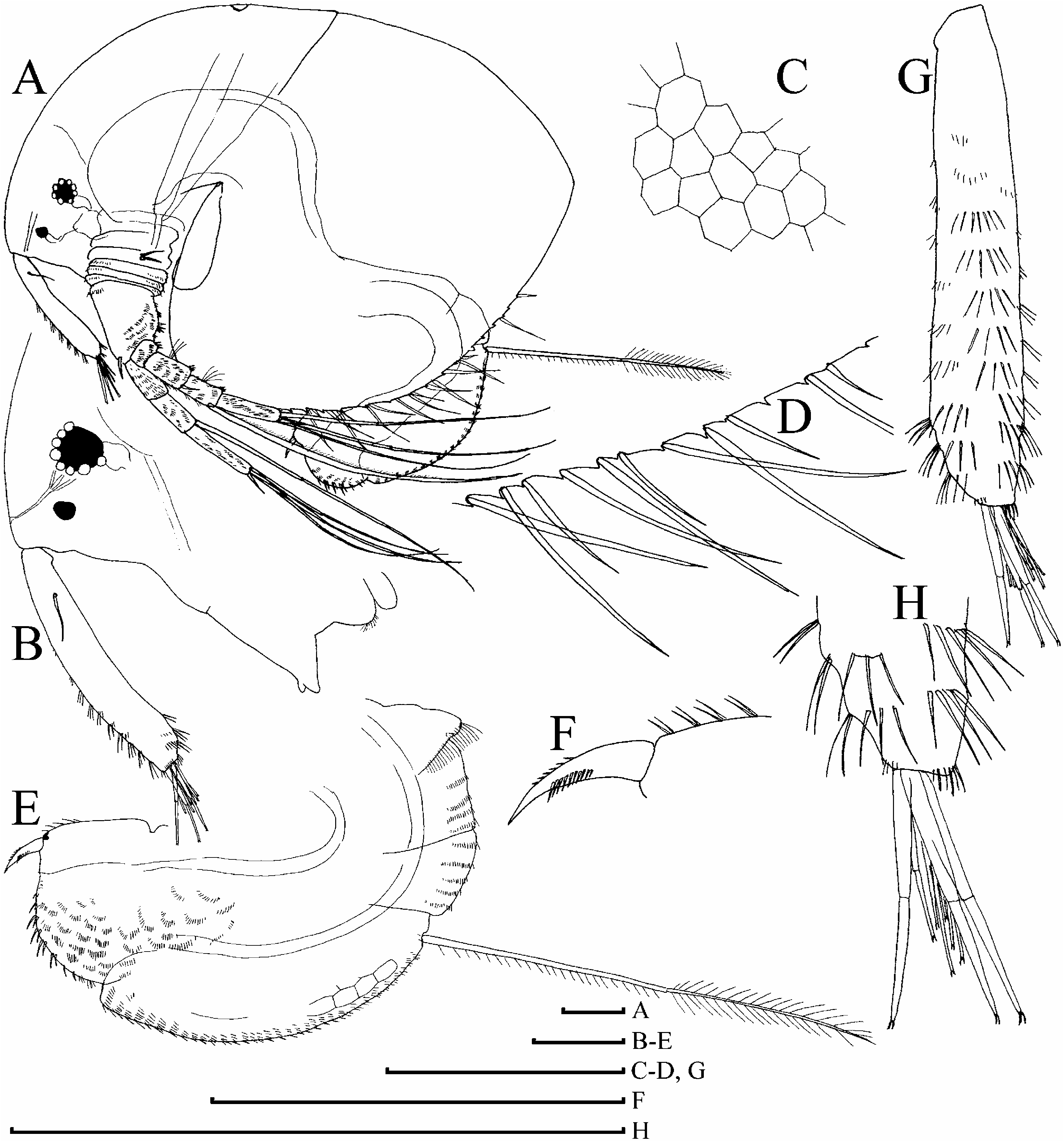
Adult parthenogenetic female. Body of large female subovoid in lateral view ( Figures 1A View Figure 1 , 2A, B View Figure 2 , 6A View Figure 6 ), with maximum height in the middle, height/length50.66–0.70. Dorsal margin in general as a regular arch from tip of rostrum to posteriormost point, without traces of cervical depression or with a slight depression, mostly breached by a ‘‘step’’ in posterior boundary of head, sometimes forming a blunt tooth ( Figure 1A View Figure 1 ). Dorsal margin of valves not elevated, or slightly elevated above dorsal margin of head. Postero-dorsal angle as a rounded triangle, lies in level of middle of body height. Whole surface of valves with fine reticulation, while head without it. No remarkable structures on valves or head. In anterior view body moderately compressed laterally ( Figure 2C View Figure 2 ).
Head large, length/body length up to 0.4. In lateral view, dorsal margin in general evenly convex, no dome above eye; ventral margin almost straight, with minute crossing ridges, no projection at base of labrum ( Figure 1B, C View Figure 1 ). A special line (fold) goes from mandibular joint anteriorly, it corresponds to a poorly expressed fornix. Compound eye large, ocellus also large (its size more than half of eye diameter), located approximately in middle of distance between tip of rostrum and eye.
‘‘Dorsal head pore’’ (‘‘dorsal organ’’ or ‘‘window’’ are more correct names, because no pore is really present here, only a window of specialized cuticle) ovoid, small, located on posterior part of head ( Figure 2E View Figure 2 ).
Labrum wide, in lateral view approximately triangular, with a moderately projected apex bearing several small tubercles, and setulated distal labral plate (term according to Kotov 1999, or anterior plug according to Dumont and Silva-Briano (2000)).
Valve surface with a distinct reticulation. Dorsal margin without any serration, but ventral margin denticulated. Marginal setae jointed to posterior sides of these denticles ( Figures 1D, G View Figure 1 , 2D View Figure 2 , 6B View Figure 6 ). These setae variable in length and size in different individuals, but there is a general order to their sequence: there are two ventrally directed setae between each larger, laterally oriented one, characteristic also of the majority of other species ( Kotov 1999; Kotov and Hollwedel 2004; Kotov et al. 2004). In anterior and posterior portion of ventral margin the order of seta alternation is not too precise.
Thorax long, while abdomen short, without projections. Intestine without convolutions.
Postabdomen subovoid in lateral view, with a rounded distal extremity, without ‘‘heel’’ (inflated base of postabdominal setae) basally ( Figures 1H View Figure 1 , 2H View Figure 2 ). Ventral margin straight, with few series of small, robust denticles. Dorsal margin distinctly bilobed, and the incision, which bilobes the margin, located at the level of proximal border of anus, separating anal and preanal margin. The latter long, slightly and regularly convex, with short transversal series of minute setules. On anal margin groups of setules with size significantly larger than those on postanal margin, laterally to them there are series of finer setules ( Figure 2G View Figure 2 ). Small postanal margin also with series of minute setules. A reticulation on sides of postabdomen fully absent.
Postabdominal seta approximately as long as postabdomen, with short distal segment, densely armed with relatively short setules; basal segment with a very few (two to three) short, sparsely located setules.
Postabdominal claw small, directed distally, slightly and regularly bent dorsally, with pointed tip and wide base in lateral view. On claw, a series of few (three to five) robust denticles on external side, medial row (immediately on ventral margin as seen laterally) of about seven to nine denticles ( Figures 1I, J View Figure 1 , 3A, B View Figure 3 ), and inner dorsal row with numerous denticles, organized in three successive series ( Figure 1K View Figure 1 ).
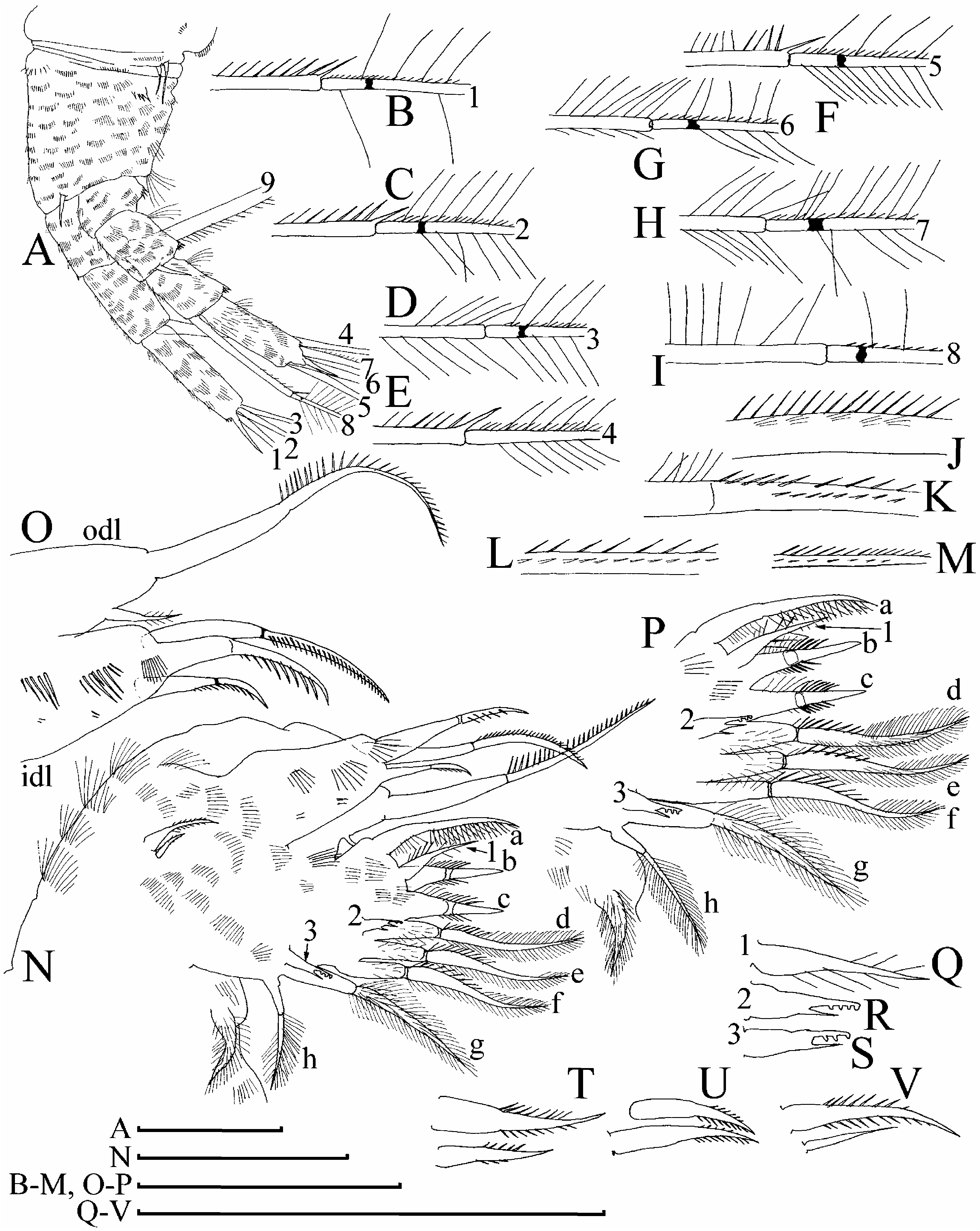
Antenna I widened distally, straight, without subapical external angulation (‘‘subapical ventral angulation’’ sensu of Silva-Briano (1998)); sensory seta located externally at distance of about two antennular diameters (at base) from antenna I joint ( Figures 1L, M View Figure 1 , 3C View Figure 3 ). About six to eight transverse rows of spinules, associated with a distinct reticulation on anterior surface of antenna I, series of fine spinules at its end. Nine relatively short terminal aesthetascs (longest about one-quarter of antenna I length), each with two minute ‘‘claws’’ at the apex ( Figure 1N, O View Figure 1 ). Three aesthetascs slightly larger than the rest, bearing unknown thin-walled, delicate structures, which could be additional sensory elements, but there is a chance that these are only epibiotic bacteria.
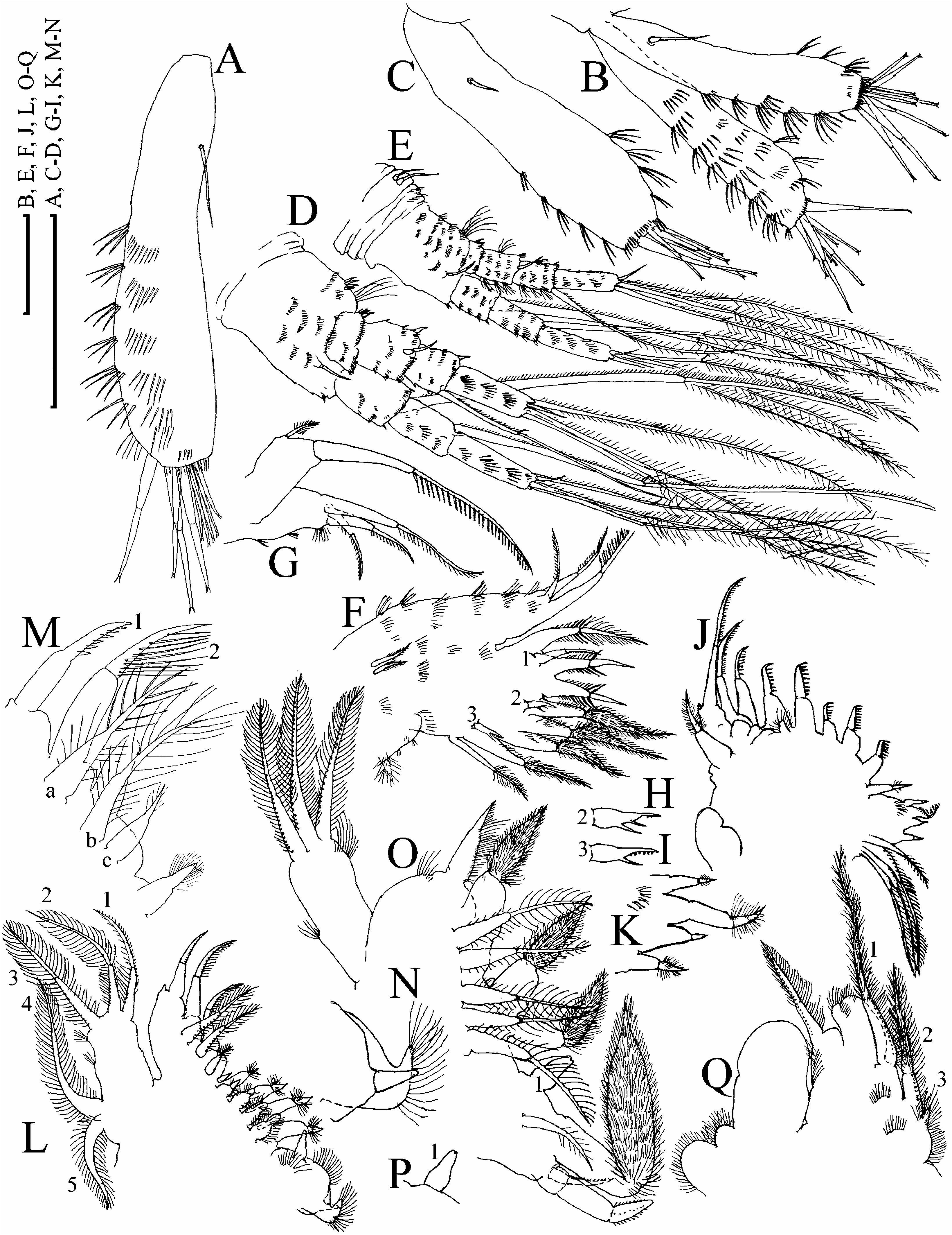
Antenna II large, coxal region folded, with two small basal sensory setae of slightly different size in middle part and rows of small setules on each fold ( Figures 3D View Figure 3 , 4A View Figure 4 , 6C View Figure 6 ). Basal segment robust, bearing numerous transverse series of spinules, and long, bisegmented distal sensory seta at inner (posterior) margin. Distal burrowing spine somewhat shorter than proximal segment of exopod, located on outer (anterior) surface, close to end of the basal segment ( Figure 3E View Figure 3 ).
Antennal branches long (about two times longer than basal segment), only proximal member of exopod shortened, all other segments elongated. Segments 2–3 of exopod subequal in size, while terminal segment as long as these two. Endopod apical segment longer than each of the other segments of this branch. All segments with transverse rows of setules. Swimming setae 0-0-1-3/1-1-3, spines 0-1-0-1/0-0-1. Length of all apical swimming setae subequal, approximately equal to length of basal segment plus length of branch. Each seta is marked with an individual number in Figure 4A View Figure 4 , armature of each seta is illustrated in Figure 4B–I View Figure 4 . Lateral seta on proximal endopod segment larger than other setae, lacking robust denticles in middle ( Figures 3F View Figure 3 , 4J–M View Figure 4 ). Apical spines short, from slightly curved to straight. A single spine on second segment of exopod, this spine half as long as next segment. On posterior side of exopod segments 1–3 there are a short series of small additional denticles (see discussion in Kotov et al. (2004)).
Mandible small, elongated, evenly dilated distally. Mandibular articulation located externally at point where valve and head come together ( Figure 2F View Figure 2 ).
Limb I large, without accessory seta; outer distal lobe cylindrical ( Figures 3H View Figure 3 , 4N, O View Figure 4 ), supplied with a long apical seta with distal segment unilaterally armed with relatively robust setules, located more rarely in proximal portion of seta distal segment, and a small lateral seta with bilaterally setulated distal segment. Inner-distal lobe massive, with median series of setules, few groups of minute setules, and three bisegmented setae of different size, unilaterally setulated in distal part, robustness of armature different in different setae, smallest one bears numerous fine setules. Endite III with a long, slightly curved, distally setulated seta a, while setae b–c short, clearly bisegmented, setulated bilaterally in middle ( Figures 3G View Figure 3 , 4N, P View Figure 4 ); anteriorly on this endite there is a short bisegmented seta 1, bearing sparse, long setules distally ( Figure 4Q View Figure 4 ). Endite II with three long bisegmented setae of subequal size ( Figure 4P View Figure 4 : d–f), each with distal segment bearing fine setules on distal parts, and more robust setules basally, and with fully setulated basal segment, and a fork-like seta 2 ( Figure 4R View Figure 4 ) anteriorly. Endite I with two bisegmented setae ( Figure 4P View Figure 4 : g–h) naked basally and supplied with long, dense setules distally; a fork-like seta 3 ( Figure 4S View Figure 4 ) anteriorly. Ejector hooks of different size, bilaterally setulated, in some specimens second hook significantly reduced in size and naked ( Figures 4T–V View Figure 4 ). A fully setulated seta at inner side of limb base, so-called maxillar process, represents a remnant of gnathobase I ( Kotov 1999).
Limb II: epipodite subglobular, exopodite as a subovoid lobe with three transverse rows of small setules and a short, bilaterally setulated seta distally ( Figure 5A, B View Figure 5 ). At inner margin of limb, eight robust scrapers, scrapers 1–2 with delicate feathering, but scrapers 3–8 with denticles specially massive for the genus ( Figures 3I View Figure 3 , 5B View Figure 5 ), a small sensillum near each scraper 1 and 4. Posteriorly to marginal scrapers, a system of low hillocks in distal limb portion, a setulated hillock near seta 4, and a solitary ‘‘soft’’ seta near gnathobase ( Figure 5C View Figure 5 ). Distal armature of gnathobase with four setae ( Figure 5C View Figure 5 : 1–4). Filter plate with four long setae, with size slightly decreasing distally.
Limb III: epipodite very small, globular, exopodite large and flat, with a distal group of three long setae ( Figure 5D View Figure 5 : 1–3), seta 1 shortest and armed with robust denticles, setae 2–3 with fine setules; there are setulated projections proximally to seta 3 and between setae 2 and 3; lateral group consists of two setae (4 and 5), similar in armature to 2 and 3. Distal endite (see discussion of its homology in Kotov (1999)) anteriorly with three bisegmented setae ( Figure 5E View Figure 5 : 1–3), unilaterally armed in distal part, seta 1 with specially short and robust denticles; small sensillum near each base of seta 1 and 3. Posteriorly, there are three soft setae: seta a with fine setules basally and robust spinules distally, setae b and c with fine setules distally, seta b characteristically long. Basal endite approximately equal in size to distal one. Anteriorly, a bottle-shaped sensillum and four setae with size increasing basally (4–7), each fully setulated distally, with distal segment longer than basal one. Posteriorly, four thick, soft setae ( d–g) subequal in size, each armed with fine setules, each has an inflated basal portion and a blunt tip. Gnathobase unclearly demarcated from basal endite, with four elements of distal armature: a large, bottle-shaped sensillum near border with basal endite, and two hooks plus a short bisegmented seta distally ( Figure 5E View Figure 5 : 1–4).
Limb IV: pre-epipodite small, with few setules, epipodite small and globular, exopodite small, with only a distal group of three bilaterally feathered setae of subequal size ( Figure 5F View Figure 5 : 1–3). Inner margin of limb with four setae ( Figure 5G View Figure 5 : 1–4), seta 1 with strong setules basally and two to three robust denticles distally ( Figure 5H View Figure 5 ), while setae 2–4 each with an inflated basal and elongated distal part, the latter pointed at the tip, fully feathered. A small sensillum near each base of seta 2 and 3. Posteriorly, a row of five long, erect, soft setae, similar in size, bilaterally setulated from base to tip ( Figure 5G View Figure 5 : a–e). Distal armature of gnathobase with four elements: a thick, bottleshaped sensillum near border with basal endite; a large seta with inflated basal segment and elongated, fully setulated distal segment; a heavy hook; and a small, naked, bisegmented and curved seta bearing minute setules distally. Posteriorly on gnathobase, a single small seta continues the posterior row of setae of the inner limb face, the sole vestige of a filter plate IV.
Limb V: pre-epipodite relatively small, flat, its margin setulated; epipodite large, subglobular. Only a small lobe with single seta represents a vestige of the exopodite. Innerdistal portion as a large flap, fringed by fine setules, on inner margin three setae with size significantly increasing distally ( Figure 5J View Figure 5 : 1–3), in some specimens seta 3 almost reduced ( Figure 5I View Figure 5 ).
Differences of juvenile female. In contrast to adult, body somewhat lower (height/ length50.63–0.67), subquadrangular, without a tooth on posterior border of head, with valve dorsal margin straight, significantly elevating beneath head ( Figure 1P View Figure 1 ), with posteroventral angle oblique, located above middle axis of body, with antennae II and swimming antennal setae longer, rows of setules on antenna I and II weakly developed.
Ephippial female, male. Unknown. Gay (1981) attempted to described some peculiarities of the adult male, but no valuable information was presented.
Size. Neotype 1.04 mm, juvenile and adult parthenogenetic females 0.41–1.06 mm.
Taxonomic comment. The only character of M. boergeni important for the differentiation from some other hirsuticornis -like species, reported by Studer (1878), is the finely setulated distal segment of the seta on the proximal segment of antenna II. During the whole of the 20th century ( Rühe 1914; Smirnov 1976, 1992), M. boergeni was regarded as a junior synonym of M. hirsuticornis ; the former is shown to be a well-differentiated valid species (see Table I), the second hirsuticornis -like taxon described for the World’s fauna.
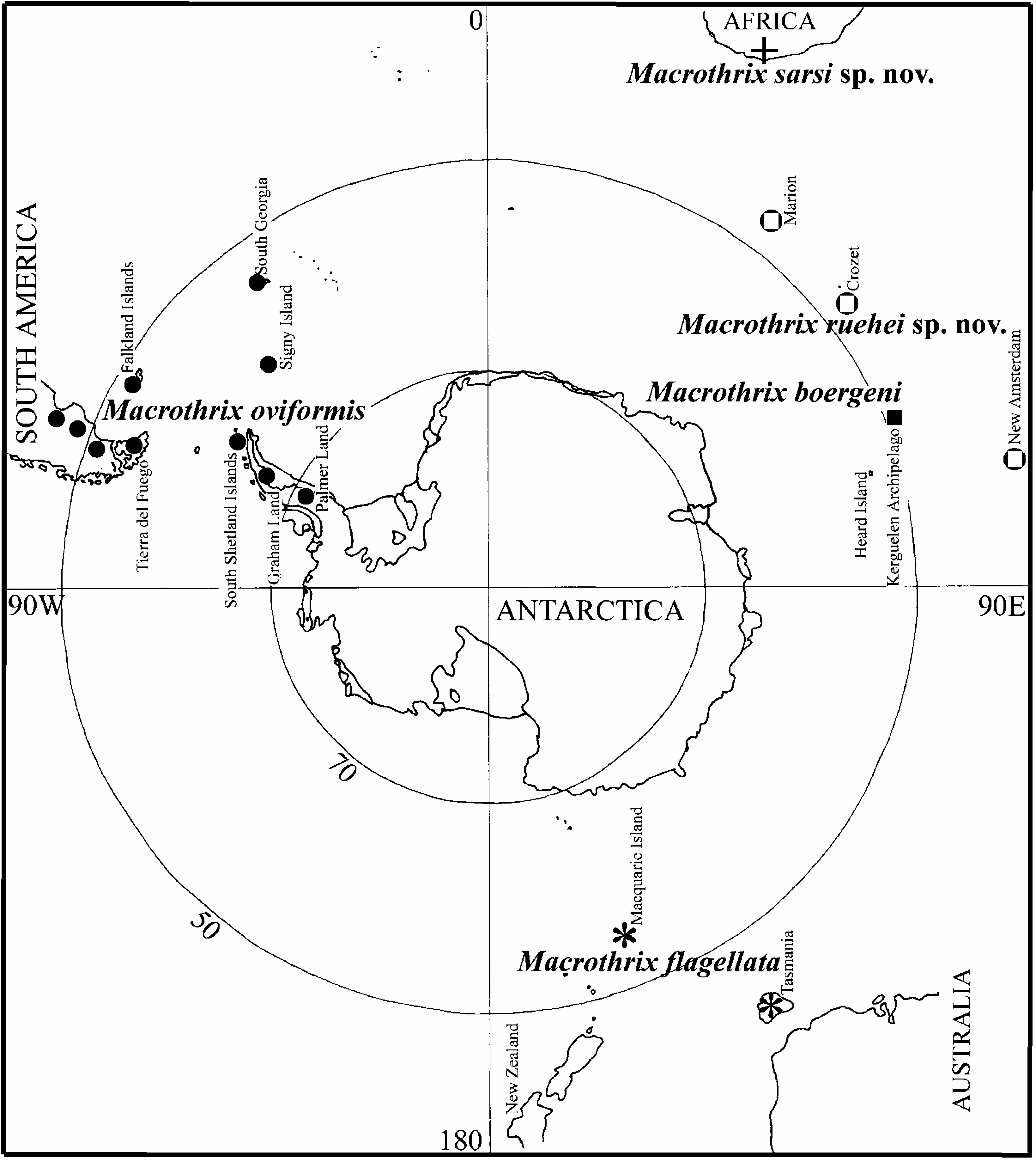
Distribution. Endemic to the Kerguelen Archipelago.


| V |
Royal British Columbia Museum - Herbarium |
No known copyright restrictions apply. See Agosti, D., Egloff, W., 2009. Taxonomic information exchange and copyright: the Plazi approach. BMC Research Notes 2009, 2:53 for further explanation.
|
Kingdom |
|
|
Phylum |
|
|
Class |
|
|
Order |
|
|
Family |
|
|
Genus |
Macrothrix boergeni Studer, 1878
| Kotov, Alexey A. 2007 |
Macrothrix hirsuticornis
| De Smet WH 2001: 263 |
| Gay C 1981: 51 |
| Brehm V 1954: 41 |
| Ruhe FE 1914: 55 |
Macrothrix Börgeni Studer 1878 , p 108
| Studer T 1878: 108 |