
Rubrocuneocoris vietnamensis, Duwal & Schwartz & Yasunaga, 2019
|
publication ID |
https://doi.org/ 10.11646/zootaxa.4652.1.10 |
|
publication LSID |
lsid:zoobank.org:pub:AC32E116-711C-4E9B-B417-1E2D8BBFC86C |
|
DOI |
https://doi.org/10.5281/zenodo.5933359 |
|
persistent identifier |
https://treatment.plazi.org/id/03E36B5B-AF52-FF95-37C2-FBE2FDC7FC60 |
|
treatment provided by |
Plazi |
|
scientific name |
Rubrocuneocoris vietnamensis |
| status |
sp. nov. |
Rubrocuneocoris vietnamensis , n. sp.
Figures 1 View FIGURE 1 E–I, 2
Type material. Holotype. ♂. Vietnam: Cao Bang Prov., Mt. Pia Oac, Deo Kolea , alt. 1250m, 14.V.1998, on light trap, M. Tomokuni (00383392) ( NMST) . Paratype: 1♂, same data as for holotype (00383393).
Diagnosis. The new species is recognized by orange-red coloration, deep red (nearly black) antennal segment I; dark base and apex of antennal segment II; red spots on the apices of the embolium and cuneus; sanguineous, reticular pattern on a wide apical region of the metafemur ( Fig. 1I View FIGURE 1 ); small, brown spots at the base of pale spines on the tibia and distinctive male genitalic structures as in Figure 2 View FIGURE 2 .
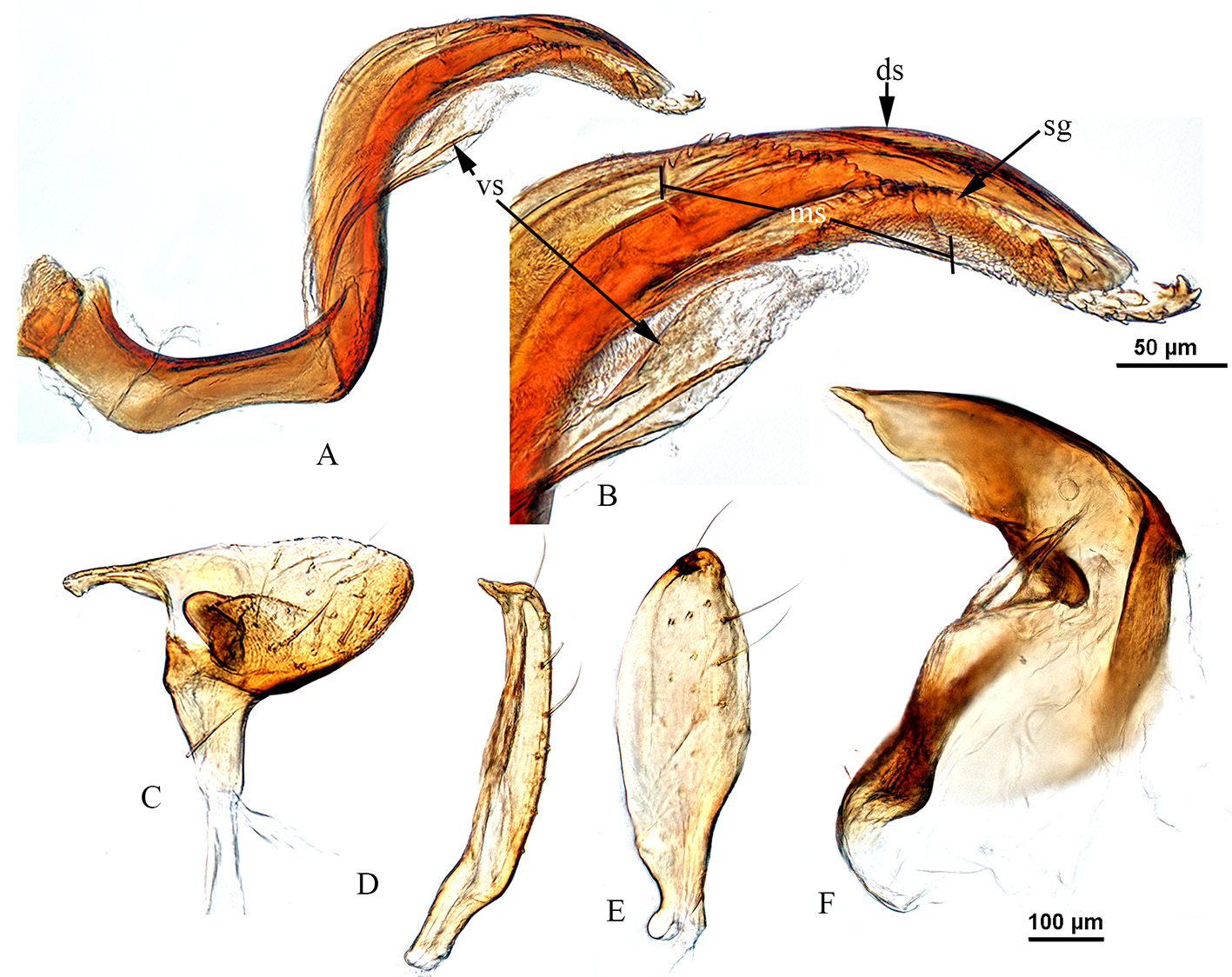
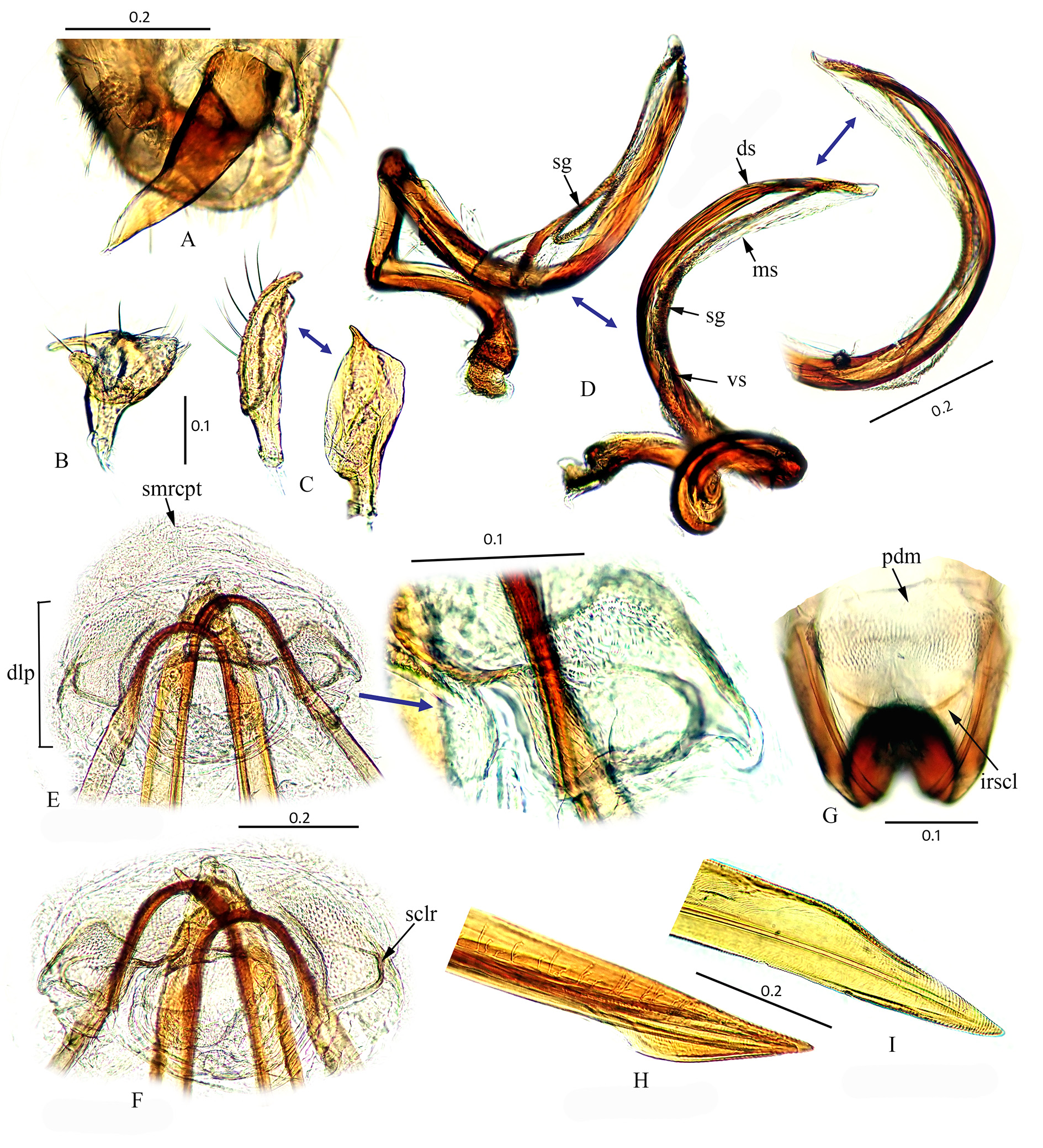
Description. Male ( Figure 1 View FIGURE 1 E–I). COLORATION: General coloration orange brown. Head: Brown, with dark along bases of antenna and clypeus ( Fig. 1G View FIGURE 1 ); clypeus and mandibular plate relatively paler than vertex and with deep red apices; maxillary plate white above and red below; bucculae pale; eyes black with red outer margins. Antenna: Segment I deep red (nearly black) except for pale extreme apex, segment II brown with dark extreme base and ¼ apical region, and segments III and IV fuscous except for brown apex of the segment IV. Labium: Basal two segments pale, and apical two segments brown. Thorax: Pronotum orange, with greyish or pale anterior region; mesocutum and scutellum same color as posterior area of pronotum except for yellow apex of scutellum; thoracic pleura red with dark margins; peritreme of scent gland evaporatory area grey ( Fig. 1H View FIGURE 1 ). Hemelytra yellow or pale brown with orange or reddish base, claval commissure, apex of clavus, lateral and inner margin of corium, and red coloration of inner margin of corium discontinuous; apices of embolium and cuneus with distinct red spots. Membrane grey with paler base; veins red but faded towards base of membrane. Legs: Brown, all coxa pale except for red (red and brown) basal regions of mesocoxa (1/4) and metacoxa (1/3), and with a small red spot on apices; bases of profemur (1/2), mesofemur (2/5) and metafemur (1/3) pale and remaining area brown, with red tinged apices and inner margins sanguineous on meso- and metafemur; and distal region of metafemur with red speckles (or pattern) ventrally ( Fig. 1I View FIGURE 1 ); tibia pale brown with red bases, and provided with brown spots at bases of pale spines. Abdomen: Deep red with dark posterior margins (on segments I–VII). SURFACE AND VESTITURE: Shining; dorsal and ventral side of body covered with uniformly distributed appressed simple pale setae; tibia with pale spines. STRUCTURE: Macropterus, oval. Head: Clypeus not observed from dorsal view; vertex weakly concave. Antenna: Segment I relatively thick; segment II cylindrical and longer than width of head. Labium: Elongate, reaching abdomen (about segment VI). Thorax: Pronotum sub-quadrate, lateral margins convexly rounded, posterior margin nearly straight and calli not developed; mesoscutum exposed; metathoracic spiracle and scent-gland system as in Figure 1H View FIGURE 1 . Pretarsus: Apex of claws strongly curved, pulvilli large and parempodia setiform (cf. Schuh, 1984: 427, Fig 1454). GENITALIA ( Fig. 2 View FIGURE 2 ): Pygophore: Small, subtriangular with apex blunt. Endosoma ( Fig. 2 View FIGURE 2 A–B): Nearly S-shaped, medially semi-coiled, dorsal strap bearing a sub-apical membranous structure with saw-toothed edge, extending apically to cover and surpass basal half of secondary gonopore ( Fig. 2 View FIGURE 2 A–B), apex of membranous structure with many spicules; ventral strap with semi-sclerotized bar arising from middle of endosoma extending to base of secondary gonopore; secondary gonopore subapically located and extended as long as apex of dorsal sclerotized strap. Phallotheca ( Fig 2F View FIGURE 2 ): Large, bent at strong right angle. Parameres: Left ( Fig. 2C View FIGURE 2 ): With typical phyline form. Right ( Fig. 2 View FIGURE 2 D–E): Small, lanceolate.
Female. Unknown.
Measurements. (n= 2♂). Total body length 2.38–2.40; length from apex of clypeus to cuneal fracture 1.6–1.64; width of head across eyes 0.58–0.60; vertex width 0.28; lengths of antennal segments I-IV 0.18, 0.68–0.70, 0.3– 0.32, 0.26–0.28; labial length 0.92–0.94; mesal length of pronotum 0.40–0.42; basal width of pronotum 0.88–0.90; maximum width across hemelytron 1.00–1.04; and lengths of metafemur, tibia and tarsus 0.86–0.90, 1.14–1.20, 0.30–0.32.
Etymology. Named for type locality of this new species, Vietnam.
Distribution. Vietnam (North)
Biology. Unknown.
Remarks. The general habitus and semi-coiled endosomal structure of Rubrocuneocoris vietnamensis are very similar to R. wudingensis Li & Liu. In the new species the endosoma has an elongated membranous structure with toothed edge, covering and surpassing the basal half of the secondary gonopore; in R. wudingensis , the membranous structure is not continuous to cover the basal half of the secondary gonopore. The orientation of the ventral strap on the anterior surface of the endosoma is similar in the two species but the apex of the strap in R. vietnamensis does not bears any spicules as in R. wudingensis ( Fig. 2 View FIGURE 2 A–B, and cf. Fig. 19 ( Li & Liu 2008)). Further, the new species is distinguished externally by pale spines and small brown spots on the tibia whereas in R. wudingensis dark spines are apparently present (Li & Lui 2008). However, we are uncertain about the true coloration of the metatibial spots in R. wudingensis ; they probably bear dark spots (brown if not black), which are obvious in the dorsal habitus image (cf. Li & Liu 2008, p. 69 Fig. 4 View FIGURE 4 ) and are larger than in the new species. The lateral margin of hemelytron in R. vietnamensis is orange red whereas in R. wudingensis the margin and corium are unicolorous.
Morphologically, Rubrocuneocoris anandros Yasunaga & Duwal differs from R. vietnamensis by red or sanguineous spots or marks or stripes on head, antennal segment II, pleura, and protibia; R. calvertae Henry by dark brown antennal segment I and yellowish-brown segments II–IV, dark brown pleura and white spots on distal half of hind femur; R. albescens Yasunaga by pale dorsal coloration and shorter labium extending to the metacoxa; R. quercicola by the reddish- or orange-yellow venter; R. bifidus Schuh by yellowish white dorsal coloration; R spiculatus Schuh by deep red abdomen laterally and reddish metafemur; and R. acuminatus Schuh and R. nigriceps Duwal et al. by dark (or fuscous) head and thorax.

| VI |
Mykotektet, National Veterinary Institute |
No known copyright restrictions apply. See Agosti, D., Egloff, W., 2009. Taxonomic information exchange and copyright: the Plazi approach. BMC Research Notes 2009, 2:53 for further explanation.