
Orthomorpha parasericata, Likhitrakarn, Natdanai, Golovatch, Sergei I. & Panha, Somsak, 2010
|
publication ID |
https://doi.org/ 10.5281/zenodo.193638 |
|
DOI |
https://doi.org/10.5281/zenodo.6199315 |
|
persistent identifier |
https://treatment.plazi.org/id/03E3878E-FF96-FFCB-E584-FB6CFB8BFB87 |
|
treatment provided by |
Plazi |
|
scientific name |
Orthomorpha parasericata |
| status |
sp. nov. |
Orthomorpha parasericata View in CoL sp. n.
Figs 11 View FIGURE 11 C & D, 12 & 13.
Material examined: Holotype male ( CUMZ), Thailand, Surat Thani Province, Khiri Rat Nikhom District, Sathit Khirirom Temple, 9°01´49˝N, 98°59´12˝E, 9 October 2008, leg. S. Panha, H. Enghoff, C. Sutcharit, N. Likhitrakarn and members of Animal Systematic Research Unit. Paratypes: 1 male, 3 females ( CUMZ), 1 male ( ZMUC), 1 male ( ZMUM), same data as holotype.
Diagnosis: Differs from the similar O. sericata in the shining tegument, coupled with a colour pattern as in O. subsericata , and a slightly shorter apical lobe of the solenophore.
Name: To emphasize the apparent similarity to O. sericata .
Description: Length 32–37 mm (male), 34–37 mm (female), width of midbody pro- and metazona 3.2– 3.3 and 4.8–5.0 mm (male), 3.5–3.8 and 4.8–5.3 mm (female), respectively. Holotype 33 mm long, 3.2 and 4.9 mm wide on pro- and metazona, respectively.
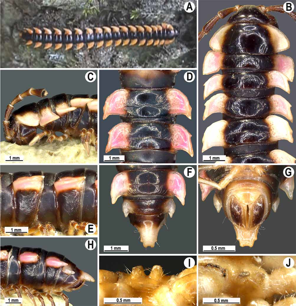
Live coloration ( Fig. 12 View FIGURE 12 A) blackish, head and antennae blackish as well, paraterga and epiproct contrastingly orange, legs and venter dark brown; coloration in alcohol somewhat faded to pale pinkish or yellowish paraterga (sometimes marbled) and epiproct, as well as to dark brown head, venter and legs after one year of preservation.
Main characters as in O. enghoffi sp. n., except as follows.
Head without contrastingly light spot above antennal socket (like in O. asticta sp. n.), vertigial region finely rugulose, shining; epicranial suture distinct. Antennae moderately long ( Fig. 12 View FIGURE 12 C), reaching end of body segment 3 (male) or 2 (female).
Head in width <collum <segments 3 and 4 <segment 2 = segments 5–16 in male, gently and gradually tapering thereafter, but head <collum <segments 3 and 4 <segment 2 <segments 5–16 in female. Collum with caudal row of setae, like both preceding rows, on a smooth, shining surface, caudal corner like a slightly obtusangular, very narrowly rounded, regularly and only slightly declined lobe ( Fig. 12 View FIGURE 12 C).
Tegument generally smooth, shining ( Figs 12 View FIGURE 12 A–H), metaterga only at places faintly rugulose, slightly more so near rear edge; surface below paraterga usually slightly more evidently rugulose. Metaterga with two transverse rows of setae: 2+ 2 in anterior (pre-sulcus) row and 3+ 3 in posterior (postsulcus) one, all mostly abraded, but still traceable as insertion points. Tergal setae short, about as long as 1/7 of metazonum. Axial line thin, but clearly visible on metazona, especially in their anterior half. Paraterga very strongly developed ( Figs 12 View FIGURE 12 A–H), especially so in male, usually slightly upturned in male or slightly declined in female, lying level to dorsum on segments 5 and 9–12, slightly above dorsum on segments 6–8, below dorsum on segments 13–19 in male, always below dorsum in female. Paraterga 2 broad, anterior edge nearly straight, lateral edge either virtually without incisions or very faintly tri- or bisinuate, caudal corner nearly pointed and slightly surpassing rear contour; posterior edge nearly straight; caudal base evidently concave ( Figs 12 View FIGURE 12 B & C). Paraterga 3 and 4 subequal, anterior edge strongly convex and slightly bordered, lateral edge with only one evident incision in anterior half, caudal corners pointed, surpassing rear contour, posterior edge evidently concave ( Figs 12 View FIGURE 12 B & C). Paraterga 5–19 increasingly surpassing rear tergal contour, especially so in male. Poreless paraterga 6, 8, 11, 12 and 14 each with single, but evident incision near anterior 1/3, pore-bearing paraterga with only slight sinuosity in its stead, another similar sinuosity in front of ozopore; posterior edge nearly unfringed ( Figs 12 View FIGURE 12 D & E). Ozopores evident, lateral. Transverse sulcus present on metaterga 5–18 (Figs B, D & F), deep, line-shaped, reaching bases of paraterga, evidently beaded at bottom, a little better developed in male than in female. Stricture between pro- and metazona narrow, heavily beaded at bottom down to base of paraterga ( Figs 12 View FIGURE 12 B, D, E–H). Pleurosternal carinae as complete and clearly arcuate crests supplied with prominent tooth caudally on segments 2–8 ( Fig. 12 View FIGURE 12 C), as low and interrupted swellings still supplied with caudal tooth on segments 9–11, retained only as increasingly reduced tooth on segments 12–16, as very small knob on segment 16, almost absent from segment 17, fully missing on segments 18 and 19 (male), or as complete and clearly arcuate crests supplied with prominent tooth caudally on segments 2–6, as low swellings still supplied with caudal tooth on segments 7–12, retained only as increasingly vestigial tooth on segments 13 and 14, thereafter missing (female). Apical papillae on epiproct well-developed, tip narrowly emarginate; preapical papillae evident ( Figs 12 View FIGURE 12 F & G).
Pair of strong, setose, deeply separated, rounded cones between male coxae 4 ( Figs 12 View FIGURE 12 I & J). Legs moderately long, slender, slightly incrassate in male, about 1.4 (male) or 0.9–1.0 times (female) as long as midbody height, tarsal brushes present on male legs 1–5, thereafter gradually thinning out.
Gonopods ( Figs 11 View FIGURE 11 C & D, 13) with femorite showing faint traces of an oblique impression in place of postfemoral lateral sulcus. Tip of solenophore axe-shaped, distally somewhat abbreviated.
Remarks. This species was observed climbing the stairs leading to the temple. The presence of a vestigial postfemoral sulcus in this species is remarkable, somewhat bridging the sericata -group with the remaining species groups of Orthomorpha . This character thus seems to be a synapomorphy of the sericata -group, the loss of a postfemoral sulcus possibly having been a secondary development.

No known copyright restrictions apply. See Agosti, D., Egloff, W., 2009. Taxonomic information exchange and copyright: the Plazi approach. BMC Research Notes 2009, 2:53 for further explanation.