
Chaulionyx paivacarvalhoi, Kihara & Huys, 2009
|
publication ID |
https://doi.org/10.3897/zookeys.17.202 |
|
publication LSID |
lsid:zoobank.org:pub:E52234CD-E65E-4F8F-95CD-04BDBDED9F39 |
|
DOI |
https://doi.org/10.5281/zenodo.3791579 |
|
persistent identifier |
https://treatment.plazi.org/id/0075F457-E809-451D-9CFB-9B2B1BE2EF19 |
|
taxon LSID |
lsid:zoobank.org:act:0075F457-E809-451D-9CFB-9B2B1BE2EF19 |
|
treatment provided by |
Plazi |
|
scientific name |
Chaulionyx paivacarvalhoi |
| status |
sp. nov. |
Chaulionyx paivacarvalhoi sp. n.
urn:lsid:zoobank.org:act:
Figs 1-37 View Figures 1-2 View Figures 3-6 View Figures 7-10 View Figures 11-15 View Figures 16-18 View Figures 19-21 View Figures 22-23 View Figures 24-26 View Figures 27-37
Type locality. Brazil, northern coast of São Paulo State, Ubatuba ( 23 ° 41.4’ S, 44 ° 58.8’ W), 44 m depth (station 17 V in Table 1 View Table 1 ).
Material examined. Holotype female in ethanol (reg. no. MZUSP 16467 View Materials ). Undissected paratypes (in ethanol) deposited in MZUSP (reg. nos 16468, 19063-19065) are 1 female and 1 male from station 17 V; 3 females and 1 male from station 16 V, 2 males from station 16I, 1 female and 1 male from station 17I. Additional undissected paratypes (in ethanol) deposited in NHM are 2 males from station 17 V (reg. nos 2009.1-2), 3 females from station 27 V (reg. nos 2009.3-5) and 1 male from station 27I (reg. no 2009.6). Dissected paratypes and other material examined are retained in the personal collection of C.E.F. da Rocha ( Departamento de Zoologia, Instituto de Biociências, Universidade de São Paulo). All material collected by T. Corbisier.
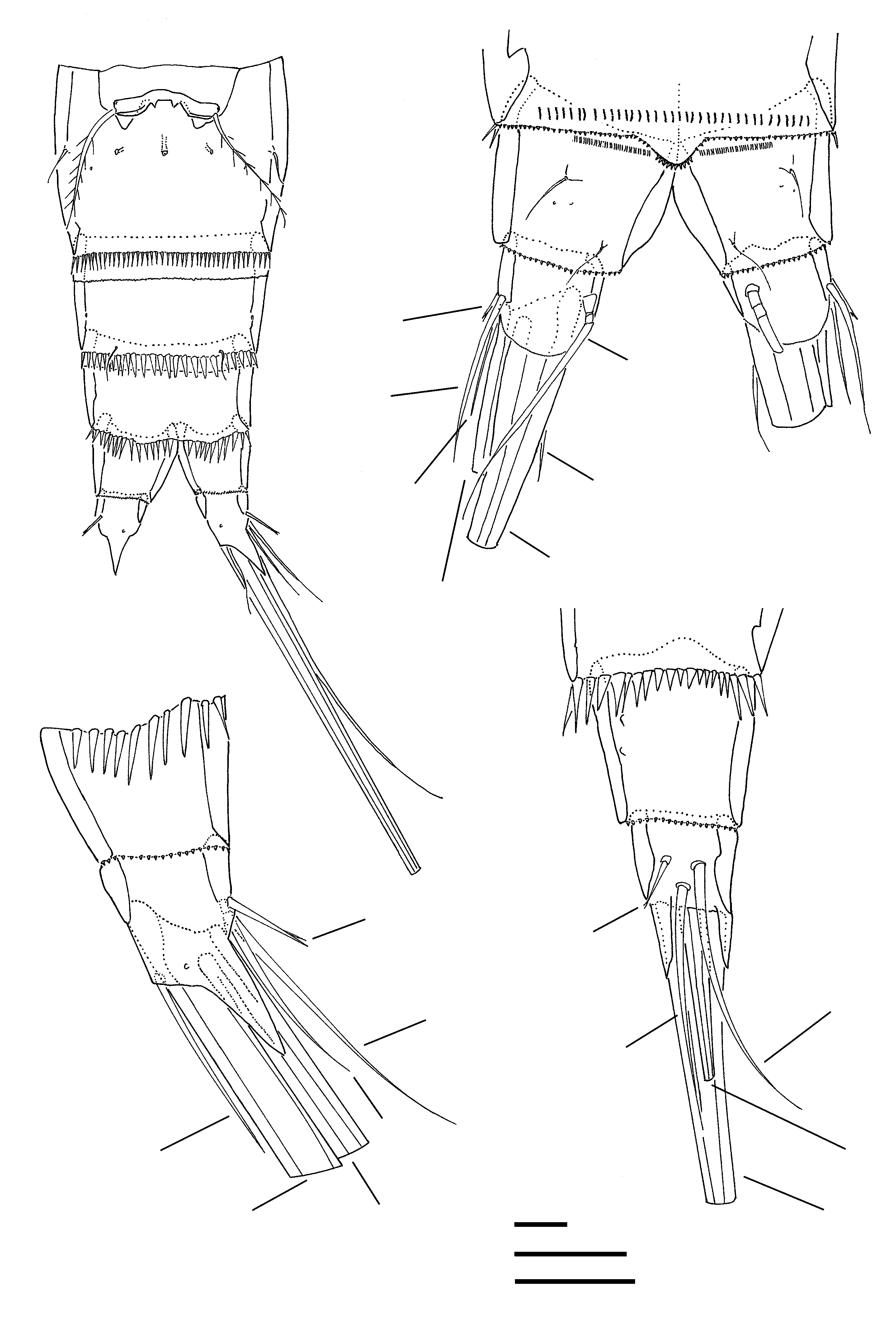
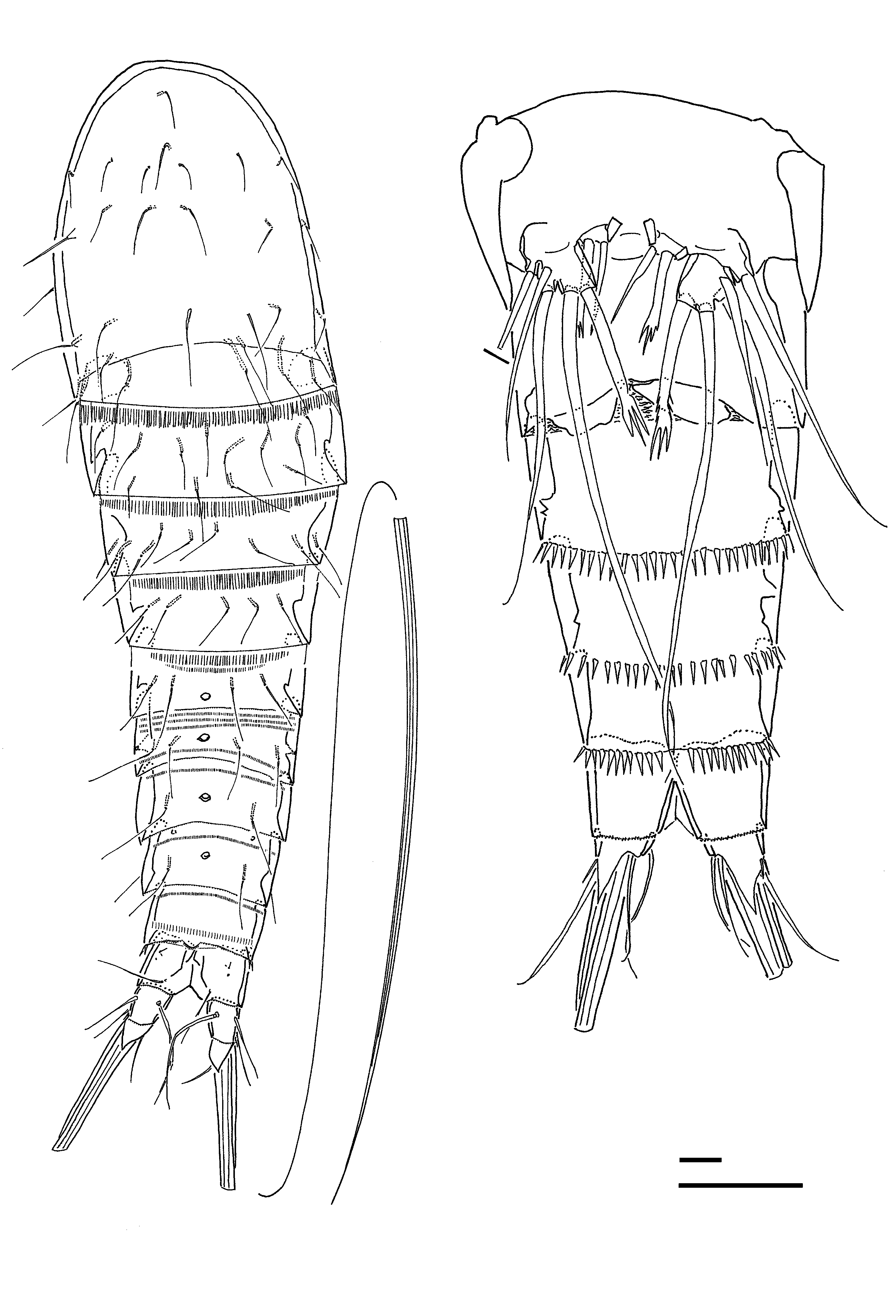
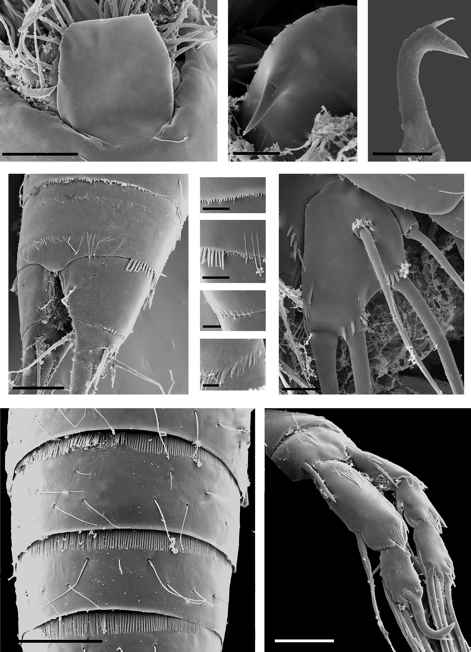
Description. FEMALE ( Figs 1-21 View Figures 1-2 View Figures 3-6 View Figures 7-10 View Figures 11-15 View Figures 16-18 View Figures 19-21 , 27-35 View Figures 27-37 ). Body length 260-290 µm (n = 4; mean = 275 µm). Body ( Fig. 1 View Figures 1-2 ) fusiform, maximum width measured at posterior margin of cephalic shield; body somites gradually tapering posteriorly. Cephalic shield with minute integumental pits and numerous pores; paired chitinous patches present dorsally in posterior half (for examples of these integumental structures see labelling in Fig. 1 View Figures 1-2 and accompanying legend). Body somites with thickly chitinized cuticle; pedigerous somites and second abdominal somite with large middorsal pore; genital double-somite with 2 pores. Sensillae long and fine, distributed as illustrated in Fig. 1 View Figures 1-2 . Hyaline frill of cephalic shield and somites bearing P2–P4 plain, partially concealing fine spinular rows located in anterior half of succeeding somite ( Fig. 1 View Figures 1-2 ); frills of urosomites minutely denticulate ( Figs 1-2 View Figures 1-2 , 30-31 View Figures 27-37 ).
Genital and first abdominal somites fused forming double-somite ( Figs 1-3 View Figures 1-2 View Figures 3-6 , 21 View Figures 19-21 ); slightly longer than broad; posterior margin with continuous spinular row; original segmentation marked by sensilla, paired dorsal chitinous patches and a middorsal pore ( Fig. 2 View Figures 1-2 ). Second and third abdominal somites with a continuous row of coarse spinules around ventral posterior margin ( Fig. 3 View Figures 3-6 ). Penultimate somite with a small pseudoper-
culum ( Figs 1-2 View Figures 1-2 , 30 View Figures 27-37 ), dorsal surface and distal margin with rows of spinules ( Figs 30, 32 View Figures 27-37 ). Anal somite ( Figs 1 View Figures 1-2 -4 View Figures 1-2 View Figures 3-6 , 30 View Figures 27-37 ) medially cleft; dorsal surface with paired anterior rows of minute spinules and pairs of sensilla and pores ( Fig. 4 View Figures 3-6 ); distal margin with small spinules ( Fig. 33 View Figures 27-37 ); anal operculum absent.
Caudal rami ( Figs 4-6 View Figures 3-6 ) about as long as wide, with 7 naked setae; bases of terminal setae covered by rounded membranous serrate extension dorsally ( Figs 4 View Figures 3-6 , 30, 34 View Figures 27-37 ) and an acuminate lappet ventrally ( Fig. 5 View Figures 3-6 ). Seta I minute, with bifid apex. Setae IV–V fused basally, without fracture planes. Seta V longest and swollen in proximal half. Seta VII tri-articulate at base.
Rostrum large ( Figs 7 View Figures 7-10 , 27 View Figures 27-37 ), ventrally deflected; broadly rounded, quadrangular; not defined at base but original demarcation marked by membranous areas bilaterally ( Fig. 27 View Figures 27-37 ); no sensilla discernible.
Antennule ( Fig. 8 View Figures 7-10 ) short, 5-segmented. Segment 1 with pinnate seta; segment 3 with conspicuous aesthetasc; distal segment long, with apical acrothek consisting of aesthetasc and 2 slender setae. Armature formula: 1-[1 pinnate], 2-[7], 3-[6 + (1 + ae), 4-[1], 5-[5 + acrothek].
Antenna ( Fig. 9 View Figures 7-10 ) consisting of coxa, basis, 2-segmented endopod and 3-segmented exopod. Coxa small, indistinctly demarcated at base, without ornamentation. Basis unarmed; with some fine spinules along abexopodal margin and coarse spinules set near outer distal corner. Proximal endopod segment with a few coarse spinules near outer distal corner. Distal endopod segment with 2 unipinnate setae laterally; distal margin with spinule row and 5 elements, 2 of which are geniculate, others bipinnate. Exopod with small proximal and middle segments and elongate apical segment; exp-1 unarmed, exp-2 with short naked seta, exp-3 with 2 pinnate apical setae.
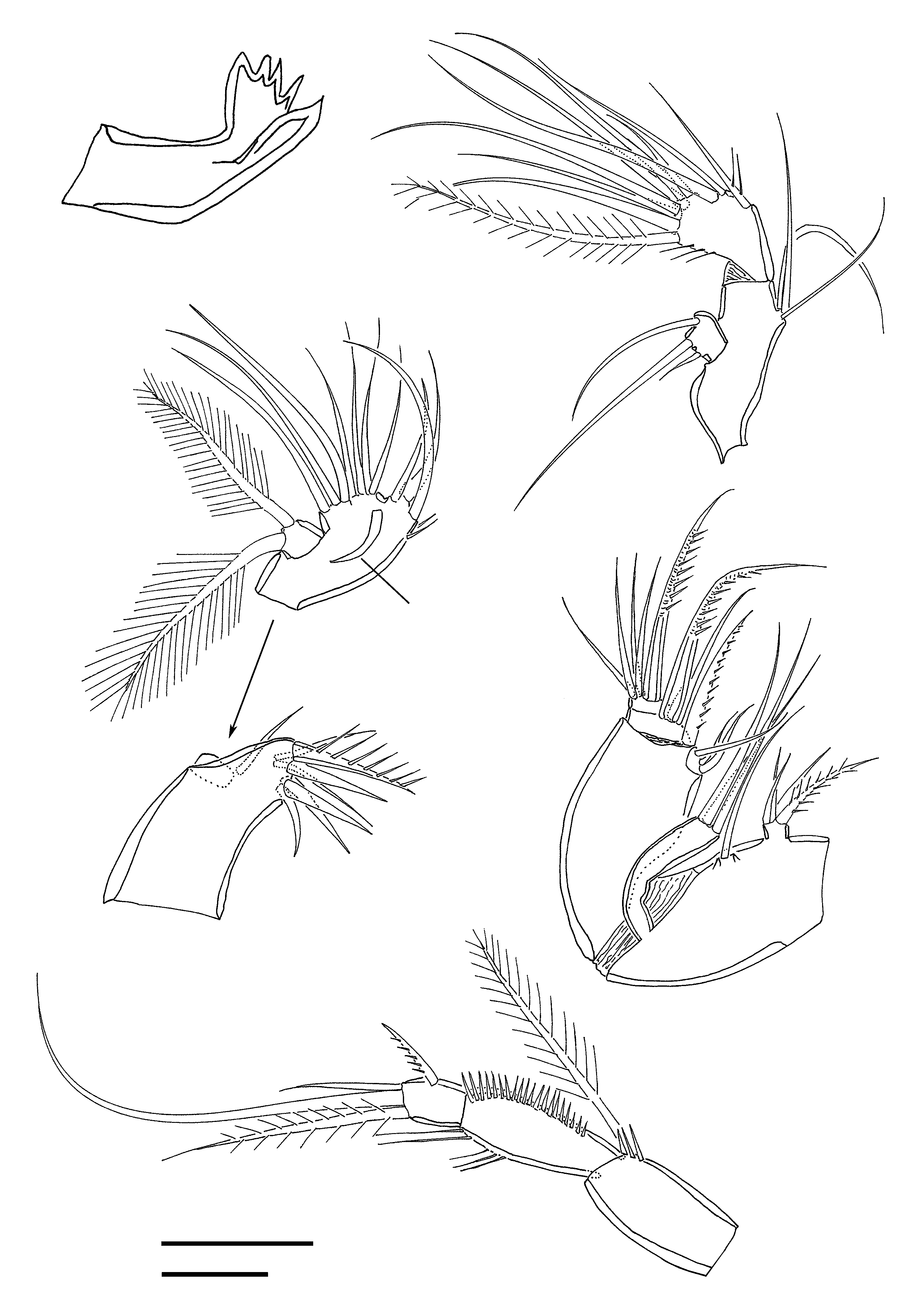
Labrum ( Figs 10 View Figures 7-10 , 28 View Figures 27-37 ) well developed, with frontal curved spinous projection bearing large anterior pore.
Mandible with small coxa ( Fig. 11 View Figures 11-15 ) and biramous palp ( Fig. 12 View Figures 11-15 ). Gnathobase reduced, with chitinized dorsal tooth and number of hyaline pointed projections. Basis elongate, with 3 lateral setae. Exopod small, 1-segmented, with 3 naked setae, outer one reduced. Endopod 1-segmented, with row of fine spinules along outer margin; armature consisting of 1 sparsely pinnate and 2 naked setae laterally and 1 bipinnate and 6 naked setae apically.
Maxillule ( Fig.13 View Figures 11-15 ) with fused praecoxa and coxa. Praecoxa with well developed arthrite bearing 4 spines and 1 seta around distal margin and 2 small setae on anterior surface; distalmost marginal spine with long spinules. Coxa represented by small seta on anterior surface near articulation with palp. Endopod incorporated into basis forming elongate segment; proximal basal endite a small protuberance bearing 3 setae; elements of distal basal endite (4) and endopod (3) forming group of 7 setae arranged around the distal margin; with cuticular reinforcement (indicated by asterisk in Fig. 13 View Figures 11-15 ) on posterior surface; distal medial margin with characteristic spinules. Exopod a free small segment; with 1 apical and 1 backwardly directed plumose seta.
Maxilla ( Fig. 14 View Figures 11-15 ) prehensile, comprising syncoxa, allobasis and 2-segmented endopod with syncoxa and allobasis directed at a right angle. Syncoxa with 3 endites; proxi-
mal endite small, with 1 bipinnate and 3 naked setae; middle endite rudimentary, with 1 long naked seta; distal endite cylindrical and recurved, located in membranous area at syncoxa-allobasis joint, with 3 long naked setae. Allobasis robust, expanding in distal half; armed with 3 setae (2 small) near inner distal corner (derived from basis) and 1 strong pinnate seta on posterior surface (derived from incorporated endopod segment). Endopod with 1 geniculate pinnate claw and 1 naked seta on enp-1; enp-2 (representing fused middle and distal segments) with 1 geniculate pinnate claw and 4 naked setae.
Maxilliped ( Fig. 15 View Figures 11-15 ) stenopodial and slender, comprising syncoxa, basis and 1-segmented endopod. Syncoxa with long pinnate seta and few spinules near distal corner. Basis unarmed; with long setules along outer margin and fine spinules along inner margin. Endopod with 1 short pinnate seta laterally and 1 short plus 2 longer (1 plumose) setae apically.
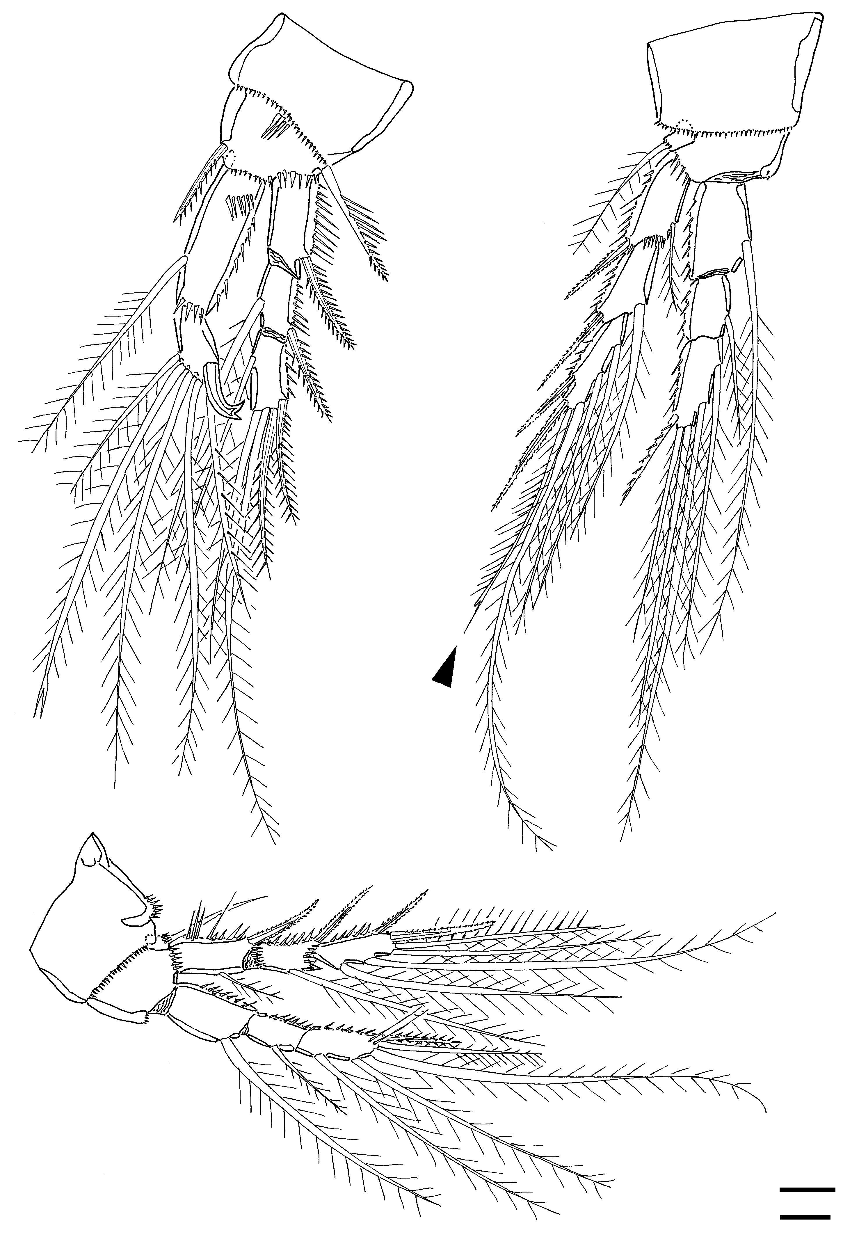
P1 ( Fig. 16 View Figures 16-18 ). Coxa with row of minute spinules along distal margin. Basis with rows of spinules on anterior surface and near insertion of exopod as illustrated; outer seta long, bipinnate and spiniform; inner spine unipinnate with spinules along outer margin. Exopod 3-segmented; with rows of spinules along outer margins; outer spines strong and bipinnate; exp-1 without inner seta; exp-2 with 1 inner plumose seta; exp-3 with 1 inner plumose seta, 2 plumose distal setae and 2 bipinnate outer spines. Endopod 2-segmented, prehensile; enp-1 elongate, with rows of spinules on anterior surface and along outer and distal margins, and 1 plumose inner seta inserted in distal third of segment; enp-2 with 3 plumose inner setae (middle one with bifid apex), 2 plumose distal setae, and 1 curved strong spine with bifid tip ( Fig. 29 View Figures 27-37 ).
P2–P4 ( Figs 17-19 View Figures 16-18 View Figures 19-21 ). Coxa with small spinules along distal margin. Basis with rows of spinules at base of exopod and around outer seta; outer seta plumose (P2) or naked (P3–P4). Exopod 3-segmented; with rows of spinules along outer margins; outer spines strong and bipinnate. Exp-1 with 1 reduced plumose inner seta; exp-2 with 1 plumose inner seta; exp-3 with 2 bipinnate outer spines, 2 plumose distal setae and 2 (P2–P3) or 3 (P4) plumose inner setae. P2 exp-3 outer distal seta plumose along outer margin and with apical flagellum (arrowed in Fig. 17 View Figures 16-18 ). P4 exp-3 proximal inner seta with bifid apex; middle inner seta extremely well developed. Endopod 3-segmented; with rows of spinules along outer margins. Enp-1 with 1 plumose inner seta; enp-2 with 1 reduced plumose inner seta; enp-3 with 4 plumose setae (2 inner and 2 distal) and 1 bipinnate outer spine (P2–P3) or 1 plumose outer seta (P4). Armature formula of swimming legs as for genus.
P5 ( Figs 20 View Figures 19-21 , 35 View Figures 27-37 ). Baseoendopod outer expansion with 1 naked seta. Endopodal lobe with spinular row and small pore on anterior surface; with 2 naked setae, inner one very long (3.2 times longer than outer one) and with bifid apex, outer one with serrate apex. Exopod with 1 pore near distal inner margin and various spinule rows as figured; anterior surface with 1 naked seta; with 3 marginal setae, innermost one with bifid apex.
Genital field ( Figs 3 View Figures 3-6 , 21 View Figures 19-21 ) with relatively small midventral copulatory pore. Sixth pair of legs ( Fig. 21 View Figures 19-21 ) vestigial, fused medially forming a common plate that covers paired genital apertures (or median slit); each P6 with 1 plumose seta. Egg-sac single.
MALE. ( Figs 22-26 View Figures 22-23 View Figures 24-26 , 36-37 View Figures 27-37 ). Body length 230-260 µm (n = 6; mean = 246 µm) ( Fig. 22 View Figures 22-23 ). Sexual dimorphism expressed in caudal ramus, antennule, P1, P5, P6, and in genital segmentation. Ornamentation of body ( Figs 22-23 View Figures 22-23 , 36 View Figures 27-37 ) generally as in female, except for small differences such as cephalic sensilla being longer and distributed differently, and pits, pores and chitin patches missing on the cephalic shield.
Caudal ramus ( Fig. 23 View Figures 22-23 ) with both dorsal and ventral posterior margin produced into triangular extension covering bases of setae IV–VI.
Antennule ( Fig. 24 View Figures 24-26 ), haplocer, 7-segmented; geniculation between segments 5 and 6; segment 5 elongated and incompletely divided. Setae and aesthetasc formula: 1-[1], 2-[7], 3-[3], 4-[1], 5-[7 + ae], 6-[0], 7-[1 + ae].
P1 ( Figs 25 View Figures 24-26 , 37 View Figures 27-37 ). Enp-2 wider than in female; outer distal spine with bifid apex.
P5 ( Figs 23 View Figures 22-23 , 26 View Figures 24-26 ) shorter than in female. Endopodal lobe small, with 2 short, stout setae, innermost with bifid apex, outer one with tridentate apex. Exopod short; with 1 seta on anterior surface and 3 marginal setae; inner distal seta with tridentate apex, outer distal seta very long.
P6 ( Fig. 23 View Figures 22-23 ) asymmetrical, without ornamentation.
Etymology. The new species is named in honour of Prof. João de Paiva Carvalho (Instituto Oceanográfico, Universidade de São Paulo) in recognition of his significant contributions to the taxonomy of Copepoda.


| V |
Royal British Columbia Museum - Herbarium |
| MZUSP |
Museu de Zoologia da Universidade de Sao Paulo |
| T |
Tavera, Department of Geology and Geophysics |
No known copyright restrictions apply. See Agosti, D., Egloff, W., 2009. Taxonomic information exchange and copyright: the Plazi approach. BMC Research Notes 2009, 2:53 for further explanation.
|
Kingdom |
|
|
Phylum |
|
|
Class |
|
|
Order |
|
|
Family |
|
|
Genus |