
Pseudoceros astrorum, Bulnes, Veronica N. & Torres, Yan, 2014
|
publication ID |
https://doi.org/ 10.11646/zootaxa.3881.1.7 |
|
publication LSID |
lsid:zoobank.org:pub:9C0FB9F8-C565-4627-87A2-D44FDFAB6781 |
|
DOI |
https://doi.org/10.5281/zenodo.6144878 |
|
persistent identifier |
https://treatment.plazi.org/id/03E487C8-1601-5E30-FF1B-FE7D5B2329C2 |
|
treatment provided by |
Plazi |
|
scientific name |
Pseudoceros astrorum |
| status |
sp. nov. |
Pseudoceros astrorum n.sp.
( Figs 1–4 View FIGURE 1 View FIGURE 2 View FIGURE 3 View FIGURE 4 )
Type material. Holotype: One mature specimen as serial sagittal sections of anterior part of body (106 slides), and the posterior part as serial transverse section (51 slides), MZUSP PL 1558. The specimen was collected on 7 October 2010 by F. Vasconcelos, from the intertidal zone.
Paratype: One mature specimen preserved in 70% ethanol, MZUSP PL 1559. The specimen was collected on 7 October 2010 by F. Vasconcelos, from the intertidal zone.
Etymology. The specific name astrorum is the Latin, genitive, plural form of astrum (star), which means “belong to the stars” and refers to the dorsal colour pattern resembling a constellation or group of stars.
Locality. Beach at Pacheco, São Gonçalo do Amarante, CE, Brazil (03° 41 'S, 38° 38' W).
Description. External morphology: Dorsally the background colour is brown with numerous dark pigment granules creating a uniform pattern. Delicate black sub-marginal band and a white marginal rim ( Figures 1 View FIGURE 1 A, C). Scattered white spots of different sizes (blotches, flecks and small spots) uniformly distributed on the dorsal surface. Pseudotentacles black, with small white spots and a single white blotch at the tip of each tentacle. The anterior-most marginal spot and the cerebral region are devoid of epidermal pigment ( Figure 1 View FIGURE 1 B). Ventrally dark brown, fading to grey at the margins and with a white marginal rim ( Figure 1 View FIGURE 1 E). Intestinal branches apparent ( Figures 1 View FIGURE 1 D, E). Body of solid appearance, from oval to elongate, 30.4 mm long and 14.9 mm wide. Scattered tentacular eyes, only visible in serial sections, 63 cerebral eyes spots arranged in a single pear-shaped cluster ( Figure 1 View FIGURE 1 B). Pharynx with numerous folds, arranged in anterior body half; the mouth opens anteriorly in the pharynx cavity. Gonopores separate, arranged distally in the anterior body half. Sucker well developed in the middle of the posterior body half ( Figure 1 View FIGURE 1 D).
Body wall: Dorsal epidermis cells are ciliated. Epidermal cells cylindrical, 32 µm high, filled with dark granular pigmentation and scattered rhabdites. Basement membrane delicate (3 µm), circular muscle fibres in bundles, longitudinal muscle layer 10 µm high. Underneath the muscular layer, granular pigmentation is apparent as well as distinct eosinophilic glands ( Figure 2 View FIGURE 2 A). Ventral body wall as thick as dorsal (50 µm), cellular, ciliated epidermis 30 µm high, the epidermal cells appear cuboidal, devoid of pigment granules and with scattered rhabdites. Basement membrane thinner than dorsally; circular muscle fibres arranged in a 5 µm deep distinct muscular layer; longitudinal muscle fibres less compact than dorsally, arranged in a 10 µm deep layer ( Figure 2 View FIGURE 2 B). Rhabdites numerous only in anterior body margin (dorsal and ventral). Body wall relatively thin in comparison to the body height. Parenchymatous muscles apparent and numerous, giving the body its solid appearance ( Figure 3 View FIGURE 3 D).
Reproductive anatomy: Spermiducal bulbs well developed ( Figures 1 View FIGURE 1 D, 1E), joining anteriorly to form a muscular common vas deferens ( Figure 3 View FIGURE 3 A). The vas deferens runs anteriorly and turns dorsal and posterior to enter the seminal vesicle on its anterior aspect. The cylindroid seminal vesicle is 1650 Μm long and 750 Μm high ( Figure 3 View FIGURE 3 C). The vesicle´s muscular wall is slightly thicker anteriorly, thinning distally. The ejaculatory duct turns ventrally and posteriorly to join the prostatic duct at the tip of the penis papillae. A short prostatic duct leads to the small ovoid prostatic vesicle (460 µm x 360 µm) with an inner smooth glandular lining, arranged free ( Figure 3 View FIGURE 3 B). The 300 µm curved stylet is supported by a short muscular papilla and housed in a muscular fold of the male atrium (penis sheath). The ciliated male atrium opens to a single male gonopore ( Figure 3 View FIGURE 3 C, D).
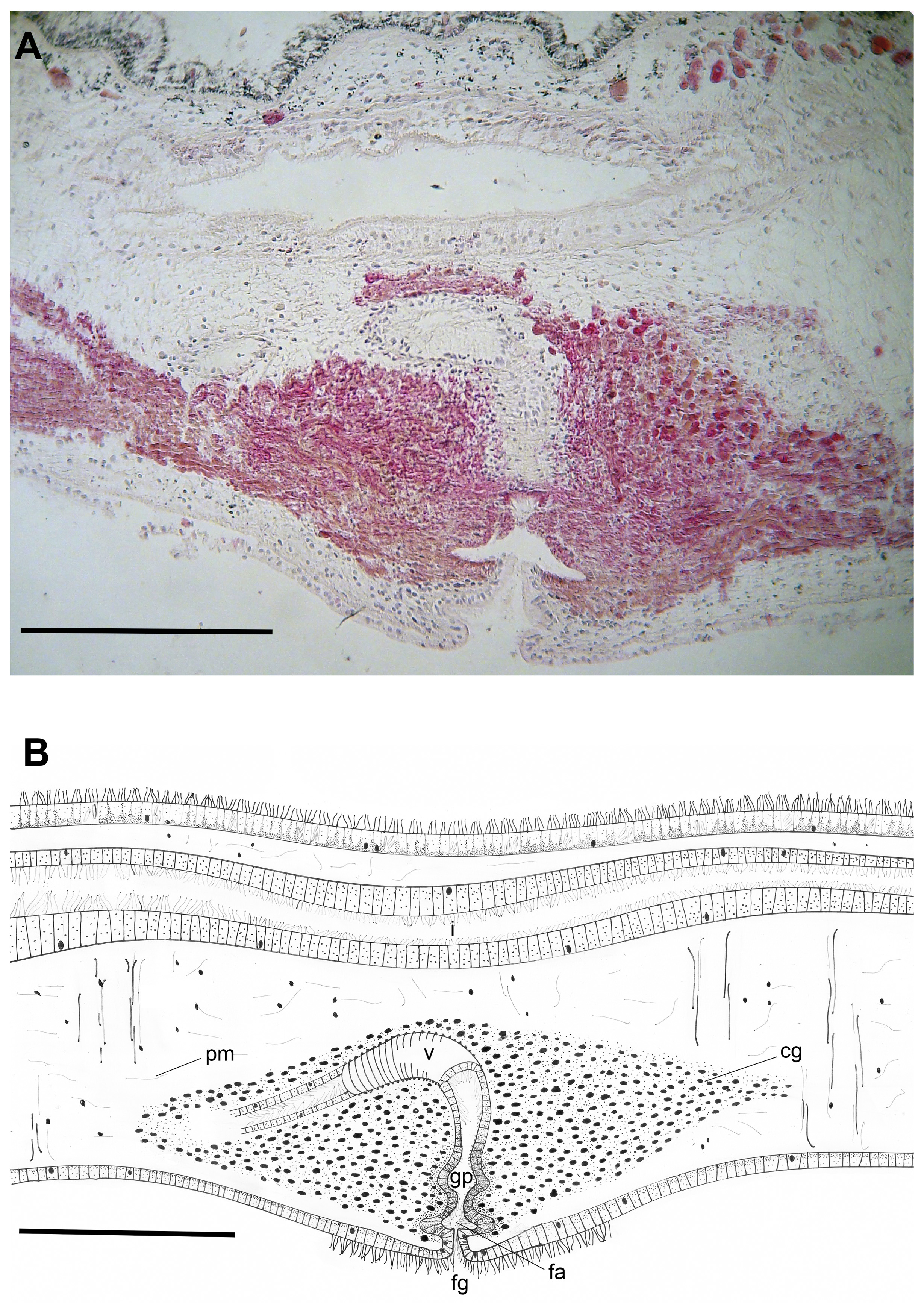
The uteri run dorsal to the spermiducal bulbs; the distal tracts of the oviducts run anteriorly and join before entering the female canal. The vagina is ciliated, surrounded by the cement gland mass ( Figure 4 View FIGURE 4 A). The vagina runs posteriorly, parallel to the body surface, then turns ventrally and opens to the small female atrium. The wall of the vagina is slightly muscular proximally, distally developing two glandular pouches ( Figure 4 View FIGURE 4 B), before entering the small female atrium and the gonopore.


| MZUSP |
Museu de Zoologia da Universidade de Sao Paulo |
No known copyright restrictions apply. See Agosti, D., Egloff, W., 2009. Taxonomic information exchange and copyright: the Plazi approach. BMC Research Notes 2009, 2:53 for further explanation.