
Trapania reticulata, RUDMAN, 1987
|
publication ID |
https://doi.org/ 10.1093/zoolinnean/zlac009 |
|
publication LSID |
lsid:zoobank.org:pub:C288BAB2-A92C-4F13-B04D-D6D4510461F5 |
|
DOI |
https://doi.org/10.5281/zenodo.7043806 |
|
persistent identifier |
https://treatment.plazi.org/id/03E487E4-FF8D-FB38-E886-F910FA85F947 |
|
treatment provided by |
Plazi |
|
scientific name |
Trapania reticulata |
| status |
|
TRAPANIA RETICULATA RUDMAN, 1987 View in CoL
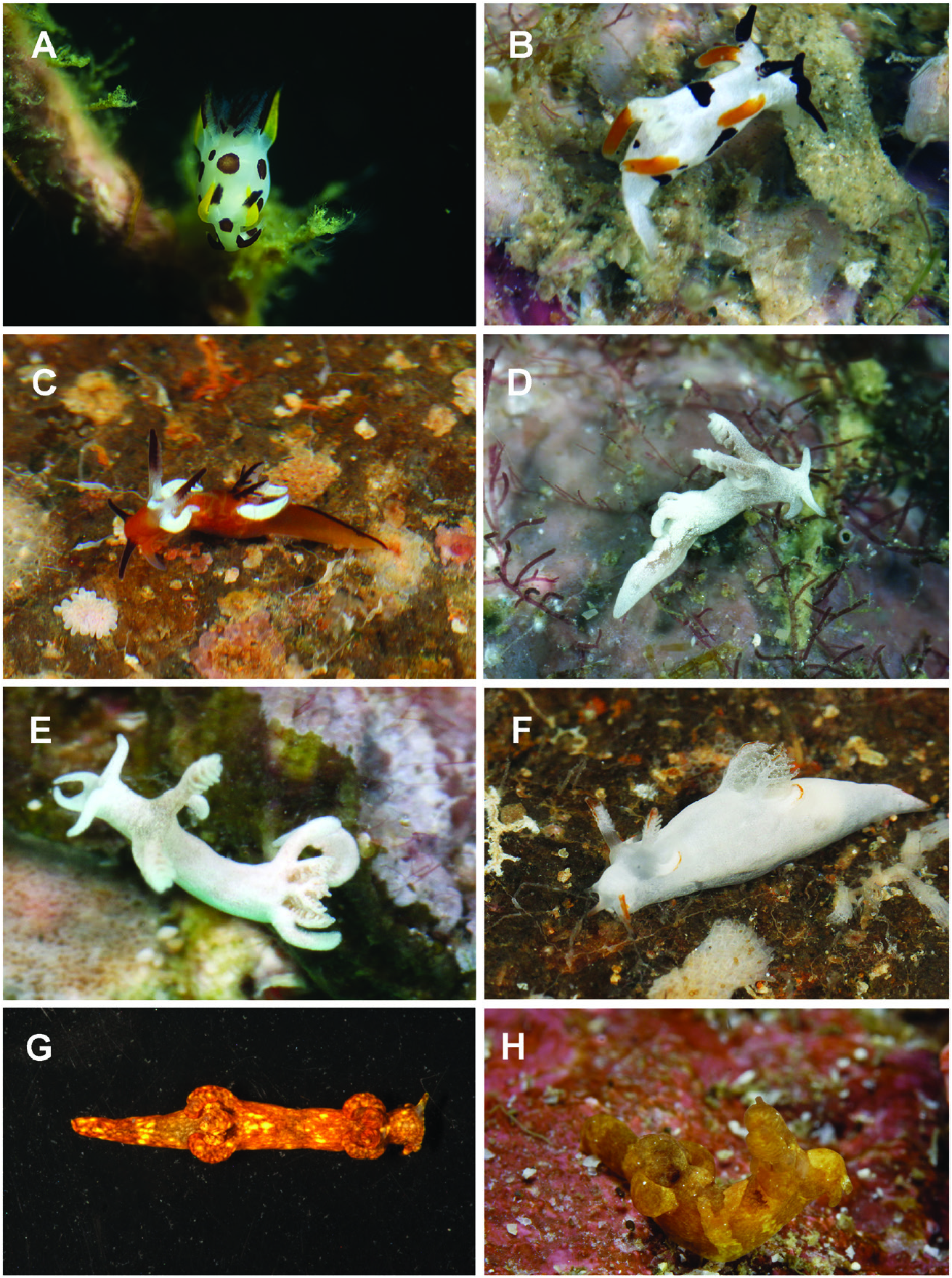
( FIGS 2G View Figure 2 , 4F View Figure 4 , 9 View Figure 9 )
Trapania reticulata Rudman, 1987: 201−203 View in CoL , figs 7C, 10; Wägele et al., 2006: fig. 12R; Gosliner et al., 2008: 138, second photograph from the bottom; Hervé, 2010: 156, middle photograph; Humann & Loach, 2010: 307, bottom-left photograph; Gosliner et al., 2015: 140, top photograph; Gosliner et al., 2018: 57, bottom photograph.
Material examined: CASIZ 191431 , one specimen, dissected and sequenced, Tab Island , Madang Province, Papua New Guinea, 9–12 m depth, 26 November 2012, collected by Vanessa Knudson.
Geographical distribution: Known from Australia ( R u d m a n, 1 9 8 7; W ä g e l e e t a l., 2 0 0 6; G o s l i n e r et al., 2015, 2018), New Caledonia ( Hervé, 2010), Thailand ( Neal, 2009) and Papua New Guinea (present study).
External morphology: The living animal has been well described previously Rudman (1987) and the specimen depicted in Figure 2G View Figure 2 closely resembles that described by him.
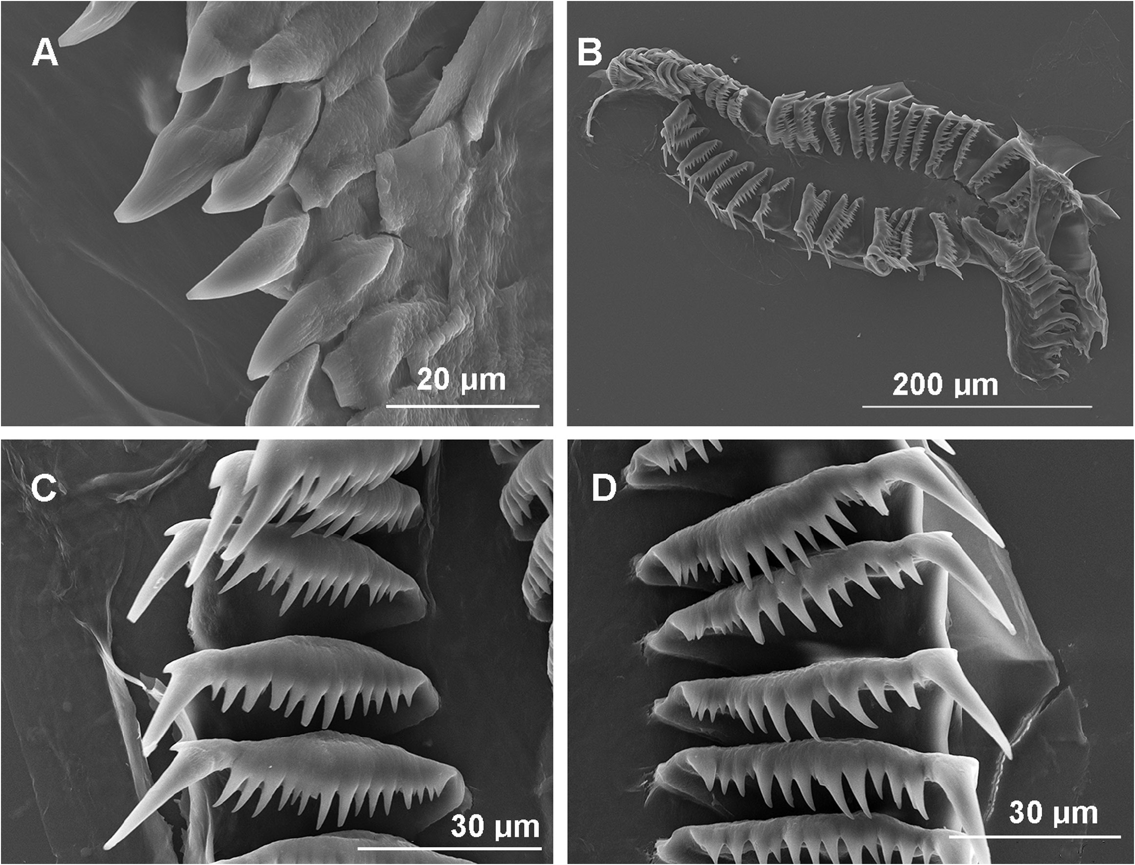
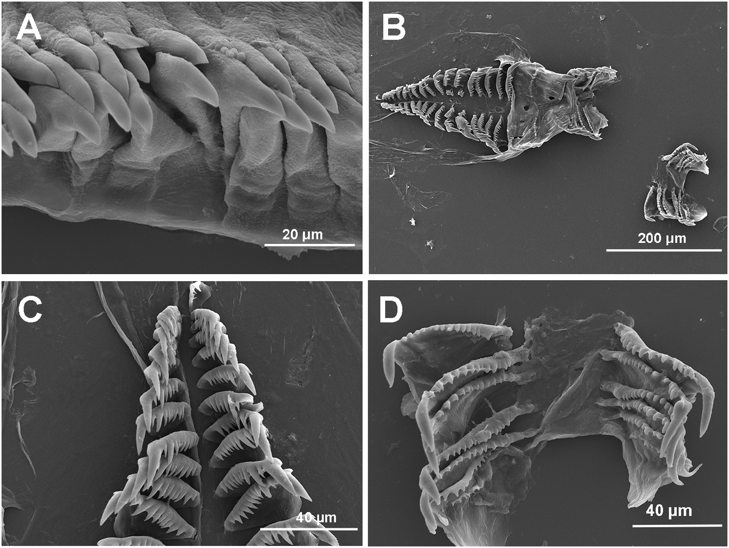
Buccal mass: The buccal mass is muscular with a moderately enlarged buccal pump on the dorsal surface. Inside the anterior portion of the mass is a pair of well-developed jaws. The jaws contain several rows of acutely pointed jaw elements of various sizes that are tightly packed together with a few gaps between them ( Fig. 9A View Figure 9 ). The radular formula is 34 × 1.0.1 ( Fig. 9B View Figure 9 ). The older teeth ( Fig. 9C View Figure 9 ) are somewhat smaller than the newer ones and the radula widens gradually towards the more newly developed teeth ( Fig. 9D View Figure 9 ). All of the teeth are narrow at the inner base and broaden out at the outer edges. The teeth bear numerous denticles, with the smallest ones being found on the inner edge of the tooth. Despite the inner denticles being smaller, the denticles are evenly graded and all much smaller than the narrow primary cusp. There are approximately 14–17 denticles on the inner side of the much larger primary cusp and one short, triangular denticle on the outer side of the cusp. The older teeth have fewer denticles than the more recently developed ones.
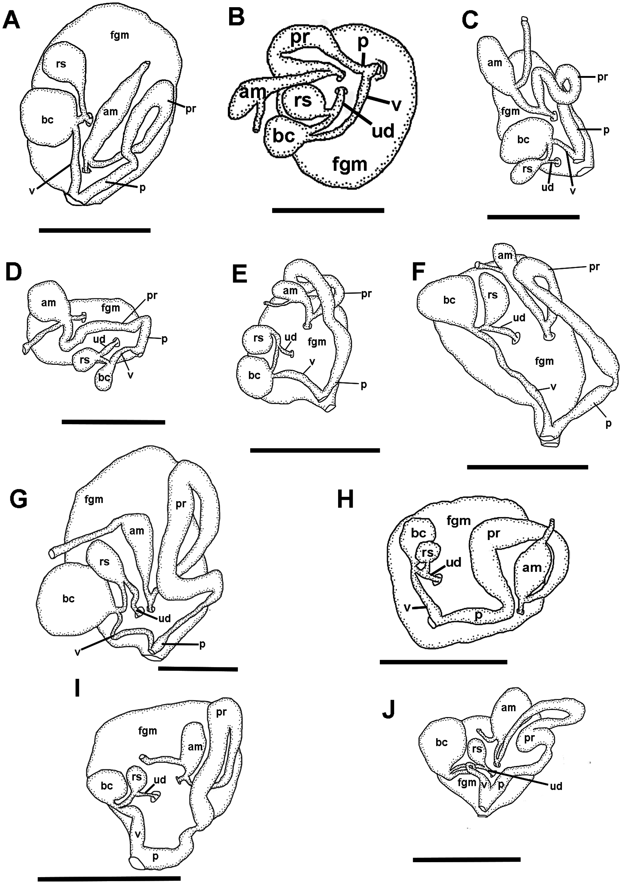
Reproductive system: The mature reproductive system is triaulic ( Fig. 4F View Figure 4 ). The narrow pre-ampullary duct enters the elongate, saccate ampulla near the proximal end of the ampulla. The ampulla narrows again at the post-ampullary duct and divides into the short, narrow oviduct, which enters the female gland mass, and the vas deferens. The vas deferens gradually widens into the thick prostatic portion that loops prior to expanding distally, immediately before it narrows abruptly into the short, narrow ejaculatory segment. The ejaculatory segment widens into an elongate penial sac. The penial sac terminates adjacent to the vagina. The width of the vagina remains uniform along a long length until it enters the base of the large, irregularly shaped bursa copulatrix adjacent to the receptaculum duct. The short receptaculum duct enters the base of the smaller, smaller slightly pyriform receptaculum seminis. The moderately long uterine duct emerges from the receptaculum duct near the base of the receptaculum and enters the female gland mass. The female gland mass is composed of the large mucous gland and the smaller albumen and membrane glands.
Remarks: Our molecular phylogeny reveals that Trapania reticulata is strongly related to a weakly supported clade of T. darvelli and the ABGD analyses reveal a low genetic divergence of 1.2–1.4% in the COI gene and 1.6% in the 16S gene between T. reticulata and T. darvelli ( Tables 3 View Table 3 , 4 View Table 4 ). The COI ABGD analysis successfully recovers T. reticulata separately from T. darvelli when the prior maximal distance was smaller than 0.003, but when the prior maximal distance was greater than 0.005, the COI ABGD analysis was unable to recover T. reticulata separately from T. darvelli . The 16S ABGD analysis also fails to recover T. reticulata as a distinct species from T. darvelli at all prior maximal distances studied here. Additionally, the bPTP analysis also successfully recovers T. reticulata separately from T.darvelli . Despite the low genetic distances between these two species, there are strong morphological characteristics that support T. reticulata and T. darvelli as distinct species. Externally, the two species have dramatically different colour patterns. In T. reticulata the body colour is translucent brown with darker brown reticulations that surround yellow spots and opaque white areas with similarly coloured oral tentacles and extra-rhinophoral and extra-branchial appendages, while in T. darvelli the body colour is solid white with brown on the oral tentacles and extra-rhinophoral and extra-branchial appendages. Both species do have proportionately thick extra-rhinophoral and extra-branchial appendages and elongate radular teeth with many denticles on the inside of the large cusp near the outer border of the tooth. The radular teeth of T. reticulata ( Fig. 10A–C View Figure 10 ; Rudman, 1987: fig. 10A– C) are distinctly straight with a narrow interior base that widens externally giving the tooth base a triangular appearance. In contrast the teeth of T. darvelli ( Rudman, 1987: fig. 15 A–C; Fig. 10 View Figure 10 ) are more broadly arched with a much wider primary cusp. The reproductive system of T. reticulata ( Fig. 4F View Figure 4 ) differs in several key features from that of T. darvelli ( Fig. 4G View Figure 4 ). The pre-ampullary duct of T. reticulata enters the proximal end of the ampulla below the proximal terminus of the ampulla, whereas it enters directly into the proximal terminus in T. darvelli . The prostate of T. reticulata is not as convoluted as that of T. darvelli . Most significantly, T. reticulata has a short, narrow ejaculatory segment that enters a long, wide penial sac, whereas T. darvelli has a long, narrow ejaculatory segment that enters a short bulbous penial sac. Both species have a long vagina, which is straight in T. reticulata and more curved in T. darvelli . The bursa copulatrix and receptaculum seminis of T. reticulata are more similar in size, whereas the bursa is much larger in T. darvelli .
| R |
Departamento de Geologia, Universidad de Chile |
No known copyright restrictions apply. See Agosti, D., Egloff, W., 2009. Taxonomic information exchange and copyright: the Plazi approach. BMC Research Notes 2009, 2:53 for further explanation.
|
Kingdom |
|
|
Phylum |
|
|
Class |
|
|
Order |
|
|
Family |
|
|
Genus |
Trapania reticulata
| Smirnoff, Dimitri S., Donohoo, Samantha A. & Gosliner, Terrence M. 2022 |
Trapania reticulata
| Gosliner TM & Valdes A & Behrens DW 2018: 57 |
| Gosliner TM & Behrens DW & Valdes A 2015: 140 |
| Herve JF 2010: 156 |
| Rudman WB 1987: 203 |