
Paraleptophlebia westoni Imanishi 1937
|
publication ID |
https://doi.org/10.11646/zootaxa.4337.1.5 |
|
publication LSID |
lsid:zoobank.org:pub:25ACCEAD-C96E-4BE0-B49C-08EB51EDE56D |
|
DOI |
https://doi.org/10.5281/zenodo.6051508 |
|
persistent identifier |
https://treatment.plazi.org/id/03E5842F-FFBE-3304-1BB5-FC7C3EF5FEAC |
|
treatment provided by |
Plazi |
|
scientific name |
Paraleptophlebia westoni Imanishi 1937 |
| status |
|
Paraleptophlebia westoni Imanishi 1937 View in CoL
( Figs 13‒37 View FIGURES 13 – 24 View FIGURES 25 – 28 View FIGURES 29 – 31 View FIGURES 32 – 37 )
Paraleptophlebia westoni Imanishi 1937: 332 View in CoL (male imago); Gose 1979 -1981 (1980): 215 (male imago); Ishiwata 2000: 79 (nymph, abdomen).
Material. RUSSIA, KHABAROSKIY KRAY, Anyui River Basin , upper reaches Kiya River , 49°28ʹ13.46ʹʹN, 137°35ʹ31.41ʹʹE, tributary Manoma River, at highway Lidoga‒Vanino, 16. VIII. 1997, T. Tiunova: 11 male and 7 female imagoes, 4 larvae .
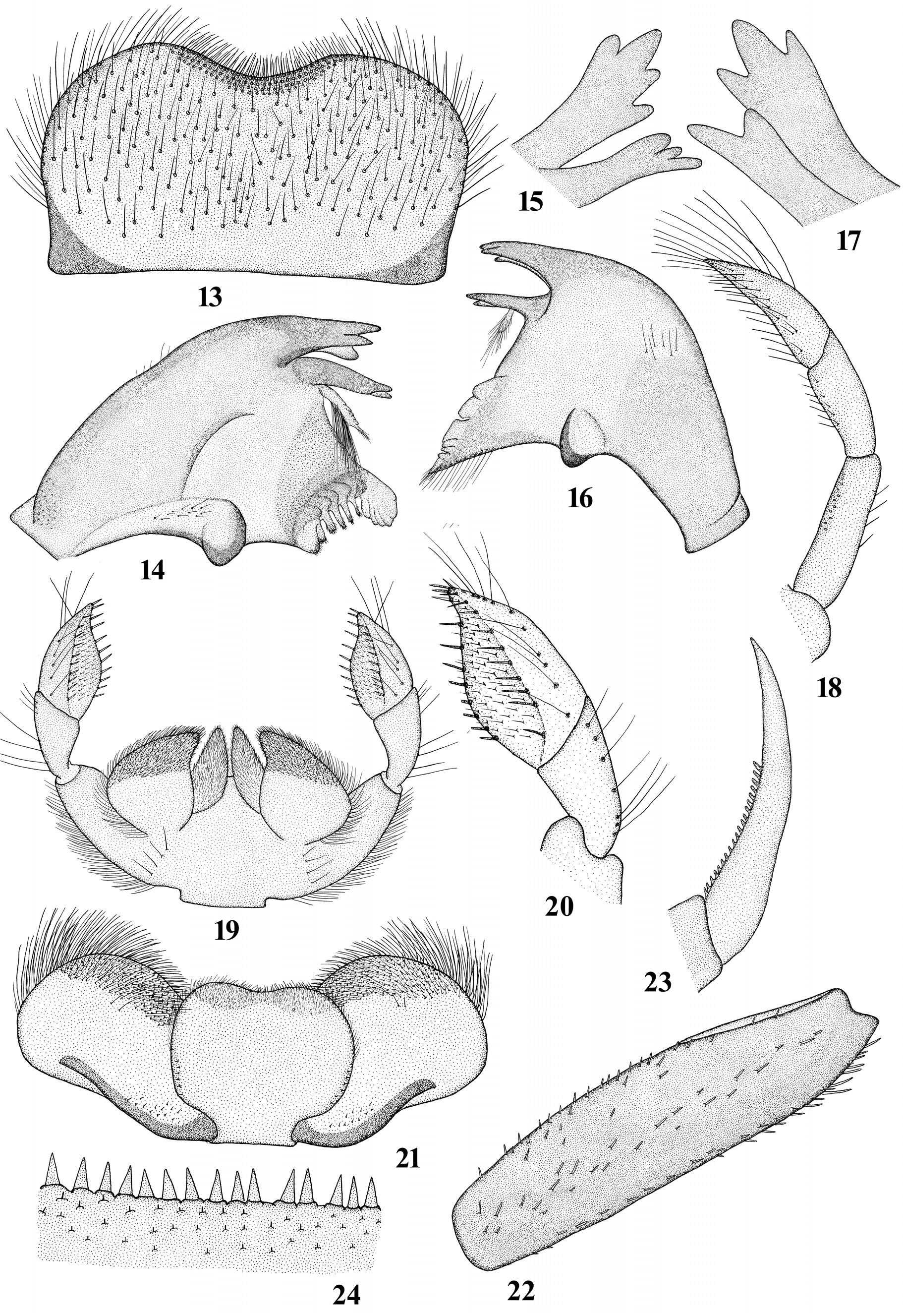
Mature larvae (in alcohol). After long fixation, the color is poorly preserved. Length (mm): body 5.8–7.0; cerci lost. Head: Dorsal surface of labrum covered with long hair-like setae; anterior margin with shallow emargination and several rows of shorter hair-like setae ( Fig. 13 View FIGURES 13 – 24 ). Mandibles brown. Incisors of left mandible with outer incisor terminated by 4 denticles and inner incisors terminated by 3 denticles ( Figs 14‒15 View FIGURES 13 – 24 ); right mandible with outer incisor terminated by 3 denticles and inner terminated by 2 denticles ( Figs 16‒17 View FIGURES 13 – 24 ). Right mandible with row of hair-like setae proximal of molar surface and long setae on dorsal surface in the middle area nearer to the outer margin ( Fig. 16 View FIGURES 13 – 24 ). Maxillary palpi with segment 3 slightly longer than segment 2, together 1.65 times longer than 1 st segment ; segment 1 with several strong setae along outer margin; segment 2 with long and short hair-like setae on inner margin; 3rd segment with very long, thin hair-like setae apically and long hair-like setae along inner margin ( Fig. 18 View FIGURES 13 – 24 ). Labium with segment 2 of labial palp shorter than segment 3, and 3rd and 2rd segments together nearly equal segment 1 ( Fig. 19 View FIGURES 13 – 24 ); segment 3 of labial palp with a tapered tip and widened in the middle part; segment 3 with row of stout setae along inner margin and middle part, area between these rows with short stout setae; row of long hair-like setae located along outer margin ( Fig. 20 View FIGURES 13 – 24 ); segment 2 with long and short hair-like setae along outer margin; segment 1 with numerous long hair-like setae along outer margin and row long hair-like setae at the base ( Fig. 20 View FIGURES 13 – 24 ). Glossae conical shape, dorsal surface densely covered with short hair-like setae; dorsal surface of paraglossae covered with small stout setae and long hair-like lateral setae ( Fig. 19 View FIGURES 13 – 24 ). Lingua of hypopharynx very slightly emarginated apically; posterolateral margins with short stout setae ( Fig. 21 View FIGURES 13 – 24 ). Thorax: Pronotum brown without visible maculation. Forefemora with row of stout pointed setae on inner and outer dorsal margins; middle area covered with stout pointed setae of identical size ( Fig. 22 View FIGURES 13 – 24 ). Foreclaw with single row of denticles, reaching middle of claw ( Fig. 23 View FIGURES 13 – 24 ). Ratio of length of femur to tibia and the length of tibia to tarsus: forelegs 1.0 and 1.8; middle legs 1.25 and 1.6. Abdomen: General color of terga brown; lateral sides with black coating; terga IX–X darker, nearly black; terga VIII–IX with light triangular spot medially. Abdominal terga III–IX with regular row of triangular stout spines on posterior margin and irregular rows of small spines on tergite surface ( Fig. 24 View FIGURES 13 – 24 ). Posterolateror corners of segment VIII and IX extended into sharp projections of equal size. Abdominal sterna light brown, without ganglion spots. Gill I forked at short distance from base, other gills forked nearer to base; tracheae without dark lateral braches ( Figs 25–28 View FIGURES 25 – 28 ); hairs very thin, pale, and break off easily; well visible only on a dark background. Cerci and paracercus lost.
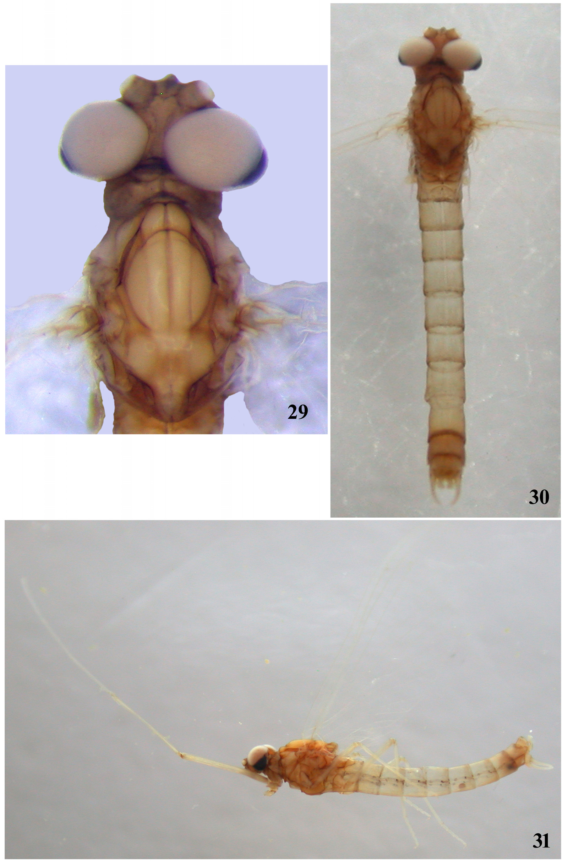
Male imago (in alcohol). Length (mm): body 5.8–7.0; forewings 5.8–6.5; cerci 10.1–11.0. Head: General color dark brown. Antennae brown; ocelli whitish apically and dirty brown basally. Upper portion of compound eyes brownish with a lilac shade; low portion black; compound eyes close to each other, non-contiguous ( Fig. 29 View FIGURES 29 – 31 ). Thorax: General color light brown with amber shade. Medioscutum light brown, submedioscutum some darker; prelateroscutum dark brown; posterior scutal protuberance light brown, scuto-scutellar impression some darker; scutellum brown; parascutellum dark brown; median longitudinal suture wide and brown. Wings pale, hyaline; longitudinal veins of fore wings yellowish or pale; transverse veins pale, almost invisible; stigmatic area milky; all veins of hind wings pale, invisible. Femora and tibia of forelegs brownish, tarsal segments yellowish ( Fig. 31 View FIGURES 29 – 31 ).
Middle and hind legs brownish, femora slightly darker. Length (mm) of foreleg segments: femora 1.7‒2.0; tibia 1.8‒2.0; tarsal segments 1.0‒1.1, 0.9‒1.1, 0.7‒0.8, 0.3, and 0.15. Abdomen: Terga pale tinged brownish with lateral black streaks; posterior margins dirty brown ( Figs 30‒31 View FIGURES 29 – 31 ); tergum I pale tinged blackish; terga II‒VI pale, translucent; terga VII‒IX brown, nontransparent. Ganglionic markings absent. Cerci lost. Genitalia: Styliger with V-shaped incision, light brown, somewhat lighter in anterior area; processes brown ( Figs 32‒33 View FIGURES 32 – 37 ). Gonostylus light brown, first segment darker, second segment slightly narrower distally, third segment widened apically, 4th segment smallest ( Fig. 32 View FIGURES 32 – 37 ). Penis lobes separated by U-shaped cleft reaching middle of penis length; each penis lobe with conical construction ( Figs 34, 36 View FIGURES 32 – 37 ), with apex curved in sides ( Figs 32, 34 View FIGURES 32 – 37 ); each penis lobe with ventral slightly curved appendage, tapering and pointed distally ( Figs 32‒35 View FIGURES 32 – 37 ), reaching proximal first segment of the forceps ( Fig. 32 View FIGURES 32 – 37 ); appendages pale with narrow longitudinal blackish stripe ( Figs 32, 36 View FIGURES 32 – 37 ). Cerci lost.
Female imago (in alcohol). Length (mm): body 6.7‒7.2; forewing 6.8‒7.5; cerci 7.0. Head: General color light brown. Antennae brownish or yellowish. Thorax: General color light brown. Medioscutum and submedioscutum light brown; all suture brown; median longitudinal suture brownish, weakly expressed; parascutellum brown or blackish. Wings pale, hyaline. All veins pale, stigmatic area milky. Femur and tibia all legs brownish, tarsal segments lighter. Length (mm) of foreleg segments: femora 1.7; tibia 1.8; tarsal segments 0.3‒0.35, 0.3‒0.35, 0.25, 0.25, and 0.15. Abdomen: Terga (without eggs) I‒VI brownish with more darker posterior margins; terga VII‒IX brown. Sterna I‒II, and VII‒IX brown, other brownish, translucent. Subanal plate with deep V-shaped incision, apexes are pointed ( Fig. 37 View FIGURES 32 – 37 ). Cerci brownish.
Distribution. Khabarovskiy Kray and Japan.
Discussion. Of all known species of the Paraleptophlebia , new species on the structure of the ventral appendages of the penis lobes are closest to the following species: Paraleptophlebia cincta (Retzius 1783) , P. debilis (Walker 1853) , P. falcula Traver 1934 , P. helena Day 1952 , P. lacustris Ikonomov 1962 , P. moerens ( McDunnough 1924) , P. ontario (McDunnough 1926) , P. packii (Needham 1927) , P. quisquilia Day 1952 , P. strandii ( Eaton 1901) , P. strigula McDunnough 1932 , P. westoni Imanishi 1937 and P. zayante Day 1952 .
New species differs from P. debilis ( Harper & Harper 1986) , P. moerens ( Harper & Harper 1986) , P. packii ( Day 1952) , P. strandii ( Eaton 1901) and P. zayante ( Day 1952) absent of large lobe at base of first segments of forceps. The same is known for the following species: P. cincta ( Ulmer 1929) , P. falcula ( Traver 1934; Tiunova & Kluge 2016), P. lacustris ( Ikonomov 1962) , P. quisquilia ( Day 1952) , P. ontario ( Burks 1953) , P. strigula ( McDunnough 1932; Burks 1953) and P. westoni ( Imanishi 1937; Gose 1979 –1980).
Male imago of Paraleptophlebia kunashirica sp. nov. can be distinguished by the following features: processes of styliger are not long (in contrast to P. falcula ) ( Traver 1934; Tiunova & Kluge, 2016); ventral appendages slightly curved, extended and blunt distally (in P. cincta , P. lacustris , P. strigula and P. westoni ventral appendages slightly curved and pointed) ( Ulmer 1929; Ikonomov 1962; Burks 1953; McDunnough 1932; Imanishi 1937); apex of each penis lobes rounded (in P. ontario and P. westoni apex of each penis lobes with conical construction, in P. quisquilia with a deep notch) ( Burks 1953; Day 1952).
Among species of Paraleptophlebia with available larval descriptions, Paraleptophlebia westoni differs from Nearctic species P. bicornuta (McDunnough 1926) , P. helena , P. packii and P. zayante by absence of tusk-like elongation of mandibular incisor; from P. altana Kilgore & Allen 1973 and P. cachea Day 1954 it differs by nonwidened lobes of gills without dark lateral branches of main trachea. The same is known for the following species of Paraleptophlebia : P. calcarica Rowbotham & Allen 1988 ( Jacobus & McCafferty2004), P. cincta ( Macan 1952; Landa 1969), P. debilis ( Ide 1930; Gordon 1933), P. falcula ( Tiunova & Kluge 2016) , P. guttata ( McDunnough 1924) ( Ide 1930; Gordon 1933), P. jeanae Berner 1955 ( Randolph & McCafferty 1996), P. lacustris ( Ikonomov 1962) , P. moerens ( Gordon 1933; Burks 1953), P. ontario ( Needham, Traver & Hsu 1935; Burks 1953), P. praepedita (Eaton 1884) ( Gordon 1933; Burks 1953), P. quisquilia ( Day 1952) , P. ruffoi Biancheri 1956 ( Belfiore & Giangrande 1979), P. strandii ( Tiensuu 1939) , P. strigula ( McDunnough 1932) , P. submarginata (Stephens 1835) ( Komarek 1921; Macan 1952; Landa 1969; Belfiore & Giangrande 1979), P. volitans ( McDunnough 1924) ( Ide 1930; Gordon 1933), P. werneri Ulmer 1920 ( Landa 1969) .
Mature larvae of Paraleptophlebia westoni can be distinguished by the following features: thorax is not hairy (in contrast to P. volitans ); hypopharynx is slightly emarginated apically ( Fig. 19 View FIGURES 13 – 24 ) (in P. calcarica , P. lacustris and P. strandii have medioapical cleft); maxillary palp segments 3 slightly longer than segment 2, together 1.65 times longer than segment 1 ( Fig. 18 View FIGURES 13 – 24 ) (in P. debilis P. moerens P. submarginata , P. guttata and P. strigula segments 2 and 3 together are equal to segment 1); labial palp segment 2 is shorter than segment 3, and together nearly equal segment 1 ( Fig. 19 View FIGURES 13 – 24 ) (in P. falcula labial palp segments 2 is shorter than segment 3, and together are longer than segment 1); legs have no brown band ( Fig. 22 View FIGURES 13 – 24 ) (in contrast to P. debilis and P. jeanae ); row of denticles reaches middle or slightly more than the middle of the claw ( Fig. 23 View FIGURES 13 – 24 ) (in P. ruffoi and P. werneri row of denticles occupies more than half of claw); abdominal segment VIII and IX with posterolateral spines (in P. guttata and P. strigula lateral spines are present on segment 9 only).


No known copyright restrictions apply. See Agosti, D., Egloff, W., 2009. Taxonomic information exchange and copyright: the Plazi approach. BMC Research Notes 2009, 2:53 for further explanation.
|
Kingdom |
|
|
Phylum |
|
|
Class |
|
|
Order |
|
|
Family |
|
|
Genus |
Paraleptophlebia westoni Imanishi 1937
| Tiunova, Tatiana M. 2017 |
Paraleptophlebia westoni
| Ishiwata 2000: 79 |
| Imanishi 1937: 332 |