
Phalangodus kuryi, Villarreal & García, 2016
|
publication ID |
https://doi.org/10.5852/ejt.2016.242 |
|
DOI |
https://doi.org/10.5281/zenodo.3854796 |
|
persistent identifier |
https://treatment.plazi.org/id/03E6D434-A124-9415-FDC0-FAC590C2D959 |
|
treatment provided by |
Valdenar |
|
scientific name |
Phalangodus kuryi |
| status |
sp. nov. |
Phalangodus kuryi View in CoL sp. nov.
urn:lsid:zoobank.org:act:
Figs 13–15 View Fig View Fig View Fig , 18 View Fig ; Tables 7–8
Diagnosis
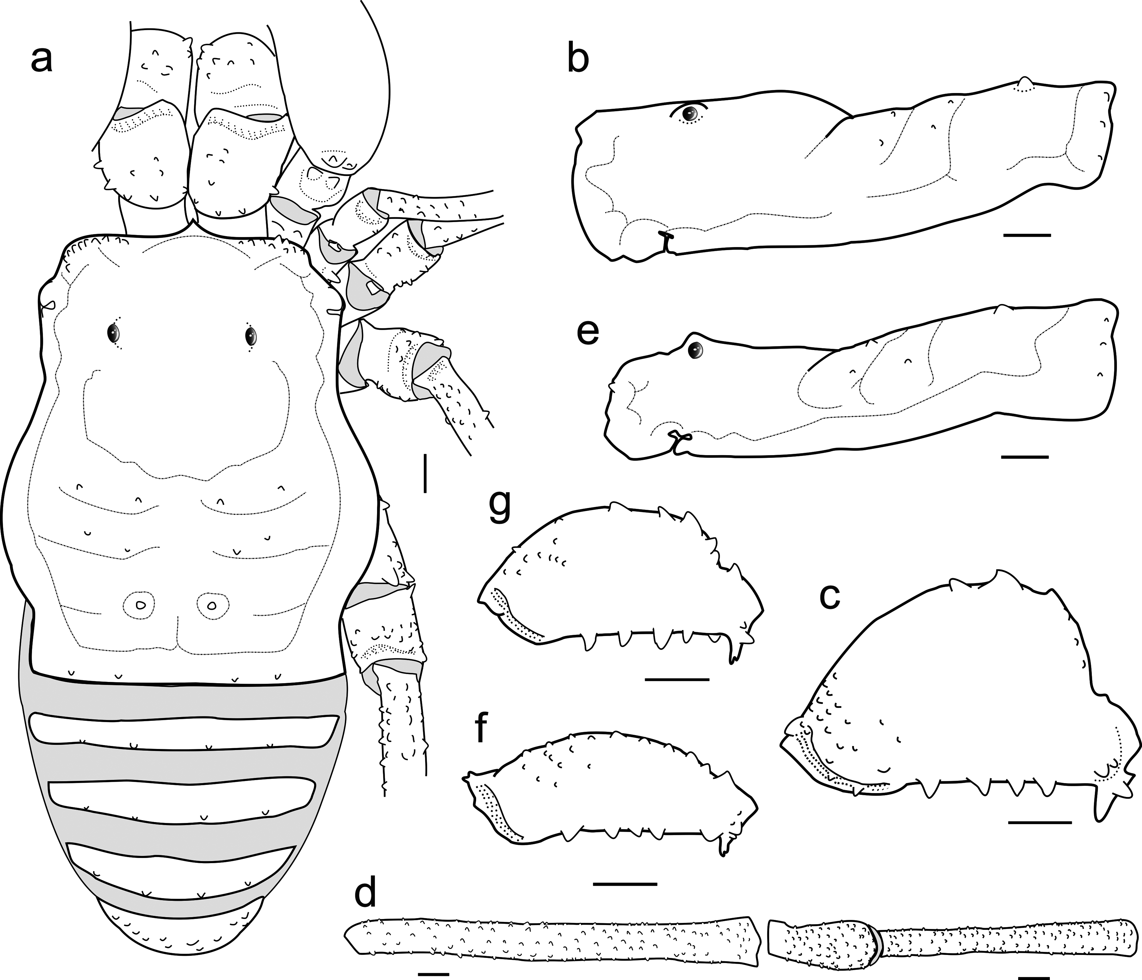
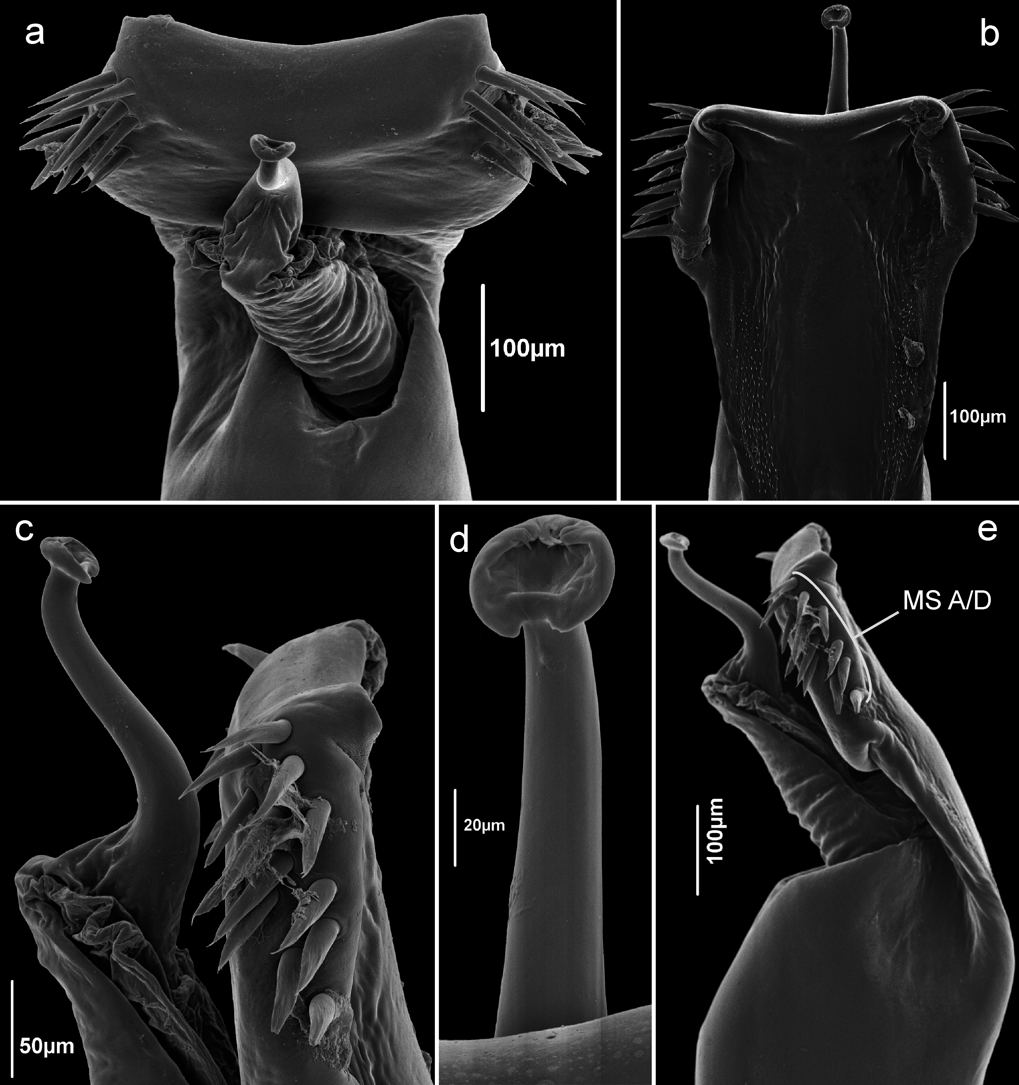
Phalangodus kuryi sp. nov. can be distinguished from all other species of the genus by the very low ocularium and paramedian paired small tubercles of area III ( Figs 13 View Fig a–b, 14a–b); ventroproximal group of tubercles of pedipalpal femora not enlarged ( Fig. 14c, g View Fig ). Femur IV unarmed ( Fig. 14d View Fig ); penis ventral plate thin in lateral view ( Fig. 15e View Fig ); MS A–D of the penis ventral plate grouped in one distal group ( Fig 15 View Fig a–c, e). Penis stylus curved ( Figs 15c View Fig ). Heteromorphism in males ( Fig. 14c, g View Fig ), evident by different sizes (length and height) of pedipalpal femora.
Etymology
The species is named after the Brazilian arachnologist Adriano B. Kury, advisor and friend, who immensely contributes to the knowledge of Opiliones worldwide.
Type material
Holotype
COLOMBIA: ♂ ( ICN AO 1436 ), Magdalena, Santa Marta, village of Minca, sector San Lorenzo , 2200 m, 30 Aug. 2014, W. Galvis and J. Moreno leg.
Paratypes
COLOMBIA: 1 ♂, 6 ♀♀ ( ICN AO 1436.1), same data as holotype; 1 ♂, 2 ♀♀, 1 juvenile ( FMNH AK 229), Magdalena, Sierra Nevada de Santa Marta, Inderena Station in Cerro San Lorenzo, under stones, 1700–2200 m, 9–12 Jul. 1970, B. Malkin and P. Burchard leg.
Description
Male (ICN AO 1436)
Measurements of body and appendage in Table 7.
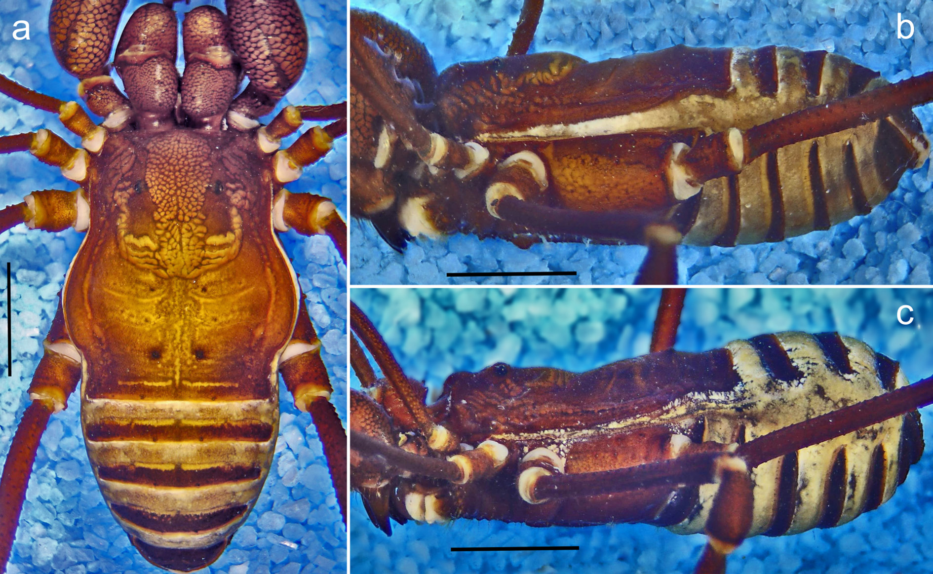
DORSUM. Dorsal scutum type alpha ( Fig. 13a View Fig ). Abdominal scutum widest at scutal groove II; lateral borders of dorsal scutum smooth. Carapace mostly smooth, with a group of granules on the anterolateral region. Ocularium unarmed, very low, without median depression. Integumentary dome of ozopore raised and conspicuous. Abdominal scutum well delimited, divided into four marked areas: scutal area I divided into left and right halves by invasion of the scutal area II; scutal areas I–II each with two pair of granules; scutal area III with a paramedian pair of low tubercles ( Fig. 13 View Fig a–b). Posterior border of scutum straight and unarmed, with a row of four minute tubercles on a row ( Figs 13a View Fig , 14a View Fig ). Free tergites I–III with few granules.
VENTER. Stigmatic area with two pairs of paramedian granules. Stigmata large, oval and slightly oblique. Coxa I densely covered with irregular rows of tubercles; coxa II longer than coxae I and III, with three rows of small tubercles, the median ones more conspicuous; coxa III with a curved median row of granules and distal minute tubercles sparsely distributed,with the posterior border sigmoid; coxa IV
strongly backward, with sparsely distributed granules, the lateral larger. Free sternites each with a row of small granules.
CHELICERA ( Fig. 13a View Fig ). Basichelicerite very swollen, bulla with sparsely distributed dorsal tubercles. Hand slightly enlarged, its frontal region covered by tubercles of different sizes. Fixed finger with a proximal minute tooth, three median teeth decreasing in size distally and one subdistal laminar tooth. Movable finger with a proximal wide and low tooth, a small gap and three teeth (the proximal one rounded and the two distal forming a lamella).
PEDIPALPUS ( Fig. 14c View Fig ). Trochanter with two tubercles on a dorsal protuberance, ventrally with two fused median tubercles and two ventrolateral tubercles. Femur strongly inflated, dorsally curved and ventrally straight in lateral view, with a dorsoproximal row of forward curved tubercles, ventrally with a proximal group of three small tubercles in a common base and two ventral rows of tubercles, ectal largest. Patella short (ratio FePp/PaPp = 2.0), subsquare in dorsal view, smooth. Tibia inflated, dorsally smooth; tibial setation: ectal (iiIi) (3> 1> 4> 2) (the two last one St share a common base), with a row of proximal tubercles and mesal (IiIi) (3> 1> 4> 2), with some ventral granules. Tarsus inflated, dorsally smooth, ectally with four distally shifted St (IiIi) (3> 1> 2> 4); mesally with three St (iii) (1> 2> 3) (teratological condition in the right side with a group of five small St). All spines of St reduced. Claw substraight, not swollen, its length approximately the same as tarsus.
LEGS. Coxa I with one prolateral and one retrolateral tubercles; II with two prolateral, two dorsal and three retrolateral tubercles; III with one prolateral and one retrolateral tubercles; IV with few lateral tubercles and a prolateral group of conical granules. Trochanters I–IV finely granulate; trochanter I with one large mesoventral tubercle and few retrolateral tubercles; trochanter II ditto, with a group of prolateral distal tubercles; trochanter III with tubercles irregularly distributed, the retrolateral one largest; trochanter IV slightly widened at base, irregularly tuberculate. Femur I slightly curved, femora II–IV straight; femora I–IV with longitudinal rows of small granules ( Fig. 14d View Fig ). Patellae I–IV unarmed; the III–IV slightly swollen. Tibia I–IV unarmed, straight ( Fig. 14d View Fig ). Metatarsus IV without clear ringed marks on the subdistal portion. Claws III–IV smooth. Ratio Fe IV/scutum = 1.14. Tarsal counts in Table 8.
GENITALIA. Ventral plate sub trapezoidal with subdistal constriction and distal parabolic concavity., Ventral surface of VP with two elongated fields of small and needle-like microsetae with a longitudinal medial well marked elevation ( Fig. 15 View Fig a–b). MS forming a single group distally positioned (A/D), composed by 12–13 pairs of acuminate and cylindrical setae ( Fig. 15 View Fig a–c, e). Two pairs of very small MS E close together in the distal region of the flange ( Fig. 15b View Fig ). Glans sac columnar elongate, with numerous proximal folders ( Fig. 15a, e View Fig ) Stylus curved, without processes ( Fig. 15c View Fig ). Stylar caps ringshaped without lateral or ventral projections ( Fig. 15d View Fig ).
COLORATION (in alcohol). Carapace with two paramedian areas of homogenous color paprika 38, three longitudinal stripes and posterior area reticulated squash yellow 83. Abdominal scutal areas reticulated between squash yellow 83 and pastel yellow 84. Paramedian tubercles of scutal area III, free tergites, anal operculum and coxae I– –IV venetian red 41. Chelicerae and pedipalps reticulated dark wine 44 on light amber 71.
Female (ICN AO 1436.1)
Similar to males, differing by: carapace shorter and lower, with ocularium and spines higher in lateral view ( Fig. 14b, e View Fig ); coda wider and longer; chelicerae smaller; pedipalpal femur, patella and tibia thinner; femur IV curved and slightly thinner. Sexual dimorphism evident in alpha males with pedipalpal femora much thicker than female ( Fig. 14c View Fig , f–g).
Ovipositor. dl and vl rounded with three and two large, acuminated, single-tippedsetae respectively. Dl with three pairs of ds without the basal located pair. and the lateral region of the ovipositor with a pair of short dorso-lateral setae.
Distribution
Known only from surroundings of Minca, Sierra Nevada de Santa Marta ( Fig. 18 View Fig ).

| ICN |
Instituto de Ciencias Naturales, Museo de Historia Natural |
| FMNH |
Field Museum of Natural History |
No known copyright restrictions apply. See Agosti, D., Egloff, W., 2009. Taxonomic information exchange and copyright: the Plazi approach. BMC Research Notes 2009, 2:53 for further explanation.
|
Kingdom |
|
|
Phylum |
|
|
Class |
|
|
Order |
|
|
InfraOrder |
Grassatores |
|
SuperFamily |
Gonyleptoidea |
|
Family |
|
|
Genus |