
Cenomanocarcinus, Van Straelen, 1936
|
publication ID |
https://doi.org/10.11646/zootaxa.4303.2.7 |
|
publication LSID |
lsid:zoobank.org:pub:99DB2319-498E-4A8B-BE10-12B53E2B7C9A |
|
DOI |
https://doi.org/10.5281/zenodo.6029999 |
|
persistent identifier |
https://treatment.plazi.org/id/03E7095C-FF8E-BF18-0EAE-FB64FC15FC4A |
|
treatment provided by |
Plazi |
|
scientific name |
Cenomanocarcinus |
| status |
|
Cenomanocarcinus View in CoL sp. 2
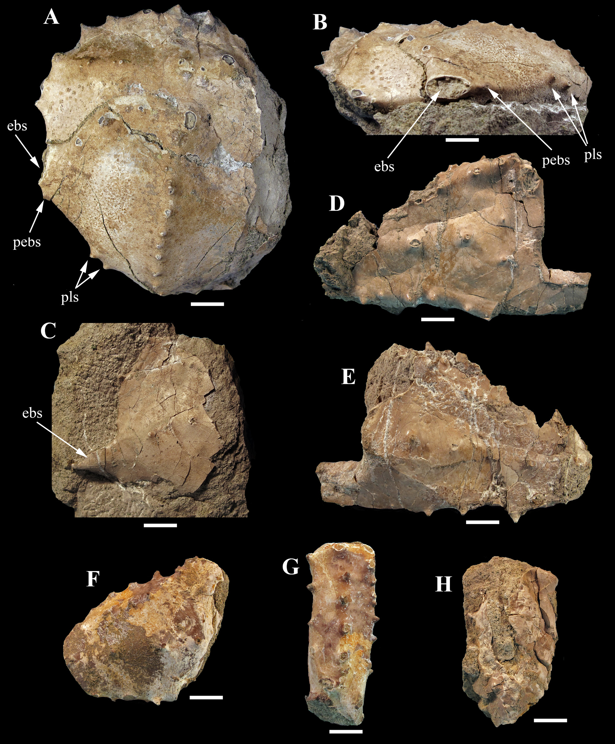
( Fig. 3 View FIGURE 3 )
Cenomanocarcinus aff. inflatus Ossó et al. 2015: 241 View in CoL , t. 1.
Locality and horizon. Trasvía ( Cantabria, Spain). Romaniceras kallesi biozone View in CoL , middle Turonian.
Material examined and measurements (in mm). One half left and one fourth left of carapaces; one incomplete crushed left propodus and isolated remains of right propodus, and a merus; MMC-BIO-CE 011240: length= 87 (rostrum absent); width= 136 ( 68x2). MMC-BIO-CE 011242: (palm of left propodus) length= 50; width= 72; thickness= 14,5.
Description. Fragments of large sized specimens with thin cuticle preserved, subhexagonal, apparently much wider than long, smooth, vaulted transverselly and longitudinally, and with a well-marked longitudinal branchial ridge; preserved dorsal carapace regions defined by shallow grooves separating the swollen epibranchial and hepatic ridges. Anterolateral margin with six short, subtriangular spines (excluding outer orbital and epibranchial spines); epibranchial spine subtriangular, slightly ovate, upper margin spiny. Posterolateral margin straight, round edged, smooth, equal to anterolateral margin, with one small post-epibranchial spine and a pair of spines at midlength. Posterolateral corner rounded. Posterior margin incomplete, appears straight. Left orbit small, rounded, complete and forward directed; outer-orbital spine subtriangular, acute; supraorbital margin with two fissures bounding a medial subtriangular tooth; inner-orbital toot acute subtriangular, smaller than the outer-orbital one. Front and rostrum not preserved. Protogastric region swollen, two prominent conical tubercles horizontally aligned, the external one granulated. Meso-, meta- and urogastric regions not preserved. Cardiac region absent. Intestinal region preserved in part, slightly swollen with a conical upward directed tubercle on the left side of the presumed position of the axial ridge. Hepatic region defined by a swollen transverse ridge with two pairs of small conical tubercles. Epibranchial region defined by a swollen sigmoidal ridge, strongly backward directed distally and with five conical tubercles; the inner one and the two outer ones the larger, and the two medial ones the smaller. Meso- and metabranchial regions swollen, smooth, crossed longitudinally by the divergent left branchial ridge that begins posterior to the post-cervical groove and aligned with the inner epibranchial tubercle; eleven conical tubercles regularly placed define the ridge, the two first and the penultimate tubercle the smallest, the last one, broken, appears to be the larger.
Propodus large, spiny. Palm subtrapezoidal, subrectangular elongate in section, higher distally; upper margin with three rows of widely spaced and upward directed spines; lower margin with two rows of widely spaced spines; outer surface smooth, some spines aligned transversally at half height; outer surface smooth, two spines transversally aligned a at half height and two longitudinally at the distal margin. Pollex of left propodus broken, flattened molariform proximal tooth.
Merus elongated, inner surface weakly concave, smooth; outer surface convex, margins armed with spaced spines.
Remarks. Albeit the remains of the Cantabrian Cenomanocarcinus (sp. 2) are well-preserved, they are incomplete, and the absence of important diagnostic characters such as the frontal margin, axial regions and the posterior margin, precludes of a specific placement of those specimens. However, their general appearance recall those of the second morphological group reported by Ossó et al. (2015: 241), in particular Cenomanocarcinus inflatus from the Cenomanian of France, more than other species of that group (see Van Straelen 1936; Van Bakel et al. 2012). Mainly in view of the dorsally inflated carapace, the curved and spiny anterolateral margin, the straight posterolateral margin with one medial spine, the spiny and axially convex left branchial ridge, and the features of the epibranchial and hepatic ridges. But, notwithstanding the aforementioned similarities, differences between both species are also present. For instance, in the Cantabrian specimens, the anterolateral margin is armed with six spines or teeth, instead of the usually five in C. inflatus ; the number of tubercles of the branchial ridge is higher and more regularly placed in the Cantabrian specimens than in C. inflatus . Furthermore, in C. inflatus the small post-epibranchial tooth is absent, but present in the Cantabrian specimens, and its medial posterolateral tooth is simple instead of double as in the Cantabrian specimen. Therefore, those differences, together with the lack of other diagnostic characters not preserved, besides the temporal span between both taxa, impede concluding that they are conspecifics. As well, the close similarity among them, rules out the belonging of the Cantabrian specimens to the other Cenomanocarcinus species (cf. Ossó et al., 2015).
No known copyright restrictions apply. See Agosti, D., Egloff, W., 2009. Taxonomic information exchange and copyright: the Plazi approach. BMC Research Notes 2009, 2:53 for further explanation.
|
Kingdom |
|
|
Phylum |
|
|
Class |
|
|
Order |
|
|
Family |
Cenomanocarcinus
| Ossó, Àlex 2017 |
Cenomanocarcinus aff. inflatus Ossó et al. 2015 : 241
| Osso 2015: 241 |