
Halecium
|
publication ID |
https://doi.org/10.5281/zenodo.205235 |
|
DOI |
https://doi.org/10.5281/zenodo.6209994 |
|
persistent identifier |
https://treatment.plazi.org/id/03E7237E-FFF6-4D59-FF77-1DDAF03DFE64 |
|
treatment provided by |
Plazi |
|
scientific name |
Halecium |
| status |
|
Halecium View in CoL ? delicatulum Coughtrey, 1876 (morphotype 2)
( fig. 3 View FIGURE 3 G–P, 4A–E)
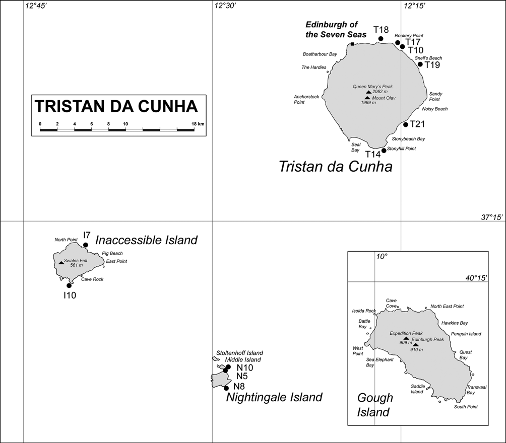
Material examined. Stn. T18, 06.iii.2005: one colony 2.5 cm high, with numerous female gonothecae, on alga ( NHM 2009.18).
Description. Stolon creeping on alga, adherent surface flattened ( fig. 3 View FIGURE 3 H) diameter ca. 125 µm; stolon mostly linear and smooth, occasionally tortuous and branched. Colony ca. 2.5 cm high ( fig. 3 View FIGURE 3 G); growth habit arborescent, in one plane, with sparingly branched stems. Stems straight, monosiphonic, with 1–3 basal constrictions above stolon ( fig. 3 View FIGURE 3 I). Auto-epizoism quite frequent, with 1–3 stolonal tubes running over basal part of primary stem, giving rise occasionally to secondary stems whose axes are (nearly) perpendicular to those of main stem. Perisarc moderately thick and pale horn basally, thinning out and becoming transparent distally. Stems divided into internodes by means of distinct oblique nodes, alternately sloping left and right ( fig. 3 View FIGURE 3 N); internodes with a distal apophysis supporting primary hydrophores, basally with a bulge above node. Internodes relatively long (455–560 µm), straight, collinear, though imperceptibly geniculate distally; diameter at node 80–110 µm. Primary hydrophores alternate, free from stem, generally very long (205–465 µm), perisarc wrinkled above base towards middle part ( fig. 3 View FIGURE 3 O), articulated; occasionally, distalmost internodes with much shorter (95–140 µm), smooth primary hydrophores ( fig. 3 View FIGURE 3 O, lower left). Pseudodiaphragm variably present on adcauline side of hydrophores, from slightly marked to obvious, and reaching abcauline side. Branches in the same plane as main stem (colony coplanar), arising irregularly from primary and secondary hydrophores ( fig. 3 View FIGURE 3 K–M); colonies with up to 2nd order branches; branches with same structure and shape as stem. Hydrotheca rather deep (55–70 µm), margin widely flaring and much everted, diameter 110–125 µm at base, 160–200 µm at rim; wall slightly asymmetrical, adaxial side tilted towards internode; desmocytes numerous, visible as refringent nodules above diaphragm. Secondary and tertiary hydrophores common and of variable length, arising from diaphragm of primary hydrothecae; pseudodiaphragm slightly marked or absent. Hydranths absent, the cnidome could not be examined. Inspected colony with mature, female gonothecae. The latter borne abundantly on stolonal tubes, along basal parts of stems ( fig. 4 View FIGURE 4 B), or arising from internode apophyses in replacement of a hydrotheca ( fig. 4A View FIGURE 4 ); exceptionally, gonothecae arising from middle part of primary hydrophores ( fig. 3 View FIGURE 3 P). Gonotheca almost oval in frontal view ( fig. 4 View FIGURE 4 C–D), laterally flattened ( fig. 4 View FIGURE 4 E), borne on smooth pedicel of variable length; 800–1000 µm long, 685– 765 µm wide in frontal view; perisarc smooth to undulate, relatively thick; aperture a shallow, circular (diameter 160–200 µm), apical depression, surrounded by thickened perisarc ( fig. 4 View FIGURE 4 C). Sporosac oval, not occupying the whole lumen of gonotheca, delimited by thin perisarc layer, containing 6–10 oval eggs, each ca. 175 × 130 µm.
Remarks. The present material is mainly characterized by its very peculiar coplanar colony, with straight stems and branches, and much long, articulated hydrophores. Similar specimens, possessing collinear internodes, were found by Hartlaub (1905) (as H. flexile Allman, 1888 ) in material from Punta Arenas, Chile. Long primary hydrophores were mentioned and/or figured by both Hartlaub (1905) (p. 613, fig. K3a) and Ralph (1958) (p. 335, fig. 11L). They were thought to belong to aberrant colonies, as stated by Vervoort & Watson (2003). Until more material is examined, it is difficult to say if these long hydrophores appear after mechanical breakage and a subsequent regeneration step.
The occurrence of two sympatric morphotypes of H. cf. delicatulum in the present collection raises the question of the morphological variability in this taxon, and its specific limitation. Coughtrey (1876) erected his new species for “very delicate”, monosiphonic, sterile specimens of a haleciid hydroid from Dunedin, New Zealand. Ralph (1958) examined material from several North and South Island localities and always found “erect monosiphonic stems with loose irregular branching”, whose female gonothecae were “more or less oval in general outline, with two distinct peaks arising from the narrow sides at the distal end”.
Schuchert (2005) gave the first accurate illustration of the female gonotheca of this species (p. 629, fig. 12B). I have had the opportunity to examine his specimen from Wellington (microslide MNHG INVE 26669). The exclusively monosiphonic stems are morphologically in agreement with the description provided by Ralph (1958), and the gonothecae clearly exhibit two prominent “ears” flanking the aperture. The gonothecae are 1335–1360 µm long, 760–790 µm wide, and contain ca. 20–22 small, rounded eggs (diameter 90–120 µm).
Ralph (1958), upon examination of types of H. gracile Bale, 1888 , H. parvulum Bale, 1888 , and H. flexile Allman, 1888 , concluded that all three species were synonyms of H. delicatulum . Previously, Bale (1888, 1894) himself did not exclude the possibility that his two species could be conspecific, and Billard (1908, 1910) recognized H. gracile as a junior synonym of H. flexile , an opinion later on shared by Bale (1915). However, both Billard (1908) and Ralph (1958) based their conclusions exclusively on characters of the trophosome, since the syntype of Allman’s (1888) species was devoid of female gonothecae, the latter being now recognized as essential for an accurate identification of Halecium species (see Schuchert 2005).
Additionally, the female gonothecae of both H. gracile and H. parvulum , as figured by Bale (1888), do not possess the characteristic perisarc projections flanking their aperture, as found in specimens from New Zealand, and Ralph (1958) did not discuss their morphological variability in H. delicatulum . Through the courtesy of J.E. Watson, I have examined a female colony from Southeastern Australia, assigned to Coughtrey’s (1876) species. All the stems were monosiphonic and the gonophores almost ripe. No apical projections could be detected, and the gonothecal aperture was situated in the middle of a slight depression.
Since Ralph’s (1958) observations, it has been shown that the South American [Blanco (1984b), p. 271, pl. 1 fig. 4 View FIGURE 4 ], South African [Millard (1975), p. 145, fig. 47K, L], sub-Antarctic [Millard (1977a), p. 7, fig. 1 View FIGURE 1 D], and Antarctic [Blanco (1984a), p. 7, pl. 5 fig. 12, as H. antarcticum Vanhöffen, 1910 ; Peña Cantero & Vervoort (2009), p. 85, fig. 1 View FIGURE 1 D–E] specimens had female gonothecae of a different shape and size, and contained a lesser number of eggs than observed in New Zealand material.
I have also examined fertile specimens referable to H. delicatulum , recently brought from southern Chile (Galea, unpublished results), and found pear-shaped female gonothecae (760–835 µm long, 560–650 µm wide) with rounded apex and the aperture situated in middle of a slight depression. Four to six large eggs (diameter 180–195 µm) were present inside each gonotheca. Their size and shape are similar to those from Antarctica [Blanco (1984a); Peña Cantero & Vervoort (2009) found gonothecae 650–1000 µm long and 750– 870 µm wide, Peña Cantero, pers. comm.], though all are smaller than Blanco’s (1984b) specimens from Mar del Plata, Argentina. On the other hand, the number of eggs in the Chilean material is in agreement with that (up to 6) reported by Blanco (1984b).
As seen from above and the available data from the literature, H. delicatulum appear as an extremely polymorphic species, with stems either mono- or polysiphonic (though both bear gonothecae), internodes of variable length and shape (straight to geniculate), a hydrothecal rim more or less everted (in mono- vs. polysiphonic colonies), and female gonothecae variable in size, shape, and number of eggs. The characters of the trophosome share, however, much more in common than the female gonothecae in the various populations from around the world. All in all, it seems likely that that we are dealing with several closely-related, cryptic species.
It is therefore imperative to accumulate more material from various localities around the world, and undertake a wide morphological study integrated with molecular data, in order to solve this problem. Meanwhile, it seems most prudent to assign some of the southern hemisphere records of H. delicatulum to Allman’s (1888) species.
Additionally, H. antarcticum Vanhöffen, 1910, occasionally ( e.g. Peña Cantero 2006) included in the synonymy of Coughtrey’s (1876) species, was recently regarded as a valid species (Watson 2008).
Distribution. See under H. cf. delicatulum , morphotype 1.


| NHM |
University of Nottingham |
No known copyright restrictions apply. See Agosti, D., Egloff, W., 2009. Taxonomic information exchange and copyright: the Plazi approach. BMC Research Notes 2009, 2:53 for further explanation.
|
Kingdom |
|
|
Phylum |
|
|
Class |
|
|
Order |
|
|
Family |