
Lepiceratus Jałoszynski, Luo, Yamamoto &, 2020
|
publication ID |
https://doi.org/ 10.1080/14772019.2020.1747561 |
|
DOI |
https://doi.org/10.5281/zenodo.3804006 |
|
persistent identifier |
https://treatment.plazi.org/id/03E7BF21-FF83-C805-FF50-F903FEC2FA0F |
|
treatment provided by |
Plazi |
|
scientific name |
Lepiceratus Jałoszynski, Luo, Yamamoto & |
| status |
gen. nov. |
Genus † Lepiceratus Jałoszynski, Luo, Yamamoto & Beutel, gen. nov.
Type species. † Lepiceratus ankylosaurus Jałoszynski, Luo, Yamamoto & Beutel , sp. nov. (here designated).
Diagnosis. Lepiceroid with heptamerous antennae; metathoracic discrimen not marked on ventral surface (but discernible on inner, dorsal surface of metaventrite); and prosternal process sub-triangular, elongate, gradually tapering toward pointed apex.
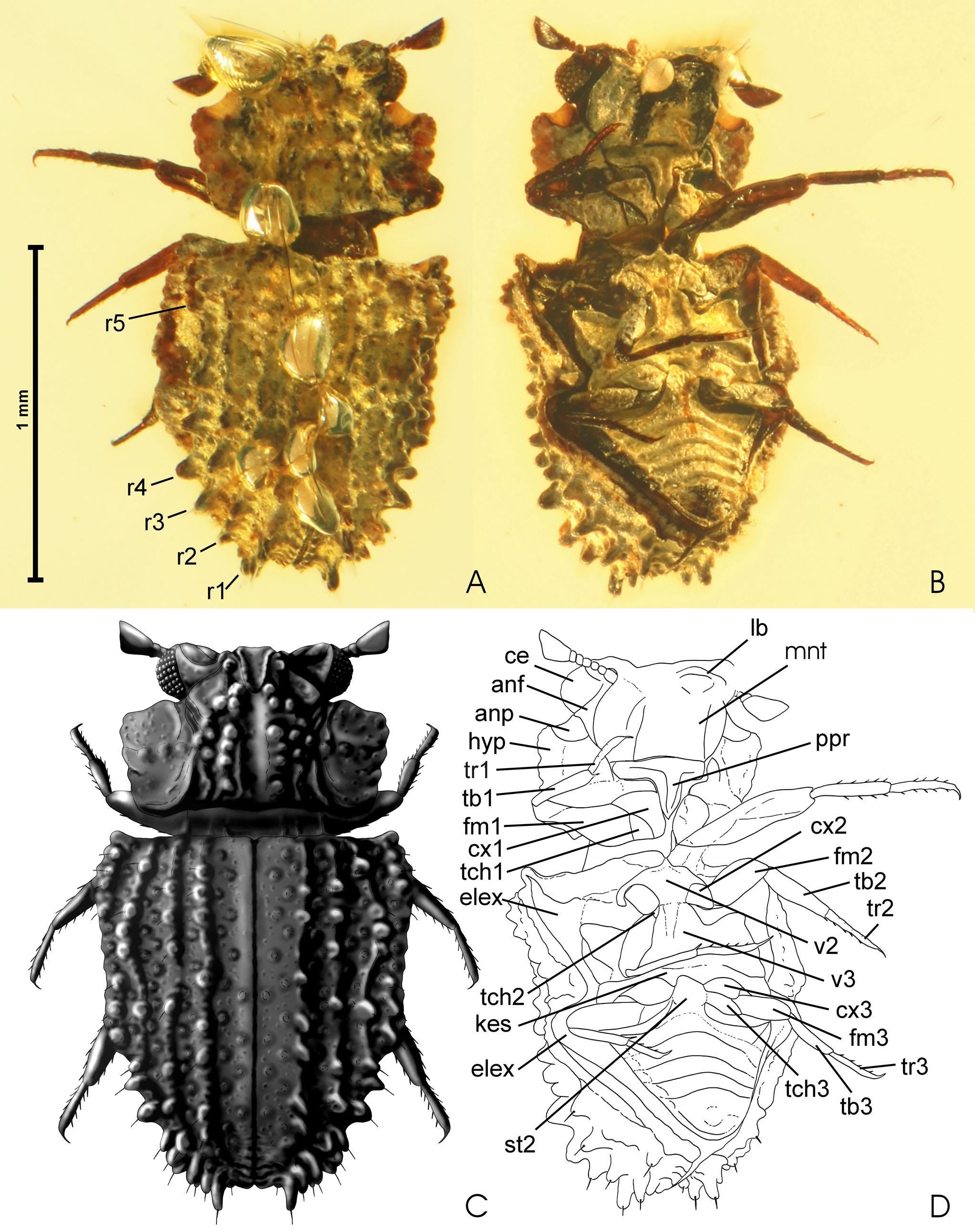
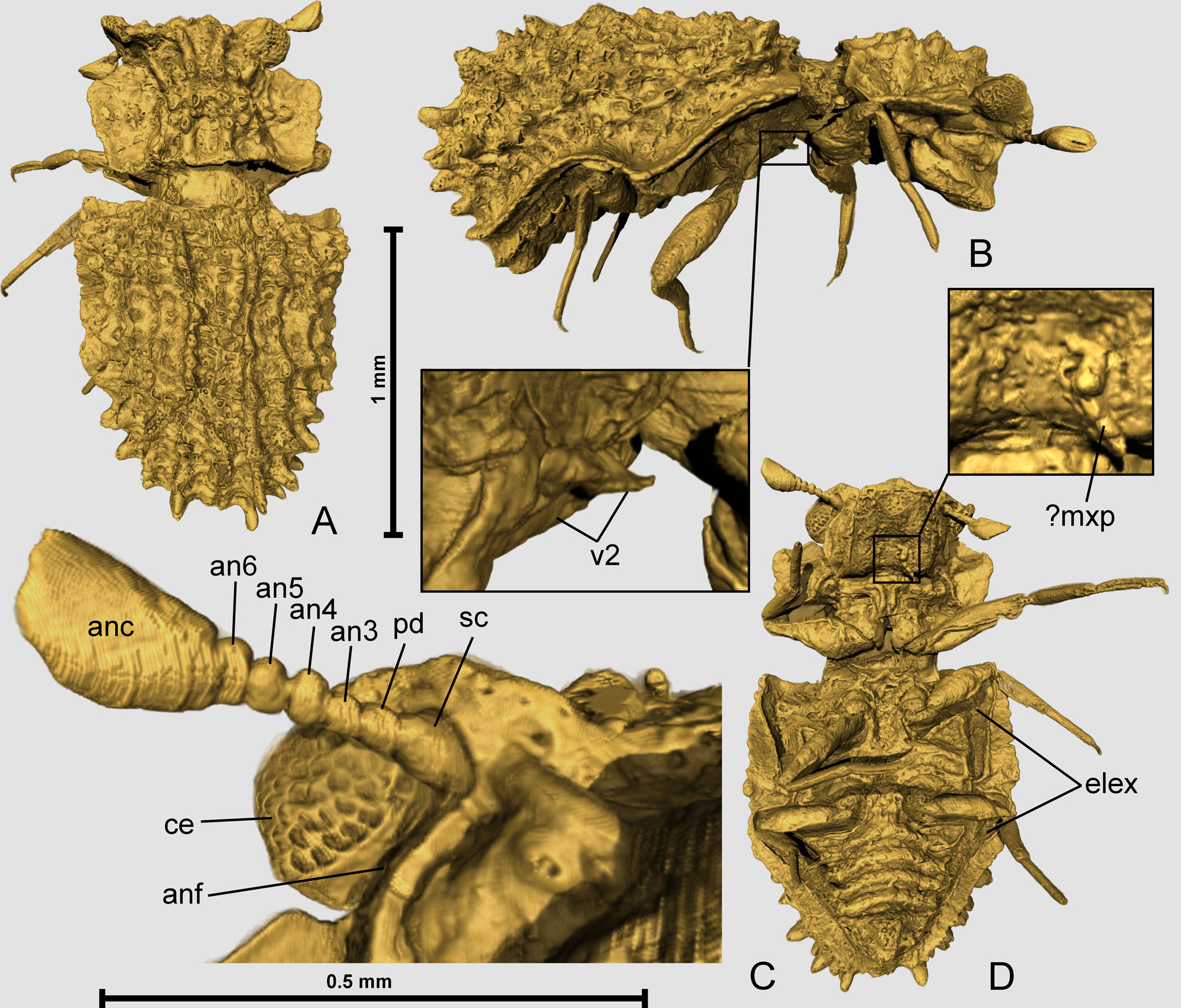
Description. Body ( Figs 1A–C View Figure 1 , 2A, B View Figure 2 , 3A View Figure 3 ) stout, compact (prothorax widely separated from posterior body as a post-mortem artefact) and moderately convex. Cuticle strongly sclerotized and conspicuously sculptured on dorsal side, sparsely setose.
Head large in relation to body, slightly inclined, only partly visible in dorsal view but largely exposed ventrally. Posterior-most part retracted into prothorax, constricted ‘neck’ region apparently absent. Dorsal side with distinct protuberances. Compound eyes ( Figs 1D View Figure 1 , 2C View Figure 2 , 3C View Figure 3 ) conspicuously large and protruding, composed of numerous small ommatidia with distinctly convex cornea lenses. Antennal furrow ( Figs 1D View Figure 1 , 2C View Figure 2 , 3C View Figure 3 ) present, sharply demarcated, as narrow as proximal flagellomeres and running along mesoventral margin of each eye. Clypeus, labrum and paired mouthparts poorly visible or not observable, except for elongated tetramerous structure that may be a posteriorly bent maxillary palp ( Fig. 2D View Figure 2 ). Mentum ( Fig. 1D View Figure 1 ) large and plate-like, almost completely covering ventral side of head.
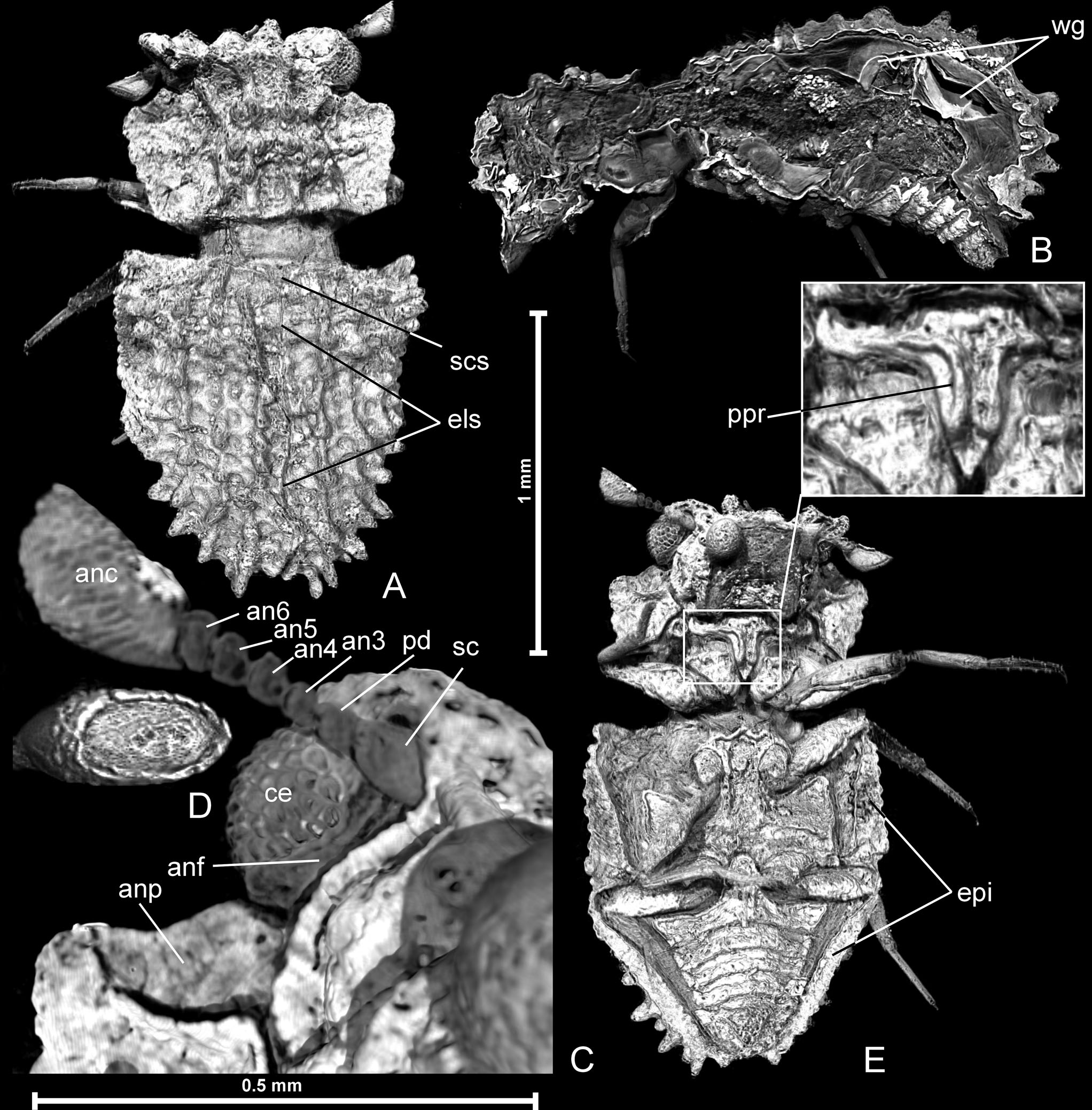
Antennae inserted in small lateroventrally open cavities; heptamerous, with broad basal part of scapus ( Figs 2C View Figure 2 , 3C View Figure 3 ) and narrower distal portion; pedicellus ( Figs 2C View Figure 2 , 3C View Figure 3 ) sub-cylindrical; antennomeres 3–6 indistinctly widening; apical antennomere 7 forming very large club ( Figs 2C View Figure 2 , 3C View Figure 3 ), broadening distad and obliquely truncated at apex; distal surface ( Fig. 3 View Figure 3 ) oval, with distinct circumferential rim.
Pronotum sub-trapezoidal, strongly transverse; anterior and posterior corners well-marked but blunt, obtuse-angled; lateral margins irregularly undulate. Anteroventral surface behind each eye with antennal pocket ( Figs 1D View Figure 1 , 3C View Figure 3 ) to accommodate antennal club. Large median area of pronotal disc strongly convex and bearing longitudinal rows of large tubercles, except for longitudinal and impressed median region; lateral portions of pronotal disc abruptly demarcated, slightly concave dorsally, laterally forming complete lateral edges.
Prosternum short and laterally distinctly demarcated from broad pronotal hypomera ( Fig. 1D View Figure 1 ); prosternal process ( Figs 1D View Figure 1 , 3E View Figure 3 ) sub-triangular and elongate, with lateral margins convergent posterad toward pointed apex, posterior process nearly reaching hind margins of procoxae, its ventral surface with pair of elongate lateral bulges which anteriorly continue along posterior prosternal margin, posterior portion of process behind bulges distinctly concave. Pleura and trochantin not discernible.
Mesonotum exposed between pronotum and elytra but details of scutum and main part of scutellum not recognizable, except for small triangular scutellar shield ( Fig. 3A View Figure 3 ) between mesal edges of elytral base. Mesoventrite ( Fig. 1D View Figure 1 ) sub-rhomboidal in shape, lacking visible discrimen and transverse suture; anterior margin sub-triangular with slightly sinuate sides, forming anteromedian process strongly projecting anterad ( Fig. 2B View Figure 2 ), which likely articulates with caudal concave portion of prosternal process when prothorax is in normal position (i.e. as in living beetles). Median area of mesoventrite between mesocoxae distinctly impressed. Mesotrochantin not recognizable.
Metanotum not visible. Metaventrite ( Fig. 1D View Figure 1 ) lacks externally visible discrimen (but visible on inner, dorsal surface of metaventrite in tomographic sections), with complete transverse suture, broad and short anteromedian intercoxal process overlapping with posterior mesoventrite. Median and lateral regions of metaventrite distinctly impressed, with anterior and lateral margins elevated. Katepisternum ( Fig. 1D View Figure 1 ) large, widening laterally, with two paramedian posterior (intercoxal) triangular processes separated by rounded median excavation.
Elytra ( Figs 1A–D View Figure 1 , 2A, B, D View Figure 2 , 3A, E View Figure 3 ) slightly broader than pronotum. Humeral angles strongly pronounced; elytral suture ( Fig. 3A View Figure 3 ) well-marked. Elytral disc with a pattern of strongly developed tubercles arranged in four complete rows ( Fig. 1A View Figure 1 ), and additionally with short row just laterad proximal portion of r4 ( Fig. 1A View Figure 1 ); continuous longitudinal costae absent. Elytra with complete lateral carina and broad epipleura ( Fig. 3E View Figure 3 ), each bearing two excavations ( Figs 1D View Figure 1 , 2D View Figure 2 ) for mesofemur (anterolaterally) and for metafemur (in median region). Elytra interlocked with metapleuron anterad metacoxae by conspicuous mesally directed process. Lateral outline of elytral edge ( Fig. 2B View Figure 2 ) strongly bisinuate, with submedian region forming broad sub-triangular ‘tooth’.
Hind wings not exposed, but well-visible in microtomographic sections, folded in sub-elytral space ( Fig. 3B View Figure 3 ).
Legs moderately long and robust; coxae ( Fig. 1D View Figure 1 ) flat, not elevated ventrad beyond ventral surface of body; trochanters ( Fig. 1D View Figure 1 ) sub-triangular, similar in size in all three pairs of legs; trochantero-femoral articulation oblique; femora ( Fig. 1D View Figure 1 ) flattened, only profemora laterally reaching beyond lateral body margins; tibiae ( Fig. 1D View Figure 1 ) sub-cylindrical, narrower than femora; tarsi ( Fig. 1D View Figure 1 ) only slightly shorter than tibiae, slender, sub-cylindrical, either indistinctly divided into four units (protarsus) or completely undivided (meso- and metatarsus), with rows of short sub-erect spines on anterior and posterior margins and each with one long and weakly curved claw.
Abdomen ( Figs 1B, D View Figure 1 , 2D View Figure 2 , 3B, E View Figure 3 ) with seven visible sternites, lacking plastron. Sternite II forms broad median process between metacoxae, anteriorly fitting with katepisternal concavity; narrowed behind posterior metacoxal margin but widening laterad. Border between sternites II and III poorly visible medially but distinct at lateral edges. Posterior sternites form distinct bulges along their posterior edges and are gradually tapering towards rounded abdominal apex; median protuberance of terminal sternite not discernible.
Derivation of name. Variation of Lepicerus . Gender masculine.
Stratigraphic range. Late Albian–early Cenomanian.
Geographical range. Northern Myanmar, Kachin, Hukawng Valley.
Remarks. The new genus is unambiguously placed in Lepiceridae on the basis of the following synapomorphies: body strongly sculptured; head large, macrophthalmous, with mouthparts partly internalized; antennae with reduced number of antennomeres, with the terminal one forming a very large undivided club; pronotum with distinctly marked anterior and posterior corners; head with antennal furrows and prothorax with antennal pockets; tarsomeres largely fused, with only indistinct septa visible on surface of protarsi; elytral epipleura with sinuate concavities to accommodate femora; elytra with longitudinal rows of convexities; prosternal process prominent and ‘U’- or ‘V’-shaped; mesoventral process anteriorly broadly sub-triangular and projecting; abdomen with seven visible sternites.
Apart from the unique characters listed in the diagnosis, the sole known species of † Lepiceratus differs from all extant and extinct Lepiceridae by rows of distinctly separated and very long tubercles, well-defined, especially on the posterior half of the elytra; this differs from other described species of the family, where the elytra have either continuous costae or rows of weakly elevated tubercles fused one to another and forming elongate bulges.
No known copyright restrictions apply. See Agosti, D., Egloff, W., 2009. Taxonomic information exchange and copyright: the Plazi approach. BMC Research Notes 2009, 2:53 for further explanation.