
Tetrastemma carneum, Hookabe & Oya & Tsuchida & Fujiwara & Ueshima, 2023
|
publication ID |
https://doi.org/ 10.12782/specdiv.28.199 |
|
publication LSID |
lsid:zoobank.org:pub:23C62CEF-53AF-4D0D-B151-8A5ED9E00372 |
|
persistent identifier |
https://treatment.plazi.org/id/EDD9D598-06CB-4590-9DE1-39B91EA2078A |
|
taxon LSID |
lsid:zoobank.org:act:EDD9D598-06CB-4590-9DE1-39B91EA2078A |
|
treatment provided by |
Felipe |
|
scientific name |
Tetrastemma carneum |
| status |
sp. nov. |
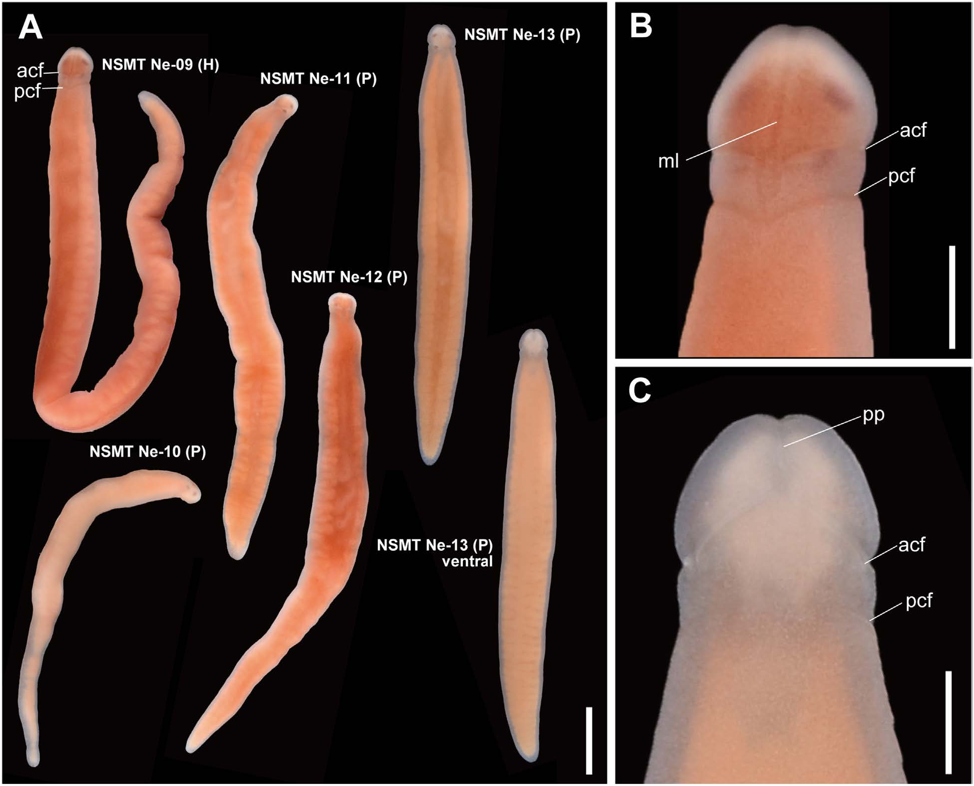
Tetrastemma carneum sp. nov. ( Figs 2 View Fig , 3 View Fig )
Tetrastemma sp. Ofunato: Hookabe et al. 2023: fig. 3, table 1.
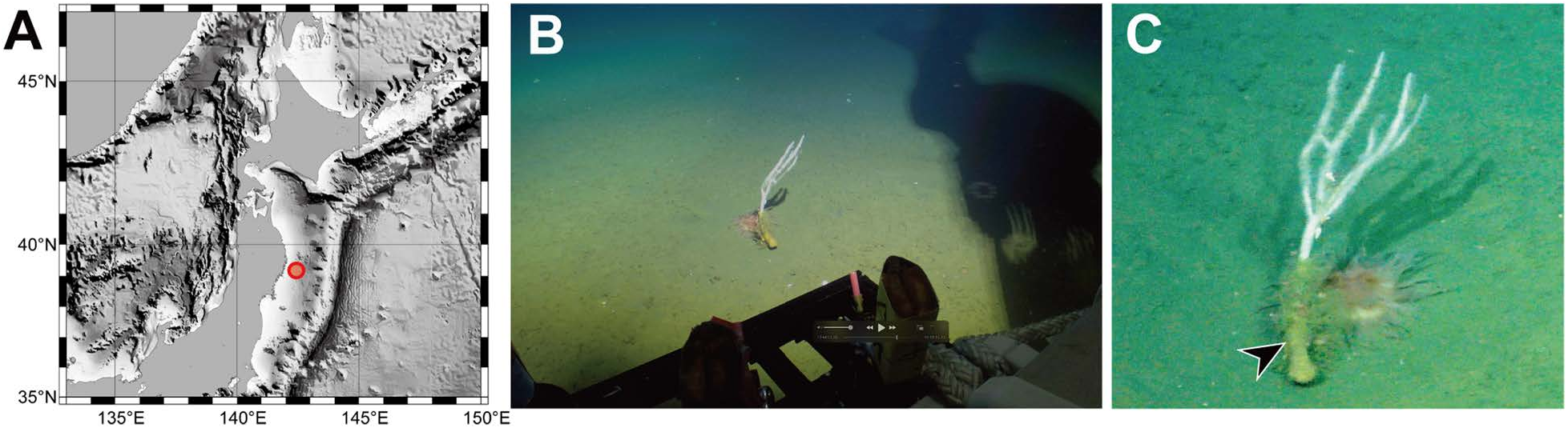
Material examined. Holotype: NSMT-Ne 09, unsectioned complete specimen except for the posterior tip, fixed in 10% formalin, posterior tip preserved in 99% ethanol, collected on 31 July 2019 by use of KM-ROV (dive #100) during KM19-05 C cruise of R / V Kaimei, at a depth of 398 m, off Ofunato (39°3.01′N, 142°9.06′E), Iwate, Japan, NW Pacific ( Fig. 1A View Fig ) GoogleMaps . Paratypes: NSMT-Ne 10–13, unsectioned complete specimen except for the posterior tip, fixed in 10% formalin, posterior tip preserved in 99% ethanol, collected on the same date and locality as the holotype.
Description. External features. Head spatulate ( Fig. 2B View Fig ) and demarcated from body by posterior cephalic furrows ( Fig. 2A View Fig ); marginal region between anterior and posterior cephalic furrows constricted ( Fig. 2B, C View Fig ). After anesthetization, live specimens 11–25mm in body length and 1.1– 1.5 mm in body width; holotype 25 mm in body length and 1.5 mm in body maximum width. Background body color generally pale tinged with flesh red, semitransparent either dorsally or ventrally ( Fig. 2A View Fig ). Head with a mid-dorsal thin dark longitudinal line; head without a cephalic patch ( Fig. 2A View Fig ). A pair of cephalic furrows present; anterior pair without meeting mid-dorsally and ventrally curving anteriorly but not reaching to proboscis pore; posterior pair shallow V-shaped and meeting mid-dorsally ( Fig. 2B View Fig ) and slightly curving anteriorly on ventral surface ( Fig. 2C View Fig ). Cerebral ganglia and blood probably not red but hard to distinguish from flesh-colored internal organs. Alimentary canals visible flesh-colored organs through body wall ( Fig. 2A View Fig ). Proboscis pale colored, extending about 1/2 of the body length ( Fig. 2A View Fig ). Four large eyes present, regular in sizes (approximately 50 µm in maximum width) ( Fig. 2B View Fig ).
Internal morphology. Epithelium approximately 50 µm in thickness, containing red and yellow gland cells ( Fig. 3A, D View Fig ). Dermis up to 15–20 µm thick ( Fig. 3B, F View Fig ). Cephalic glands well developed; acidophilic glands in anterior region ( Fig. 3A, B View Fig ) posteriorly replaced with basophilic glands ( Fig. 3C View Fig ). Cephalic blood lacuna extending beneath rhynchodaeum and posteriorly bifurcated ( Fig. 3B View Fig ). Stomach without a ventral blind outgrowth; stomach wall with ciliated cells, red acidophilic cells and blue-stained basophilic cells ( Fig. 3C, F View Fig ). Intestinal caecum anteriorly branched ( Fig. 3G View Fig ).
Proboscis pore slit-like, ventrally opening ( Fig. 2C View Fig ). Rhynchocoel long, extending about 5/6 of body length, nearly reaching to the posterior end of body ( Fig. 3H View Fig ). Rhynchocoel wall two-layered, with outer circular and inner longitudinal muscle fibers ( Fig. 3D–F View Fig ). Proboscis with outer circular, middle longitudinal, and inner circular muscle layers; proboscis epithelium with developed papillae containing red acidophilic and blue basophilic gland cells ( Fig. 3E View Fig ); 12 proboscis nerves present ( Fig. 3E View Fig ). Stylet apparatus with two accessory stylet pouches; at least two accessory stylets observed in serial histological sections ( Fig. 3I View Fig ).
A single frontal organ present ( Fig. 3A View Fig ). Cerebral organ laterally opening ( Fig. 3C View Fig ), posteriorly running without branching. Dorsal cerebral ganglia with glomerular structures ( Fig. 3D View Fig ). Accessory nerve not detected in lateral nerve cords.
Etymology. The species name is derived from the Latin carneum (flesh-colored), referring to the body coloration of the new species.
Type locality and distribution. The species is only known from the type locality, off Ofunato , Iwate Prefecture, four large eyes (up to 50 µm in the widths) and pale-yellow to flesh-colored body . However , the present species is distinguished from the latter two species by the presence of a narrow mid-dorsal longitudinal line on its head . Furthermore , the new species is well distinguished from T . appendiculatum by the presence of 12 proboscis nerves while T. appendiculatum has 10 ( Chernyshev 1998) as in most of Tetrastemma species. Tetrastemma appendiculatum and T. carneum sp. nov. share similarities not only in external morphology but also in their habitats within demosponges; T. appendiculatum was described based on two specimens collected from demosponge A. digitatus at depths of 40–60 m ( Chernyshev 1998).
An interspecific genetic distance between T. carneum sp. nov. and Tetrastemma sp. IP Iturup, calculated as 6.0% in uncorrected p -distance, exceeds a threshold commonly used for species distinction in Nemertea (e.g., Sundberg et al. 2016; Hookabe et al. 2022).

| R |
Departamento de Geologia, Universidad de Chile |
| V |
Royal British Columbia Museum - Herbarium |
No known copyright restrictions apply. See Agosti, D., Egloff, W., 2009. Taxonomic information exchange and copyright: the Plazi approach. BMC Research Notes 2009, 2:53 for further explanation.
|
Kingdom |
|
|
Phylum |
|
|
Class |
|
|
Order |
|
|
Family |
|
|
Genus |