
CROCODYLIDAE, Cuvier, 1807
|
publication ID |
https://doi.org/ 10.5281/zenodo.5374025 |
|
persistent identifier |
https://treatment.plazi.org/id/03E85855-FFDD-3217-8C64-FB94FC7AAB8E |
|
treatment provided by |
Marcus |
|
scientific name |
CROCODYLIDAE |
| status |
|
CROCODYLIDAE indet. 1
MATERIAL EXAMINED. — BRSUG 27360 and MNHN.LBE.311.
DESCRIPTION
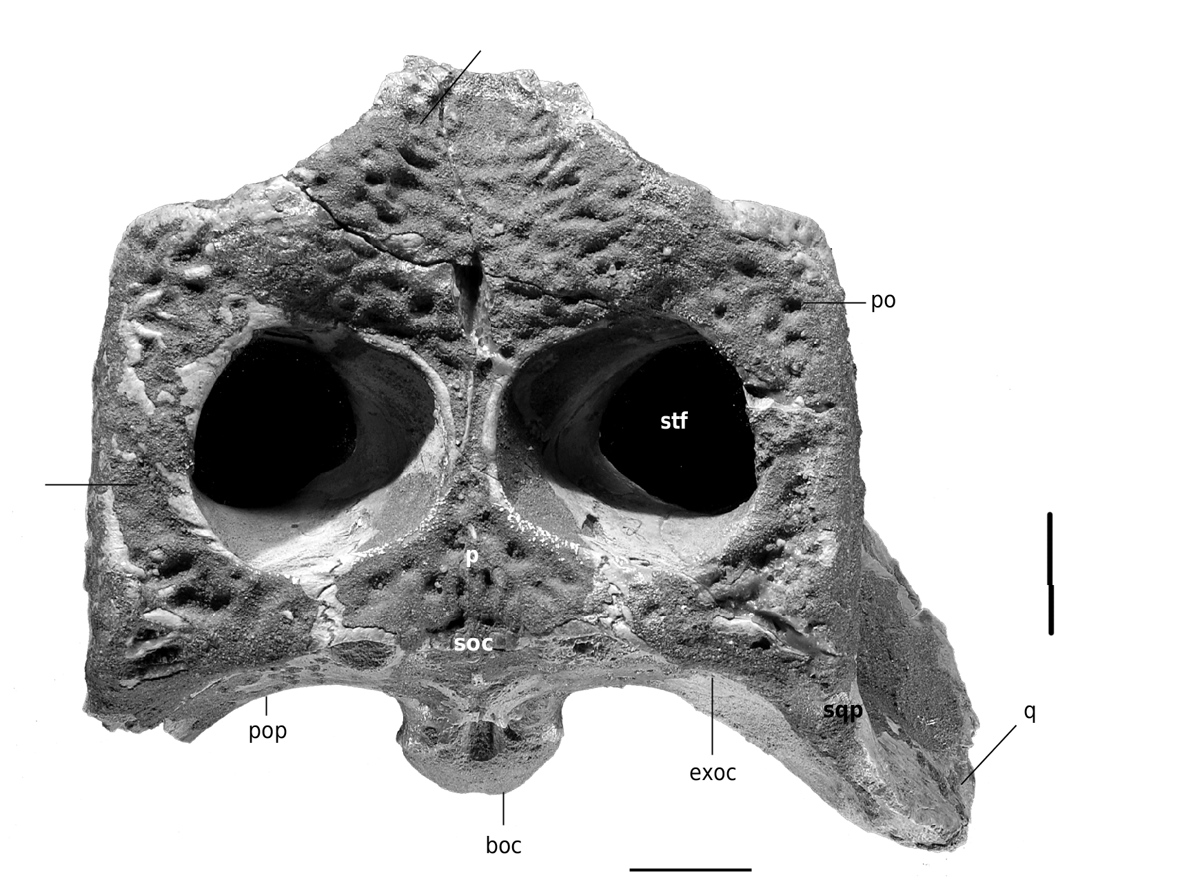
Skull table ( Fig. 3 View FIG )
The skull table is rectangular in shape, wider than long. Its surface is transversely concave and its ornamentation consists of small irregular pits. The posterior margin of the skull table is almost linear and horizontal. Its lateral margins are parallel to subparallel, their anterior (postorbital) margins deflect medially toward the orbits. The bars formed by the postorbitals and squamosals are narrow. The plate between the supratemporal fossae is so narrow that the ornamentation on its surface is formed by a low longitudinal ridge. This ridge runs backward to the posterior margin of the skull table over the dorsal surface of the parietal and supraoccipital. The dermal bones of the skull table do not overhang the borders of the supratemporal fossae.
The supratemporal fenestrae are wide and subrectangular in shape. The orbito-temporal foramen, situated in the posterior wall of the supratemporal fenestra, is large and is bordered by the squamosal, quadrate and parietal.
The postorbital constitutes about half the length of the lateral border of the supratemporal fenestra, and extends posteriorly about half the length of the lateral borders of the skull table. Its anterolateral corner is rounded and overhangs the base of the postorbital bar. The postorbital bar is partially preserved. There is a large nutrient foramen on its base.
The squamosal is also large. Its dorsal surface has a depression on its posterolateral corner separating it from the large posterolateral squamosal projection. On its lateral aspect, the squamosal forms the dorsal wall of the otic meatus. It has a shallow longitudinal groove for the attachment of the external ear valve musculature whose borders remain parallel before flaring anteriorly toward the postorbital. In lateral view, the postorbitalsquamosal suture is oriented ventrally and it does not pass medially ventral to the skull table. The squamosal-quadrate suture extends dorsally along the posterior margin of the external auditory meatus.
The parietal is posteriorly wide. It narrows at the interfenestral space and its anteriormost portion has a short length. Only the posterior part of the frontal is preserved. It is excluded from the anterior border of the supratemporal fossa by the parietal and the postorbitals. The fronto-parietal suture is concavo-convex, with the convex side toward the parietal. The dorsal surface of the posterior part of the frontal is concave, especially in the interorbital plate, which is wider than the interfenestral plate. Only the posterior and posteromedial borders of the orbits are preserved. They were larger than the supratemporal fenestrae and directed upward and slightly outward.
f
sq
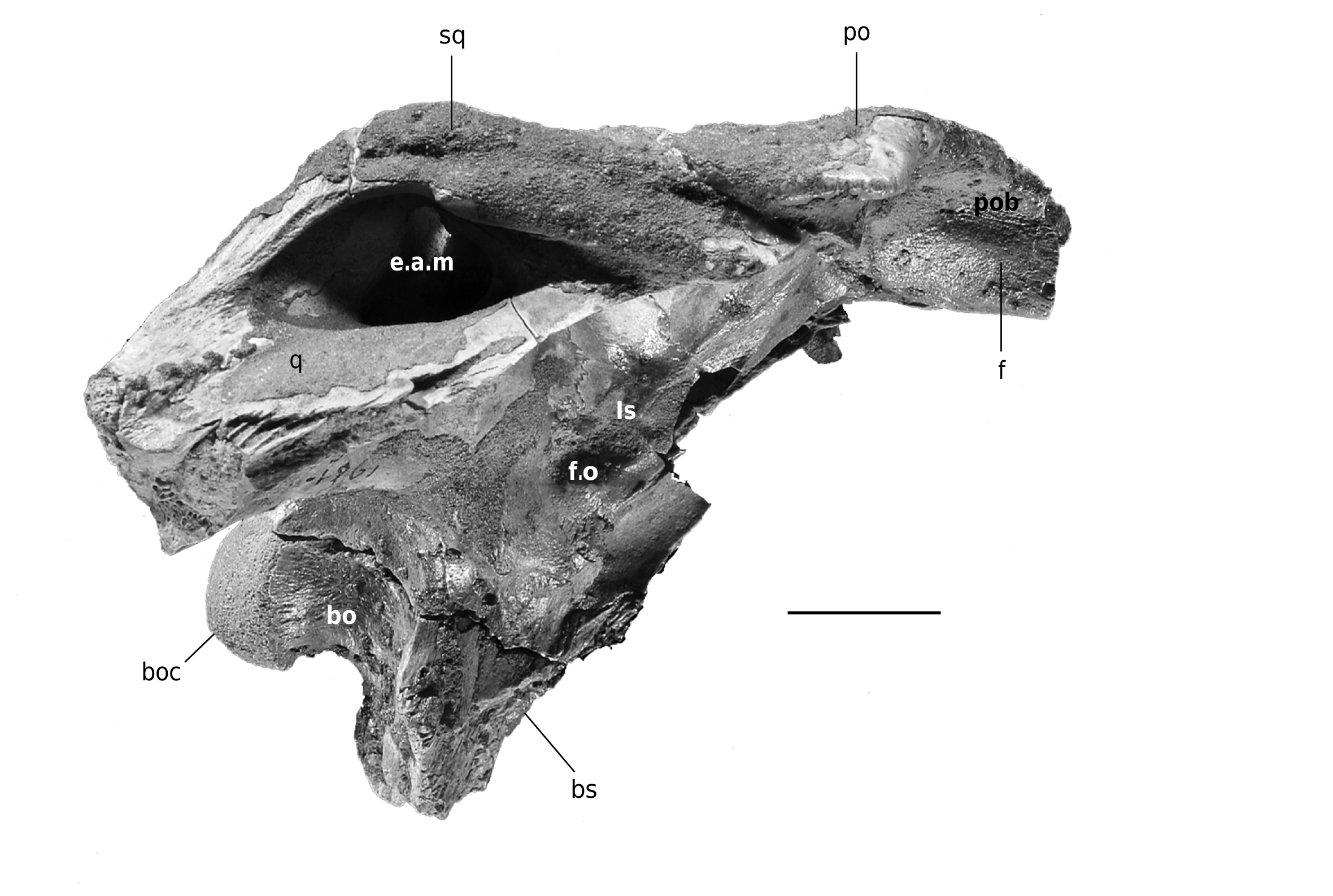
Quadrate region ( Figs 4 View FIG ; 6 View FIG )
The articular surface of the quadrate is badly eroded but it seems to have been divided in two hemicondyles (medial and lateral). The quadrate forms the floor and much of the anterior and posterior walls of the external auditory meatus, where it contacts the squamosal. The posterior margin of the otic aperture is invaginate. There is a foramen preoticum just in front of the external otic aperture. The distal end of the quadratojugal is also preserved. It abuts the lateral border of the quadrate and reaches backward just to the level of the lateral articular hemicondyle. Its lateral surface seems to have been sparsely sculptured.
The quadratojugal did not extend dorsally to contact the squamosal and the quadrate that formed the dorsocaudal border of the infratem- poral fenestra. The quadrate contacted the postorbital at the medial aspect of the base of the postorbital bar. The foramen aereum cannot be observed; however, a shallow groove is visible on its usual position on the dorsomedial surface of the quadrate. On its ventral surface, the quadrate presents a longitudinal crest running for about three fourths of its length and reaching its distal end, near which it thickens. The quadrate abuts the lateral wall of the braincase and shows a sharp ridge near the basisphenoid.
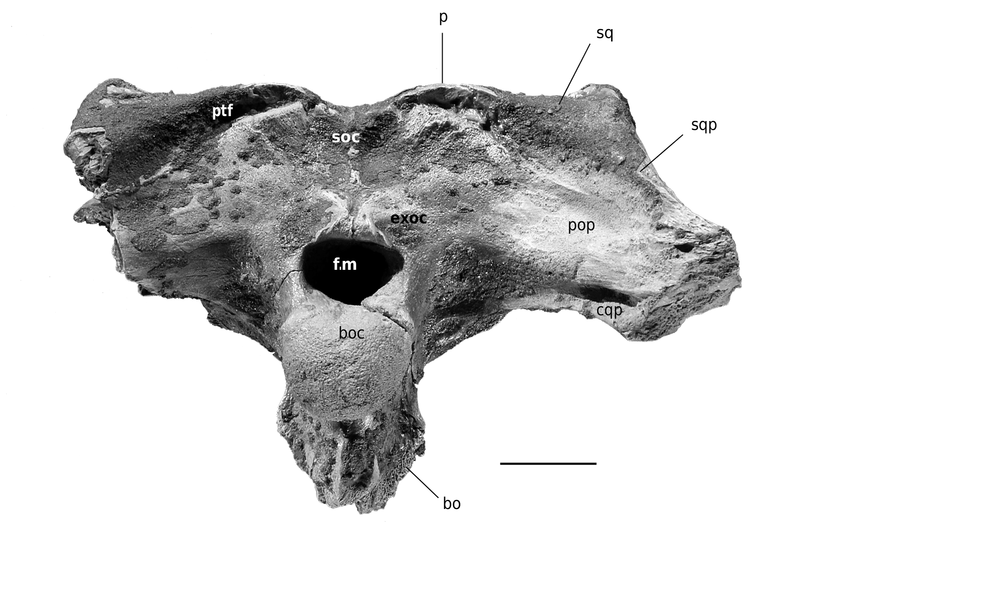
Occipital region ( Fig. 5 View FIG )
The occipital surface is slightly inclined backward, but not to the degree seen in gavialids. Dorsal to the foramen magnum, its posterior surface is vertical. The exoccipital borders of the pob foramen magnum and the basioccipital are visible in dorsal view.
The supraoccipital is large and subtriangular in shape, being far wider than high. Its dorsal bor- der is clearly concave. The supraoccipital extends downward to about two thirds the distance to the dorsal border of the foramen magnum, from which it is excluded by the exoccipitals. The supraoccipital has a low median vertical crest accompanied by a shallow depression on each side of it. Toward both of its dorsolateral ends, the supraoccipital shows the two large tuberosities of the ventromedial borders of the postemporal fenestrae. The postemporal fenestrae are bordered by the squamosals, the supraoccipital, the exoccipitals and the parietal bone. In dorsal view, the supraoccipital is triangular in outline, being approximately as long as wide, and it occupies the central quarter of the posterior margin of the skull table.
The squamosals also participate in the occiput. The squamosals form the posterolateral projections over the paraoccipital processes of the exoccipitals, although these projections do not reach their distal extremities. The occipital view of the squamosal extends along its contact with the exoccipital to the ventrolateral corner of the supratemporal fenestra.
The exoccipitals have long paraoccipital processes that are devoid of sculpture on their surface and extend for a long distance lateral to the entrance of the cranio-quadrate passage. The foramen magnum is elliptical in shape, wider than tall. The exoccipitals form the dorsal and lateral borders of that foramen. The ventral processes of the exoccipitals are short and cover only the dorsalmost part of the lateral borders of the exoccipital. The hypoglossal foramen is small and is placed dorsomedially to the foramen vagus. It is oval-shaped with acute ends and very deep. The foramen caroticum posterious is also oval-shaped and deep and it is situated immediately ventral to the former and over the suture with the quadrate.
The basioccipital forms the basioccipital condyle and the ventral border of the foramen magnum. The basioccipital condyle is subcircular in outline. The basioccipital shelf is tall and does not form the prominent tuberosities seen in gavialoids. It presents a prominent vertical median crest. The lateral eustachian canals opened dorsolaterally to the medial one. They are placed at the level of the lowest point of the ventral process of each exoccipital. The median eustachian canal opened in the ventral aspect of the basisphenoid, just anterior to the suture with the basioccipital.
Braincase ( Figs 4 View FIG ; 6 View FIG )
Its walls are formed by the parietal, the laterosphenoids, the quadrates, the exoccipitals, the prootics, the basisphenoid and the basioccipital. The foramen ovale is wide and its dorsal and posterior borders extend posterodorsally as a shallow recess over the posterior margin of the laterosphenoid and the anterior margin of the quadrate. The laterosphenoids are relatively well preserved. They form the anterior border of the foramina ovale. Their capitate processes are oriented anteroposteriorly toward the middline and they do not seem to have been very long. The lateral wing-like process of the laterosphenoid contacts the base of the postorbital bar medially and its anterior border is slightly constricted.
The basisphenoid is incompletely preserved, lacking the basisphenoid rostrum. Only a very small part of the pterygoids is preserved, covering the lateral surfaces of the basisphenoid. The caudal part of the basisphenoid placed between the posterior border of the pterygoids and the anteri- or border of the basioccipital is thick and not reduced to a thin lamina, but it is not extensively exposed on the braincase wall anterior to the median eustachian foramen. The basisphenoid is not broadly exposed ventral to the basioccipital. The quadrate-pterygoid suture is linear from the basisphenoid to the foramen ovale and there is not significant ventral quadrate process on the lateral wall of the braincase.
CROCODYLIDAE indet. 2
MATERIAL EXAMINED. — MNHN.LBE.306, MNHN.LBE.307 and MNHN.LBE.308.
DESCRIPTION
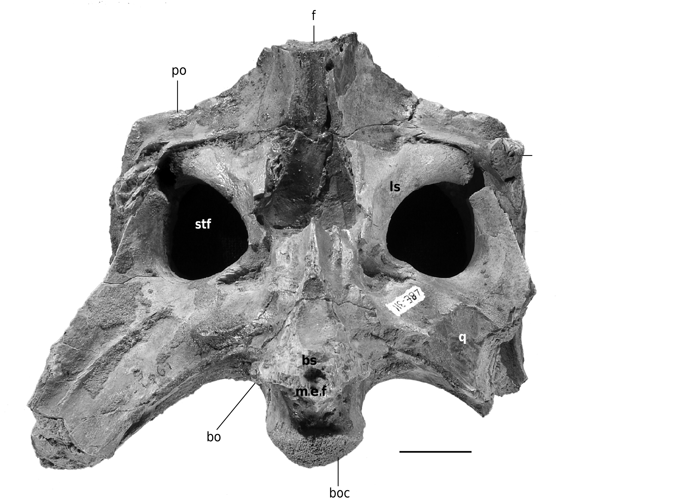
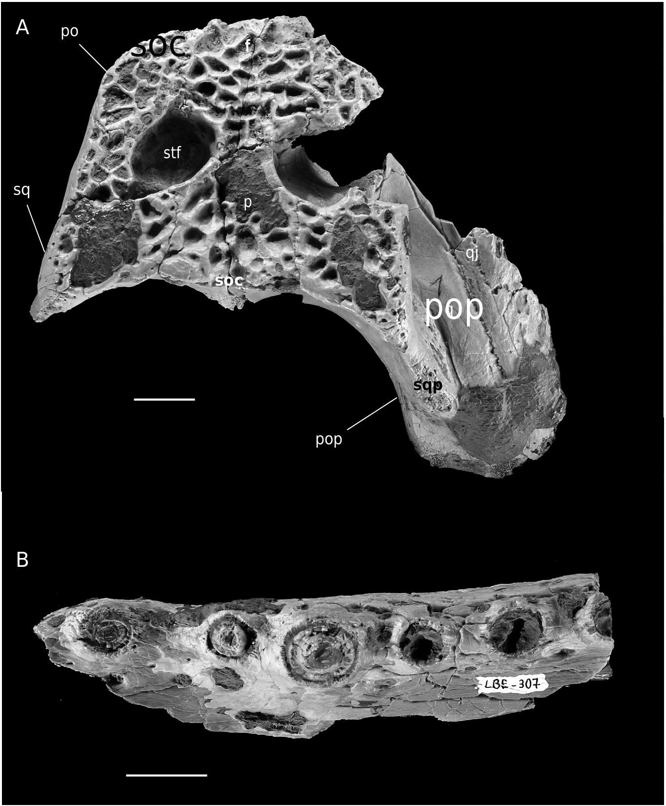
Skull table and associated regions ( Fig. 7A View FIG )
The skull table is almost as wide as long. Its lateral borders converge anteriorly. The surface of the skull table is nearly flat and sculptured by numerous wide but shallow pits that are ovate in shape. The supratemporal fenestrae are small and suboval. Their lateral margins are clearly longer than the medial ones. The interfenestral plate and the lateral borders of the skull table are broad. The squamosals form about two thirds of the length of the lateral borders of the skull table, the postorbitals making up the other third. The anterolateral margin of the postorbital is broadly rounded. The frontal is excluded from the anteri- or border of the supratemporal fenestrae by the parietal and the postorbitals. The fronto-parietal suture is concavo-convex, with the convexity facing toward the parietal. The frontal is slightly depressed with respect to the postorbitals.
The posterior margin of the skull table is slightly concave. The parietal and the supraoccipital make up its central half and the squamosals make up the other. The posterior part of the parietal is broad and almost quadrangular in outline. The exposure of the supraoccipital on the dorsal surface of the skull table is reduced. In occipital view, the supraoccipital is large and subtriangular in outline. The occipital surface is planar and fully verticalized. Only the right quadrate region is preserved. The articular surface of the quadrate is differentiated into a medial and a lateral hemicondyle. The foramen aereum is close to the median margin of the quadrate. The posterior margin of the external otic meatus is invaginate. There is a foramen preoticum in front of the external auditory aperture. The posterior part of the quadratojugal is also preserved. It reaches backward to the level of the articular surface of the quadrate but it does not participate in that surface. Its lateral surface is ornamented with large and shallow pits, although the ornamentation has somewhat been eroded.
Lower jaw ( Fig. 7B View FIG )
Only the right dentary and splenial are fragmentary preserved. The mandibular ramus is robust. The total dentary tooth count cannot be determined with certainty. The dentary symphysis is short and its rear part is damaged, so its exact posterior extent cannot be determined, although it probably extended backward to the level of the fourth or fifth dentary tooth. It is unclear whether the splenial participated in the symphysis, although this seems unlikely. Although short, the dentary symphysis is clearly more elongated than that of R. lloydi or C. niloticus . The ventral border of the symphysis is nearly flat and horizontal, resembling that of R. lloydi . The anterior fragment of the lower jaw preserves six alveoli (second-seventh), all of which are subcircular in outline. The second alveolus was apart from the first one, which is not preserved. It is also clearly apart from the third, this being the greatest interalveolar distance. From the third alveolus backward, all the alveoli are clearly apart from each other and regularly spaced, including those from the fourth to the seventh. This is in sharp contrast with the condition observed in R. lloydi or C. niloticus , in which these alveoli are close together (in some individuals of C. niloticus these alveoli are slightly apart and there are pits for the reception of the first maxillary teeth slightly labially to them; there are no such pits in the present specimen).
The posterior fragment of the lower jaw preserves seven alveoli, most probably those from the ninth to the 15th. All are apart from each other but the distance is not too great in any case. They are circular to subcircular in outline and there are no great differences in size among them. The splenial forms the medial border of the last three alveoli, unlike in R. lloydi or C. niloticus . The lateral border of the lower jaw is strongly festooned. The fourth dentary alveolus occupies the highest point in the first convexity, whereas the 11th occupies that of the second. The ventrolateral surface of the dentary is sculptured with longitudinal grooves and small pits.
| MNHN |
Museum National d'Histoire Naturelle |
No known copyright restrictions apply. See Agosti, D., Egloff, W., 2009. Taxonomic information exchange and copyright: the Plazi approach. BMC Research Notes 2009, 2:53 for further explanation.
|
Kingdom |
|
|
Phylum |
|
|
Class |
|
|
Order |
|
|
Family |