
Oloncholaimus piipi, Mordukhovich & Zograf & Saulenko & Fadeeva, 2020
|
publication ID |
https://doi.org/ 10.11646/zootaxa.4802.3.10 |
|
publication LSID |
lsid:zoobank.org:pub:ADA3638F-A28E-4BB6-8D20-25FE42E51AC2 |
|
persistent identifier |
https://treatment.plazi.org/id/61C64D5C-B879-4F4F-9B51-DD1613941533 |
|
taxon LSID |
lsid:zoobank.org:act:61C64D5C-B879-4F4F-9B51-DD1613941533 |
|
treatment provided by |
Plazi |
|
scientific name |
Oloncholaimus piipi |
| status |
gen. et sp. nov. |
Oloncholaimus piipi View in CoL gen. et sp. nov.
( Figs 2–7 View FIGURE 2 View FIGURE 3 View FIGURE 4 View FIGURE 5 View FIGURE 6 View FIGURE 7 ; Table 1)
urn:lsid:zoobank.org:act:61C64D5C-B879-4F4F-9B51-DD1613941533
Diagnosis. Oloncholaimus . Body large, 5960–6820 μm long in males and 6497–7045 μm in females. Six outer labial and four cephalic setae equal in size (5–7 μm). Buccal cavity 40–45 μm long and 17–21 μm wide in males, 44–49 μm long and 20–23 μm wide in females. Excretory pore situated at a distance of 96–108 μm (males) and 101 μm (female) from cephalic apex. Spicules equal, 110–131 μm long, without gubernaculum. In front of cloacal open- ing one complex supplementary organ composed of 8–9 cylindrical processes. Tail conico-cylindrical, 131–143 μm long in males and 167–175 μm in females.
Type material. Whole mount specimens of holotype male and three paratype males ( MN BS 1178 Ol1), and three paratype females ( MN BS 1178 Ol2) are deposited in the Zoological Museum of Far Eastern Federal University , Vladivostok, Russia .
Other material. Au-coated SEM specimens.
Etymology. The species is named in reference to the area it was found.
Type locality. Sandy sediments at the South Summit of the Piip volcano in the Bering Sea (55.382° N, 167.261° E), outside the area with hydrothermal activity, water depth 470 m. GoogleMaps
Measurements. See Table 1.
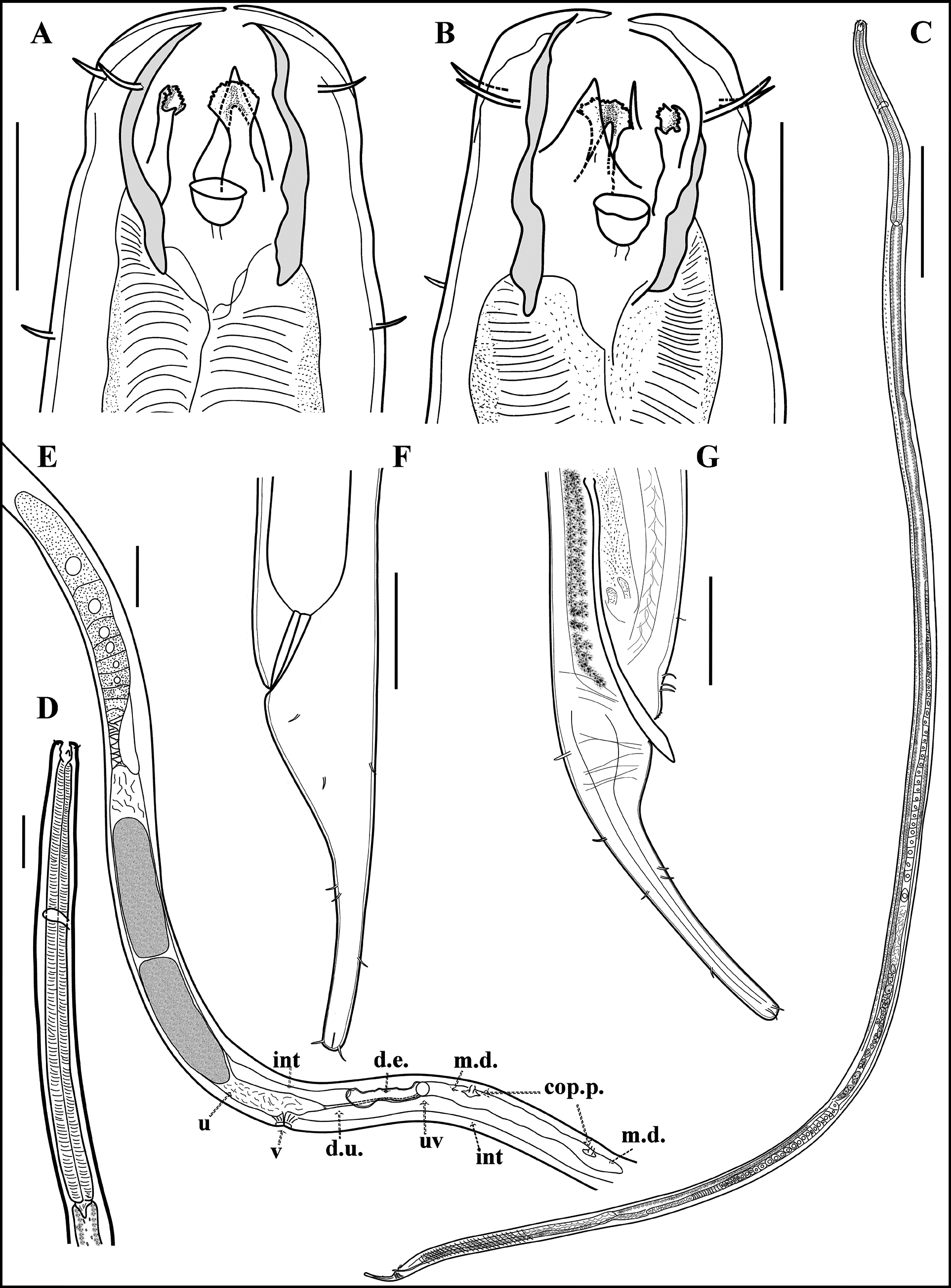
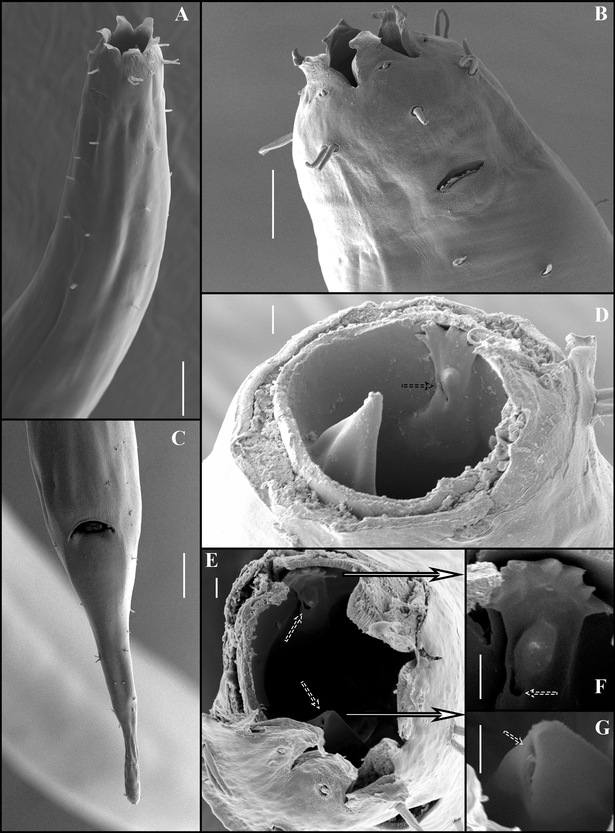
Description. Males: body length 5960–6820 μm; maximum diameter 78–91 μm. Cuticle looks smooth under optical microscope, but with fine transverse striation under SEM. Six triangular lips with rounded tips, each lip bearing a minute rounded inner labial papilla ( Fig. 3B View FIGURE 3 ). Six outer labial and four cephalic setae in one circle, equal in size (5–7 μm). Head diameter 35–36 μm. Large buccal cavity with sclerotized walls, 40–45 μm deep and 17–21 μm wide ( Fig. 4 View FIGURE 4 B–E). Three teeth present, left ventrosublateral typical oncholaimoid tooth is the largest (30–33 μm). Right ventrosublateral and dorsal teeth almost equal in size (24–27 μm) and have complex structure with onchium and api- cal antler-shaped extension (5–6 μm width) ( Figs. 3D View FIGURE 3 , E–G, 4B, C). Amphids pocket-like with elliptical openings, 11–14 μm wide (0.3–0.4 corresponding body diameter), and 7–11 μm in height, located 17–23 μm from anterior end ( Figs 3B View FIGURE 3 , 4A View FIGURE 4 ). The left ventrosublateral tooth is perforated by the subterminal outlet of the corresponding pharyngeal gland. In the right ventrosublateral and dorsal teeth the pore of the pharyngeal gland lies in the middle part of the tooth ( Figs 3D View FIGURE 3 , E–G, 4B, C, E).
Pharynx muscular and cylindrical, 0.1 times body length. Excretory pore opening at about 0.12–0.13 times pharynx length from anterior end (2.0–2.7 times buccal cavity length, 96–108 μm), nerve ring at 0.36–0.42 times pharynx length. Short somatic setae are sparsely distributed throughout the body, but mainly near both ends ( Fig. 3A View FIGURE 3 ). Glandular organs occur at irregular intervals, mainly in the lateral chords. Orthometanemes present. Tail conico-cylindrical, ventrally bent, 131–143 μm long (2.8–4.1 cloacal body diameter) ( Fig. 4F View FIGURE 4 ). Two pairs of short ventral and one pair of dorsal setae in the middle of tail. In addition, about 3 scattered subdorsal caudal setae. Tail tip slightly swollen and rounded with four small terminal setae ( Fig. 4I View FIGURE 4 ). One circle of circumcloacal setae, 7–8 pairs. In front of cloacal opening one complex supplementary organ composed of 8–9 cylindrical processes ( Fig. 4G, H View FIGURE 4 ).
Genital musculature represents a long series of preanal and postanal dorsoventral copulatory muscle bundles ( Fig. 5G View FIGURE 5 ). Two opposed testes to the right side of intestine. Anterior one 1457–2003 μm long, posterior one 1442– 1687 μm. Three zones can be distinguished along the testis: germinal zone, growth zone, and ripening zone. The germinal zone is filled with small, tightly packed cells near the blind end of the testes. The growth zone is composed of very big cells, each with a large nucleus, arranged in a single row ( Fig. 5A, D View FIGURE 5 ). The ripening zone is filled with smaller cells containing condensed chromatin and arranged in two rows ( Fig. 5A View FIGURE 5 ). Both testes join at the seminal vesicle that contains a number of broad oval spermatozoa ( Fig. 5B View FIGURE 5 ) and that further leads to the vas deferens The latter is set off from ductus ejaculatorius by a sphincter about 805–1143 μm anterior to cloaca ( Fig. 5C View FIGURE 5 ). This ductus joins the rectum from the ventral side. The whole vas deferens is situated on the right and ventral side of the intestine. Spicules equal, 110–131 μm (2.6–3.7 cloacal body diameter), slightly ventrally curved, with some striation in the middle part, proximally slightly cephalate ( Fig. 5F View FIGURE 5 ).
Females: very similar to males but differ in several dimensions and sexual characteristics. Body length 6497– 7045 μm, maximum body diameter 84–110 μm. Tail somewhat longer than in males, 167–175 μm long (3.6–3.7 anal body diameter). Only anterior reproductive branch is developed with reflexed ovary, lying to the right side of the intestine ( Fig. 6B View FIGURE 6 ). Two or three intra-uterine eggs in the paratypes, 183–249 μm long and 63–76 μm wide. Vulva at 80–84% of body length. Demanian system well-developed ( Fig. 6B, C View FIGURE 6 , E–H). Ductus uterinus posterior to uterus (102–196 μm long) connecting to the main duct through uvette. Ductus entericus anterior to uvette (about 135 μm long) connecting to dorsal side of intestine through osmosium. Main duct branched to the two long blind sacs pierced by the multiple terminal ducts ending in terminal pores. The edge of the sacs situated at a distance of 458–656 μm from uvetta and at 188–203 μm from the anus.
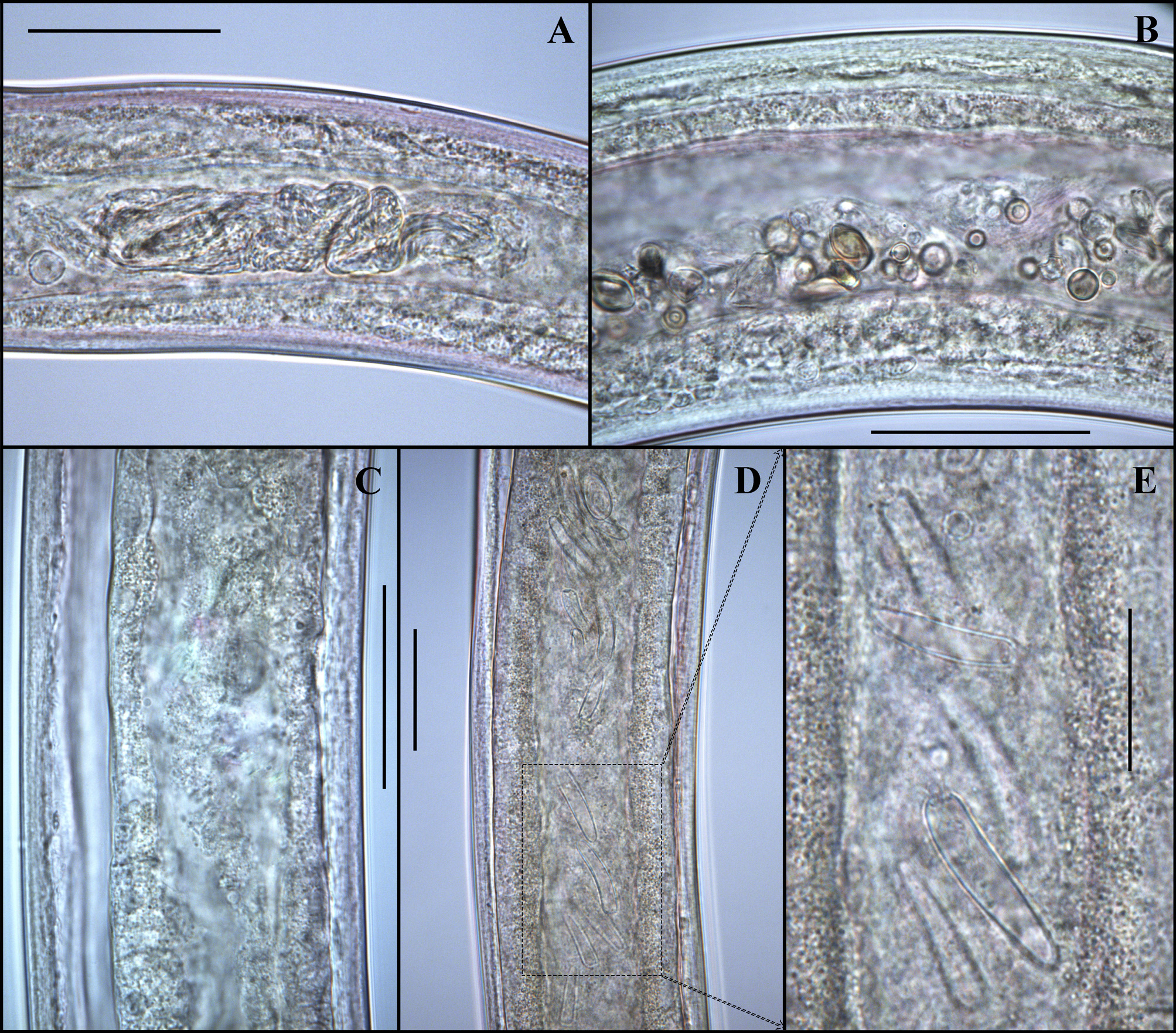
Gut content. About 80% of studied male and 50% of female specimens have empty intestine. The intestinal contents of other specimens consist of usually difficult to recognize material ( Fig. 7A, C View FIGURE 7 ) and in two females intestine contains material that looks like some protozoa shells ( Fig. 7B, D, E View FIGURE 7 ).


| MN |
Museu Nacional, Universidade Federal do Rio de Janeiro |
No known copyright restrictions apply. See Agosti, D., Egloff, W., 2009. Taxonomic information exchange and copyright: the Plazi approach. BMC Research Notes 2009, 2:53 for further explanation.
|
Kingdom |
|
|
Phylum |
|
|
Class |
|
|
Order |
|
|
Family |
|
|
Genus |