
Microlaimus paraglobiceps, Revkova, 2017
|
publication ID |
https://doi.org/ 10.11646/zootaxa.4344.2.13 |
|
publication LSID |
lsid:zoobank.org:pub:BF66D84F-4686-4305-8DB9-22F6D0632CA9 |
|
DOI |
https://doi.org/10.5281/zenodo.6042083 |
|
persistent identifier |
https://treatment.plazi.org/id/03E887A1-8913-641F-649E-FE6213A390F5 |
|
treatment provided by |
Plazi |
|
scientific name |
Microlaimus paraglobiceps |
| status |
sp. nov. |
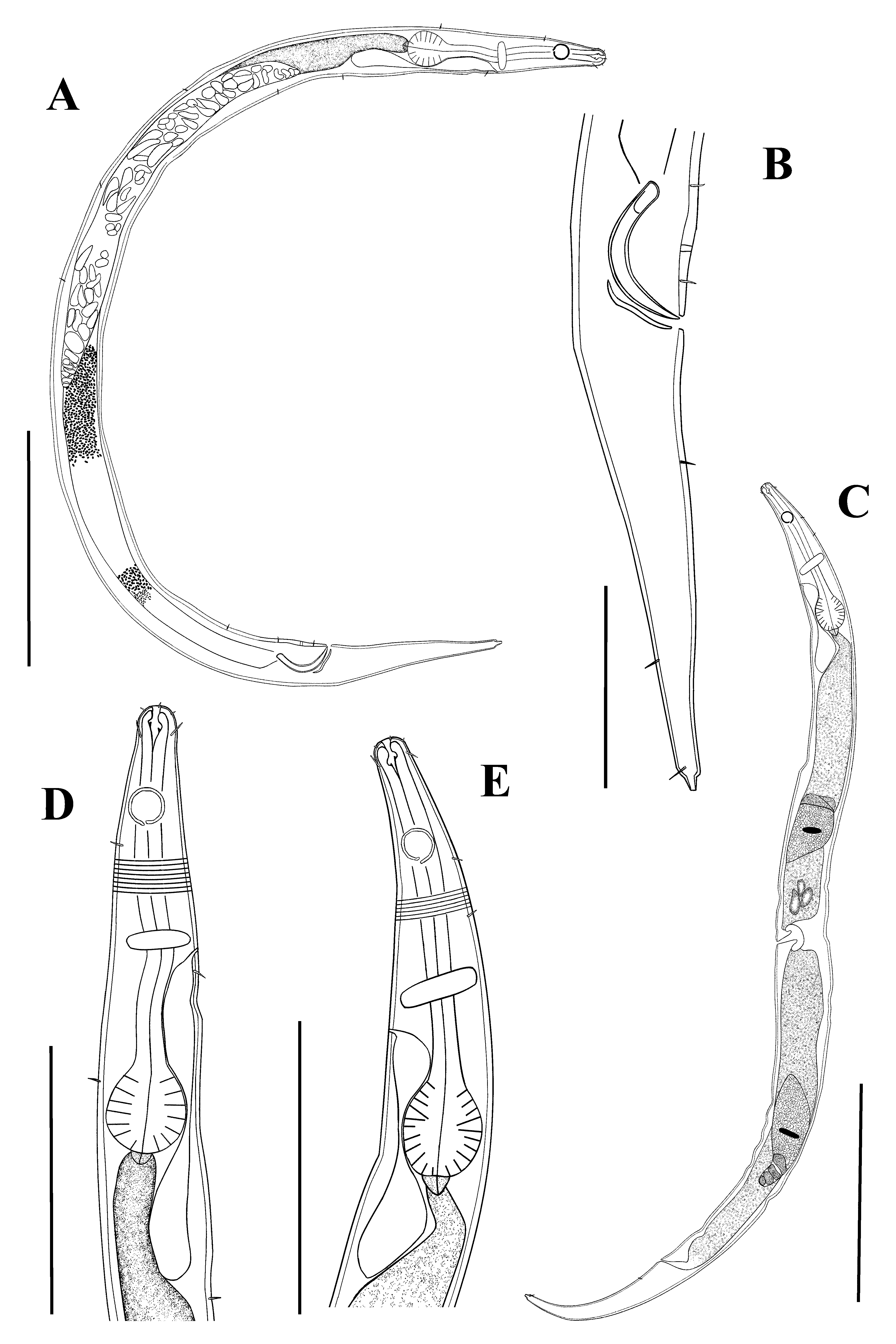
Microlaimus paraglobiceps sp. n.
Figure 2 View FIGURE 2 , Table 2
Material examined: two males, four females. Glycerin-gelatin slides. Holotype, male in slide # FlN/BS–10 and paratypes, male, FlN/BS–11; same for females, from FlN/BS–12 to FlN/BS–15.
Type locality: Black Sea, Crimea shelf, oxic/anoxic interface at the region of the submarine Dnieper Canyon, depth: 120 (0–5 cm horizontal layer), 130 (0–3 cm), 140 (0–3 cm), 150 (0–5 cm), 160 (0–3 cm), 170 (0–1, 2– 3 cm), 180 (0–1 cm), 190 (0–1 cm), 210 (0–1 cm), 240 m (0–2 cm). Depths 120–140 m—sandy silt, 150–240 m—silt and bacterial aggregations.
Etymology. The specific name paraglobiceps refers to the similarity to M. globiceps: Greek para = similar; globiceps = M. globiceps .
Description. Male Body short, slender. Cuticle finely annulated (annules 1 µm wide) all over body. Annulation often difficult to observe under the light microscope. Sensillae of cephalic end arranged in three circles: six inner labial papillae indistinct, six outer labial papillae 1 µm long and four cephalic setae 3 µm long. Somatic setae 2 µm long, scattered along the body. Head not set off from the rest of the body. Cryptocircular amphidial fovea 60̄66.7% of the corresponding body diameter with interruption in the posterior part, situated 13̄16 µm from the anterior extremity. Buccal cavity narrow, strongly sclerotized with small dorsal tooth and slightly below two smaller ventrosublateral teeth. Dorsal wall of the buccal cavity more sclerotized than the ventral wall. Pharynx cylindrical and muscular, posteriorly forming a pronounced spherical bulb, 14 µm in width. Nerve ring situated at level of middle of pharynx. Secretory-excretory system present; renette cell situated posterior to cardia. Secretoryexcretory pore situated between the nerve ring and the bulb, posterior to nerve ring. Cardia small.
Reproductive system diorchic; testes opposite and outstretched. Anterior testis situated to the right of intestine, posterior testis to the left of intestine. Posterior testis shorter (65 µm) than anterior (107 µm). Spicules arcuate, equal to 1.5̄1.7 of the corresponding body diameter in length. Gubernaculum simple, lying parallel to the spicules. One precloacal pore, difficult to discern under light microscope and one precloacal setae 2 µm long, located 5 µm from anterior of anus. Tail conical, 4.2̄4.3 of the corresponding body diameter, with short subventral setae present in post-cloacal regions. One caudal seta, 3 µm long, present on the terminal part of the tail. Caudal glands not visible. Spinneret present.
Female Similar to male in general characteristic. Amphidial fovea 6 µm in diameter (60̄64.5%). Reproductive system didelphic, amphidelphic, with outstretched ovaries. Anterior branch shorter (62 µm long) than posterior (112 µm). Vulval lips not sclerotized and not protruding outside the body contour, located at 43.3% from anterior end. Spermatozoa elongate and rounded, 3×12 µm in size visible in uterus. Vagina short, not thickened. The diameter of the large oocytes is 40 µm. Precloacal setae not observed.
Diagnosis. Body short (436̄504 µm) and thin. Cuticle finely annulated. Amphidial fovea cryptocircular with interruption in the posterior part. Head not set off from the rest of body. Sexual dimorphism in size of amphidial fovea absent. Female reproductive system didelphic-amphidelphic, ovaries outstretched; anterior branch shorter than posterior. Male reproductive system diorchic, outstretched and opposite; anterior testis longer than posterior. One precloacal pore and setae present. Tail in females is longer than in males (c’= 6.4̄6.8 cbd vs 7.7̄8 cbd).
Discussion. Microlaimus paraglobiceps sp. n. differs from most species of the genus in having short body and finely annulated cuticle. New species morphologically resembles M. globiceps de Man, 1880 as described by Gerlach (1950, 1953), Jensen (1979) and Turpeenniemi (1997) in the shape of the body, structure of the male sexual organs and presence of precloacal pore (described in Turpeenniemi, 1997). However, the new species differs from M. globiceps in the absence of sexual dimorphism in the size of amphidial fovea (6 µm (60̄66.7%) vs in female 3̄4 µm (30̄33%), in male 4̄5 µm (40̄42%) in M. globiceps ), head not set off from rest of body and width of cuticular annules (1 µm vs 2̄3 µm in M. globiceps ).
Microlaimus paraglobiceps sp. n. is also similar to M. naidinae Tchesunov, 1978 , but differs in having shorter body (436̄504 µm vs 578̄770 µm in M. naidinae ), the size of the amphidial fovea (6 µm (60̄66.7%) vs 4̄4.3 µm (33.3̄43%) in M. naidinae ), low value ‘c’ (6.4̄8 vs 9.1̄ 11.3 in M. naidinae ) and the distance from the cephalic edge to the anterior rim of the amphidial fovea (13̄16 µm vs 7.4 µm in females and 9 µm in males in M. naidinae ).
This work was carried out as a part of the state project of IMBR ffi 0828-2014-0014. I am grateful to Prof. N.G. Sergeeva for critical reading of the manuscript and valuable suggestions. Special thanks to our colleagues L.F. Lukyanova for processing of bottom sediments samples and K.A. Ivanova for sample collecting during cruise 72/2 of the R/V “Meteor”. Author is very grateful to anonymous reviewers for providing constructive criticisms and improving on the manuscript.
No known copyright restrictions apply. See Agosti, D., Egloff, W., 2009. Taxonomic information exchange and copyright: the Plazi approach. BMC Research Notes 2009, 2:53 for further explanation.