
Seiria mesophotica A.R. Sherwood, 2021
|
publication ID |
https://doi.org/ 10.11646/phytotaxa.524.1.2 |
|
DOI |
https://doi.org/10.5281/zenodo.5606532 |
|
persistent identifier |
https://treatment.plazi.org/id/03E96803-FFD3-E008-B1AB-A05CFE9AFE4F |
|
treatment provided by |
Plazi |
|
scientific name |
Seiria mesophotica A.R. Sherwood |
| status |
sp. nov. |
Seiria mesophotica A.R. Sherwood sp. nov. ( Fig. 4A–J View FIGURE 4 )
Type: — USA, Hawai‘i, off West Moloka‘i , 21.0455°N, 157.3524°W, 80 m depth, 28 November 2006, H. Spalding (holotype BISH 780873 View Materials ; ARS 09666; field code P4-189 -cont.8-#328) GoogleMaps .
Description: Thallus crustose, prostrate, on coral rubble, brick red to dark, brownish maroon in color ( Fig. 4A,B View FIGURE 4 ). Thallus moderately to heavily calcified, somewhat lobed or entire, mostly to completely adherent to substratum. Crusts thin, typically approximately 100 µm, ranging from 80–180 µm ( Fig. 4C–E View FIGURE 4 ). Hypothallus filaments consisting of parallel files of cells ( Fig. 4G View FIGURE 4 ), mostly unbranched, cells 4.6–8.8 µm in width and 18.2–27.7 µm in height, giving rise to perithallial assurgent filaments above and rhizoids below. Perithallial assurgent filaments arising at 60–90º angle from hypothallus ( Fig. 4C–E View FIGURE 4 ). Perithallus thin, composed of short filaments that are mostly 4–8 cells long ( Fig. 4C–E View FIGURE 4 ). Lower perithallial cells taller than broad, 14.5–24.8 µm in height × 5.9–8.0 µm in width ( Fig. 4D View FIGURE 4 ). Perithallial cells becoming shorter towards upper portion of perithallus. Mid-perithallial fusion cells absent. Uppermost perithallial cells quasi-isodiametric, 7.8–15.8 µm width × 5.0–9.3 µm height ( Fig. 4E View FIGURE 4 ). Cuticle on surface of perithallus 4–8 µm thick ( Fig. 4C–E View FIGURE 4 ). Rhizoids 5.4–8.8 µm diameter, mostly short to medium length, unbranched, and unicellular ( Fig. 4F View FIGURE 4 ). Hair cells absent ( Fig. 4H View FIGURE 4 ). Gametangial reproduction not observed. Mature tetrasporangia 43–99 × 22–44 µm, cruciate decussate to irregularly cruciate ( Fig. 4I View FIGURE 4 ), borne in raised, subcircular, scattered gelatinous nemathecia, nemathecial paraphyses up to 9 cells in length, 130 µm long ( Fig. 4J View FIGURE 4 ).
Other examined material:— USA, Hawai‘i: Papahānaumokuākea Marine National Monument, Manawai (Pearl and Hermes Atoll), 27.9625°N, 175.8361°W, 67 m depth, 31 July 2019, S. Matadobra ( BISH 780874 View Materials ; ARS 09967; field code NWHI-833) GoogleMaps .
Distribution and Habitat:— Known from the off West Moloka‘i in the Main Hawaiian Islands and Manawai of the Papahānaumokuākea Marine National Monument at mesophotic depths of 67- 80 m.
Etymology:— Named for the mesophotic depths at which the specimens were collected.
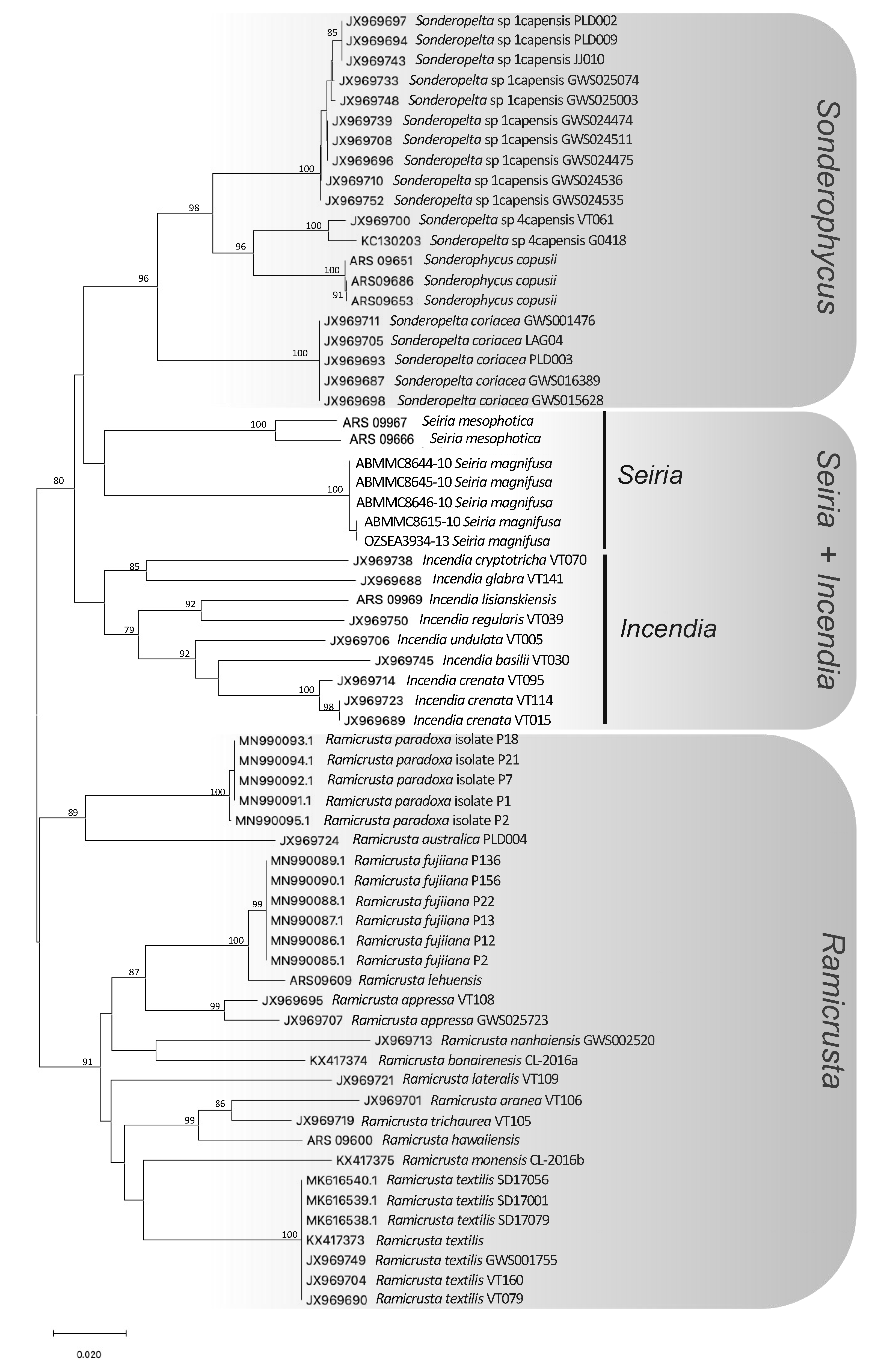
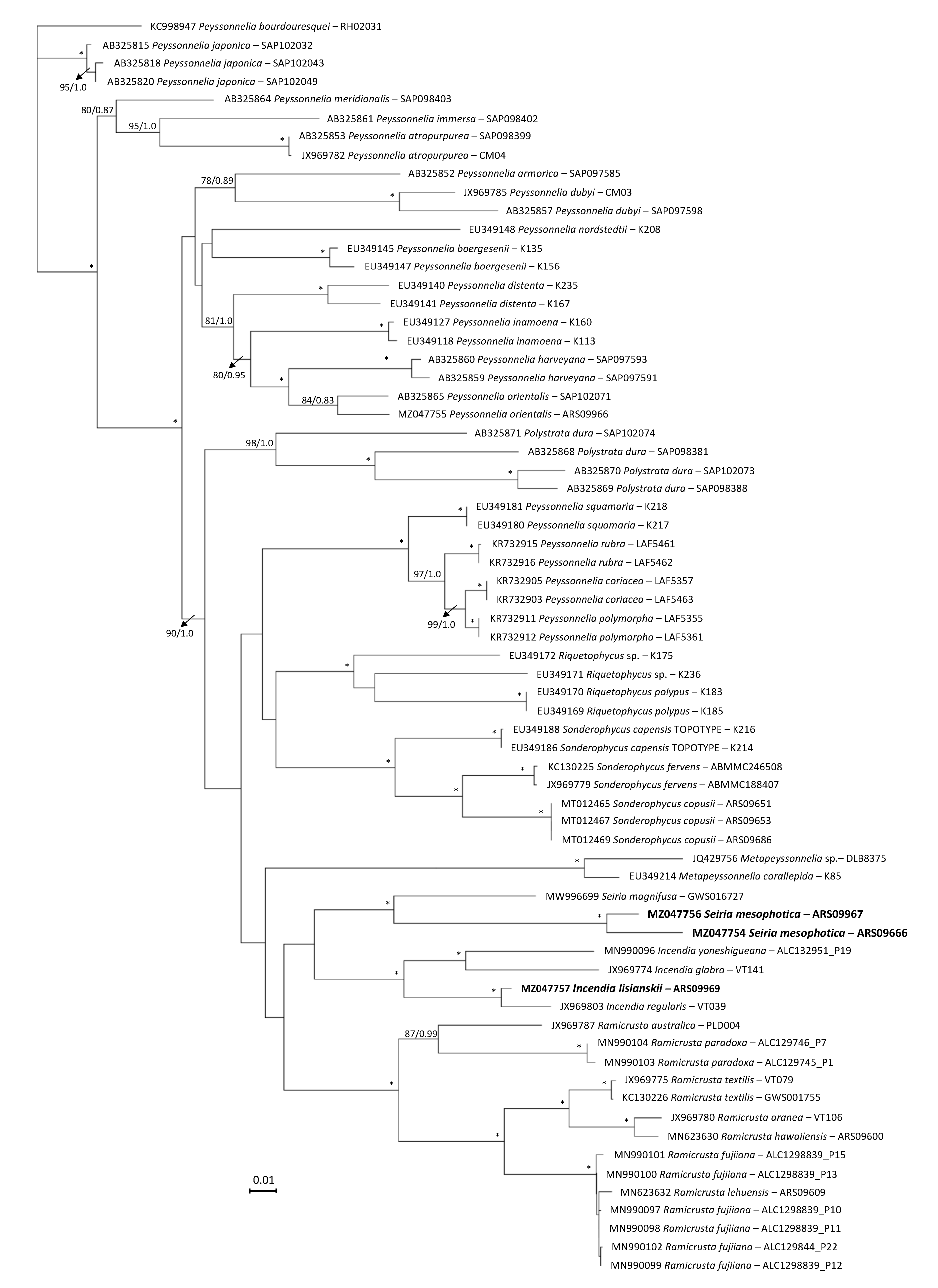
Identification using DNA sequence data:— A DNA barcoding analysis of COI sequences for the two specimens of Hawaiian Seiria ( MZ043099 View Materials and MZ043100 View Materials ) and available COI data for related taxa demonstrated less than 4% sequence divergence between the two Hawaiian specimens (3.3% difference between ARS 09666 and ARS 09967), and much greater distance to the type species, S. magnifusa K.R.Dixon (12.0-12.2%; Fig. 2 View FIGURE 2 ). The rbcL phylogeny supported S. magnifusa as the closest relative to S. mesophotica , with 12.1-13.0% difference in the sequences of the two taxa ( Fig. 3 View FIGURE 3 ).

| H |
University of Helsinki |
| S |
Department of Botany, Swedish Museum of Natural History |
No known copyright restrictions apply. See Agosti, D., Egloff, W., 2009. Taxonomic information exchange and copyright: the Plazi approach. BMC Research Notes 2009, 2:53 for further explanation.
|
Kingdom |
|
|
Phylum |
|
|
Class |
|
|
Order |
|
|
Family |
|
|
Genus |