
Lasiodiplodia chiangraiensis N. Wu, A.J. Dissanayake & Jian K. Liu, 2021
|
publication ID |
https://doi.org/ 10.11646/phytotaxa.508.2.3 |
|
DOI |
https://doi.org/10.5281/zenodo.5485130 |
|
persistent identifier |
https://treatment.plazi.org/id/03E987E0-7936-4A66-65D3-622E1264FE26 |
|
treatment provided by |
Marcus |
|
scientific name |
Lasiodiplodia chiangraiensis N. Wu, A.J. Dissanayake & Jian K. Liu |
| status |
sp. nov. |
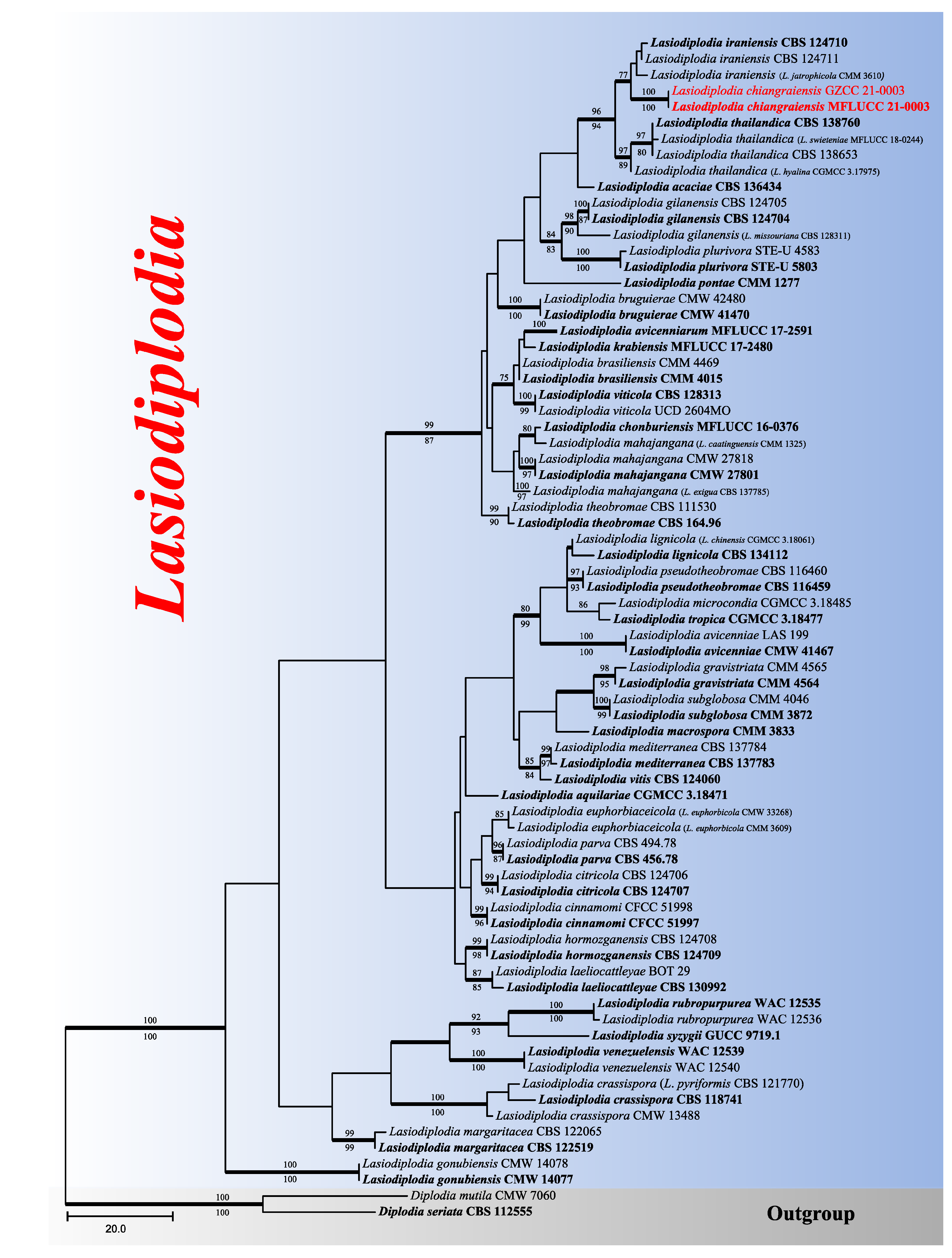
Lasiodiplodia chiangraiensis N. Wu, A.J. Dissanayake & Jian K. Liu View in CoL sp. nov. ( FIG. 2 View FIGURE 2 )
MycoBank number: MB839203, Facesoffungi number: FoF09518.
Etymology: —Named after Chiang Rai Province in Thailand, where the fungus was collected.
Holotype:— MFLU 21-0003 View Materials .
Saprobic on the bark of an unidentified host, forming conspicuous, black spots on the host surface. Sexual morph: not observed. Asexual morph: Conidiomata 170–190 μm diam., 160–190 μm high, semi-immersed or immersed in the substrate, solitary, gregarious or confluent, globose to subglobose, short neck, dark brown. Peridium up to 21–35 μm wide, consisting of brown, small cells of textura angularis, becoming thin-walled and hyaline towards the inner region. Ostiole 30–70 μm diam., centrally located, papillate. Paraphyses 2–5 μm wide, hyaline, cylindrical, aseptate, not branched, rounded at apex. Conidiophores reduced to conidiogenous cells. Conidiogenous cells 7–11 μm long, 3.5–5 μm wide, hyaline, cylindrical. Conidia (21–)22–27(–30) × (12–)13–15(–17) μm (av. = 25 × 14 μm, n = 30), subglobose to oval, rounded at the apex, frequently constricted in the middle, hyaline, aseptate or one-septate, guttulate, without longitudinal striations or mucilaginous sheath.
Culture characteristics: —Conidia germinating on PDA within 12 h. Colonies reaching 90 mm diam. after 4–5 days at 20–23 ° C, circular, white during the first few days, sparse, aerial, surface smooth with crenate edge, filamentous, after 2 weeks becoming black.
Material examined: — THAILAND. Chiang Rai: Amphoe Mueang, Tambon Nang Lae, Mae Fah Luang University , Botanical Garden , 20°02’22.7’’N, 99°53’38.1’’E, on unidentified dead wood, 17 July 2019, Na Wu, YW 113 ( MFLU 21-0003 View Materials , holotype; GZAAS 21-0003 , isotype), ex-type living culture MFLUCC 21-0003 View Materials GoogleMaps ; ibid., on decaying wood, 12 December 2019, Na Wu, YW 401 ( GZAAS 21-0014 ), living culture GZCC 21-0003 GoogleMaps .
Known distribution:— Chiang Rai, Thailand.
Notes:— Lasiodiplodia chiangraiensis is phylogenetically closely related to L. iranensis but formed a distinct linage ( FIG. 1 View FIGURE 1 ), and can be recognized as a new species. Morphologically, these species can be distinguished from the dimensions of their conidia ( TABLE 3). In addition, conidia of L. chiangraiensis are hyaline without longitudinal striations, while those of L. iraniensis become dark brown with age. In terms of the nucleotides comparison, L. chiangraiensis ( MFLUCC 21-0003) and L. iraniensis ( CBS 124710, ex-type) differed in one base pair (bp) in ITS region, seven in tef region and two in tub2.

No known copyright restrictions apply. See Agosti, D., Egloff, W., 2009. Taxonomic information exchange and copyright: the Plazi approach. BMC Research Notes 2009, 2:53 for further explanation.
|
Kingdom |
|
|
Phylum |
|
|
Class |
|
|
Order |
|
|
Family |
|
|
Genus |