
Moruseodina cusucoensis Bravo & Cordeiro
|
publication ID |
https://doi.org/ 10.11646/zootaxa.3841.3.6 |
|
publication LSID |
lsid:zoobank.org:pub:5308906B-BA1E-47AB-819C-9B449ACD9D4B |
|
DOI |
https://doi.org/10.5281/zenodo.5691283 |
|
persistent identifier |
https://treatment.plazi.org/id/03E98C2F-FFD0-EE2C-5AEE-DA93B6B6A209 |
|
treatment provided by |
Plazi |
|
scientific name |
Moruseodina cusucoensis Bravo & Cordeiro |
| status |
sp. nov. |
Moruseodina cusucoensis Bravo & Cordeiro sp. nov.
(Figs. 2–23)
Type material. HONDURAS, Cortés province, Parque Nacional el Cusuco, VII–VIII.2008, Jocque M. and Mendes H. col., holotype male with pupa exuvia (44774, MZFS); three paratypes, all male with pupa exuviae, same locality, date and collectors as holotype (44775, 44776, 44777, MZFS). The specimens were collected from a single tree hole in cloud forest close to the visitors center at an elevation of 1557 masl. (N15 29 ,824, W88 12,682). Larvae are mounted on a separate slide.
Etymology. The epithet refers to the name of the National Park in Honduras where the species was collected.
Diagnosis. Eye bridge separated by 4.0 facet diameters; R2 with basal swelling; R5 ending at wing apex; cercus with nine apical tenacula; aedeagus with two branches, one simple, the other longer and bifurcated.
Description. Male (Figs. 2–12): Head (Fig. 2, 3) with eye bridge with 4 facet rows, separated by 4.0 facet diameters; interocular suture present, concave, nearly V-shaped; hair patch of frons undivided, extending narrowly to frontal suture. Antenna with 14 flagellomeres (Fig. 2), scape subcylindrical 1.3 times as long as pedicel; pedicel spherical (Fig. 4); flagellomeres nodiform (Figs. 4, 5); apiculus of last flagellomere as long as the internode of 13th flagellomere (Fig. 5); flagellomeres 1–14 with pair of digitiform ascoids (Figs. 4, 5). Palpus formula = 1.0:1.9:1.7:2.4 (Fig. 6). Wing (Fig. 7). Length, 2.28 mm; maximum width, 1.05 mm. R2 with basal swelling; other longitudinal veins without swellings; radial fork little basal to medial fork, which is basal to CuA2; R2+3 and R4+5 completes; R5 ending at wing apex. Male terminalia with epandrium fused to hypandrium ( Fig. 8 View FIGURES 8 – 15 ). Epandrium wider than long with distal U-shaped cleft, with clothed setae only in posterior area ( Fig. 9 View FIGURES 8 – 15 ); presence of a single large, oval foramen ( Fig. 9 View FIGURES 8 – 15 ). Cercus pilose, approximately 1.3 times as long as gonostylus, basally approximately 3 times wider than apically; with 9 subapical tenacula ( Fig. 8 View FIGURES 8 – 15 ). Hypandrium bent, strip-like ( Fig. 10 View FIGURES 8 – 15 ). Gonocoxite pilose, as long as gonostylus, but approximately 3.7 times the width of gonostylus, in dorsal view ( Fig. 10 View FIGURES 8 – 15 ). Gonostylus slightly pilose. Epiproct and hypoproct tongue-shaped, both with apical micropilosity ( Fig. 10 View FIGURES 8 – 15 ). Aedeagal complex asymmetric, with a dorsal plate covering aedeagus ( Fig. 8, 10, 11 View FIGURES 8 – 15 ); aedeagus with two branches, one simple, slender, the other longer, bifurcated in dorsal and ventral pieces, dorsal piece bilobed at apex and ventral piece with acute apex ( Figs. 10–12 View FIGURES 8 – 15 ). Aedeagal apodeme simple, as long as aedeagal complex ( Figs. 8, 10–12 View FIGURES 8 – 15 ). Female unknown.
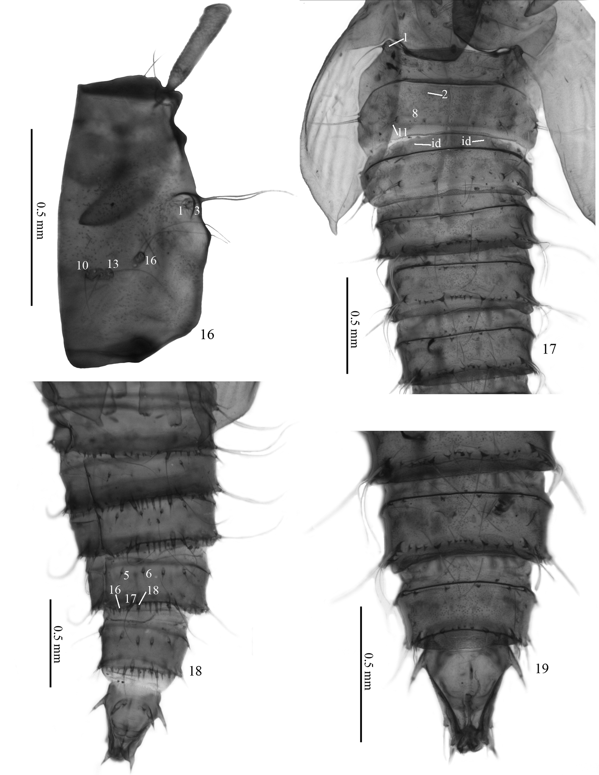
Pupa ( Figs. 13–19 View FIGURES 8 – 15 View FIGURES 16 – 19 ): Color light brown; body conical ( Fig. 13 View FIGURES 8 – 15 ); total length: 3,55–4,0 mm; tips of sheats of legs not extending beyond the apices of the wing covers; respiratory horn approximately six times as long as its medial width, with small group of pits in the basal 1/5 and irregular longitudinal double rows of pits terminally ( Fig. 14 View FIGURES 8 – 15 ). Abdomen with spiniform protuberances (generally with bifid apex) present on the posterior margin of the tergites of abdominal segments III–VII. Spiniform protuberances also present on the posterior margin of the sternites of abdominal segments III–VII, but in this case the protuberances are larger and have two to five acute endings. Following the segmentation, these protuberances reduce in number but increase in size. Anal division conical, less than 2X as long as its width at base, carrying protuberances with spiniform setae at the apex, as in the pattern shown by Vaillant (1971).
Chaetotaxy. Head with setae h very reduced in comparison to the nearby setae f and g. At the place of setae c, two long setae are present. Other setae are present at ocular and frontoclypeal sclerites, as illustrated ( Fig. 15 View FIGURES 8 – 15 ). Thorax with setae 1, 11 and 14 very small ( Fig. 16 View FIGURES 16 – 19 ). Abdomen with setae 1 of anterior tergite, and setae 2, 8 and 11 of posterior tergite, small ( Fig. 17 View FIGURES 16 – 19 ). 2nd to 7th segment with same pattern of chaetotaxy, as follows: each side of anterior tergite with only one very small setae (id) and of posterior tergite with four setae on anterior margin and nine on posterior margin ( Fig. 17 View FIGURES 16 – 19 ) and with the same pattern of reduction of the 1st segment (setae 2, 8 and 11 very small). From abdominal segment III–VII all setae of posterior tergal plate are associated with a protuberance. The five sternites ( Fig. 18 View FIGURES 16 – 19 ) with three setae (16, 17 and 18) on posterior margin, and two setae (5 and 6) anteriorly (except the first sternite, where the anterior setae are missing). Anal division ( Fig. 19 View FIGURES 16 – 19 ) cone shaped, dorsally with a uniform plate carrying setae 1–5.
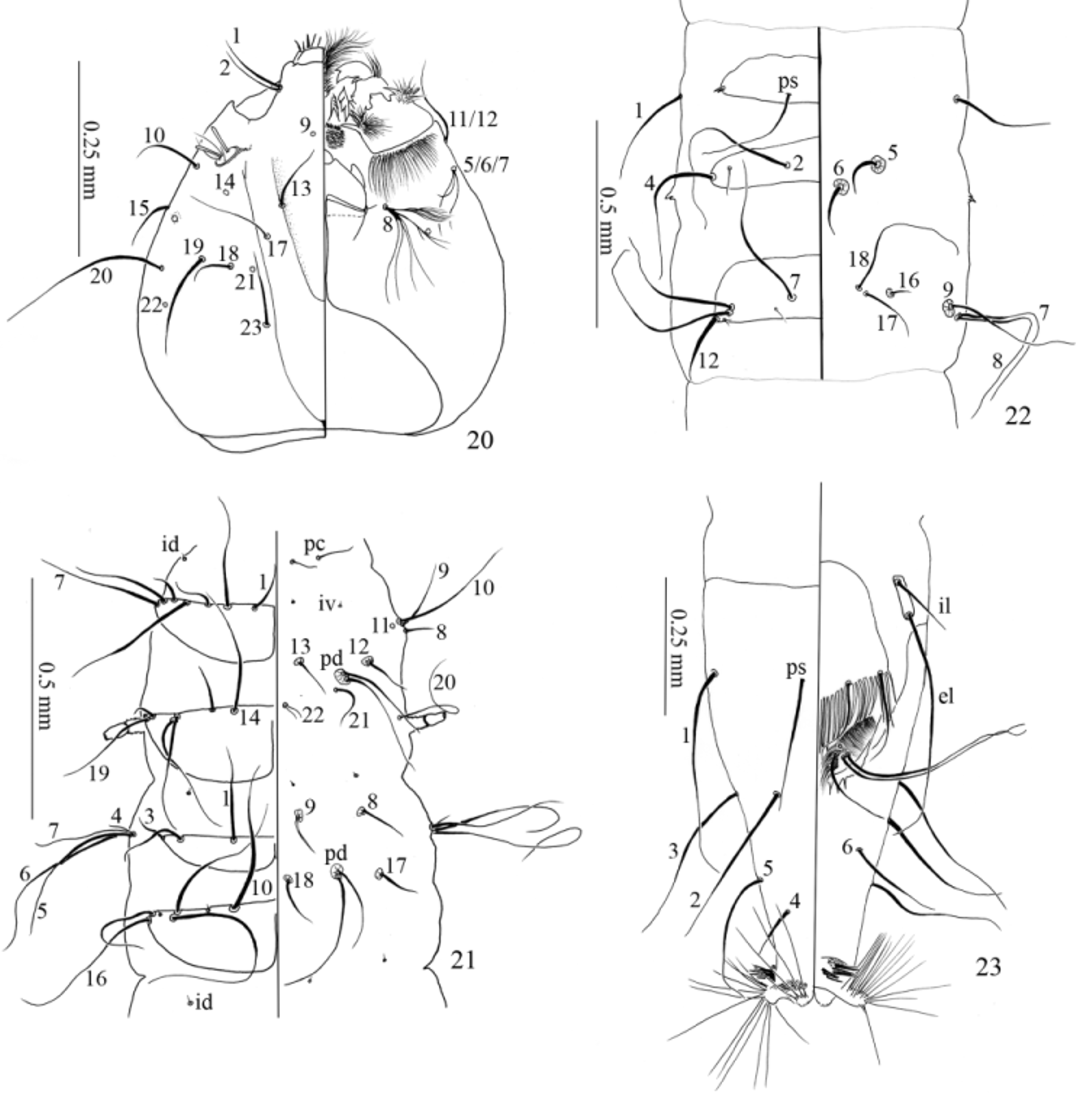
Larva ( Figs. 20–23 View FIGURES 20 – 23 ): Measurements, total length 8.15–8.55mm; head length 0,49–0,53 mm; head width 0,45–0,48 mm. Larvae of usual shape in Psychodinae : head oval; three thoracic segments, two tergal plates on each; seven abdominal segments, first with two tergal plates and II–VII with three tergal plates, and elongate anal division. Head ( Fig. 20 View FIGURES 20 – 23 ) with dorsal epicranial suture V shaped. Frons with only five setae (setae 3 and 4 absent) on each side. Gena with only 13 setae (one trifid setae instead of setae 5, 6 and 7; one setae on the place of 11 and 12, one of them may be absent on this species). Setae 8 brush-like. Labrum with digitiform and setiform setae anteriorly. Hypostomium with three apical teeth. Thorax ( Fig. 21 View FIGURES 20 – 23 ) with setae on all segments as described in Vaillant (1971) for the larvae of 4th instar of Clogmia albipunctata (Williston) , arranged as illustrated. Meso- and metathorax with the same number and arrangement of setae, differing only on the presence of a medial longitudinal suture on tergites of mesothorax, as on the prothorax. Ventrally, a transversal row of small spines formed by the cuticle is found on the line of setae 8 and 9, and nearby pedichaetes of meso- and metathorax. The anterior spiracle is short, without ornamentation. Abdomen ( Fig. 22 View FIGURES 20 – 23 ) with protergite present on segments II–VII, meso- and metatergites present on segments I–VII, all showing same pattern of chaetotaxy of illustration ( Fig. 22 View FIGURES 20 – 23 ). Small spines formed by the cuticle present on the side of each segment, on the level of mesotergite. Ventrally, each segment has a transversal row of spines on pro- and mesothorax, and an arched row on the metathorax. The spines on these rows are gradually smaller following the body segmentation, until the rows of spines of meso- and metathorax are completely indistinguishable on abdominal segment VII. Anal division ( Fig. 23 View FIGURES 20 – 23 ) with dorsal sclerite around 2X longer than width at base. Number and arrangement of setae in accordance with the Vaillant (1971), as illustrated. Preanal plate subtrapezoidal, apical margin slightly convex, with four true setae on apical margin, together with a row of accessory setae.
Ecological observations. Moruseodina cusucoensis is commonly observed in water puddles in tree holes and in bromeliad leaf axils ( Tillandsia guatemalensis and Catopsis sp.). They often occur in high densities (30+ individuals) in small amounts of water (<200ml), together with several species of Chironomidae , Culicidae and Tipulidae . The species is also commonly found in experimental set-ups (unpublished information) consisting of plastic cups tied to trees in the forest, indicating a fast colonization rate and general common presence in this forest.

No known copyright restrictions apply. See Agosti, D., Egloff, W., 2009. Taxonomic information exchange and copyright: the Plazi approach. BMC Research Notes 2009, 2:53 for further explanation.