
Guimaraesiella (Cicchinella) iuga Gustafsson, Clayton
|
publication ID |
https://doi.org/ 10.11646/zootaxa.4543.4.1 |
|
publication LSID |
lsid:zoobank.org:pub:4F591303-AF92-4BBB-8B68-EDD27AA229DE |
|
DOI |
https://doi.org/10.5281/zenodo.5936040 |
|
persistent identifier |
https://treatment.plazi.org/id/AE635C47-2E45-4CE8-8722-B49CA3B6753B |
|
taxon LSID |
lsid:zoobank.org:act:AE635C47-2E45-4CE8-8722-B49CA3B6753B |
|
treatment provided by |
Plazi |
|
scientific name |
Guimaraesiella (Cicchinella) iuga Gustafsson, Clayton |
| status |
new species |
Guimaraesiella (Cicchinella) iuga Gustafsson, Clayton & Bush, new species
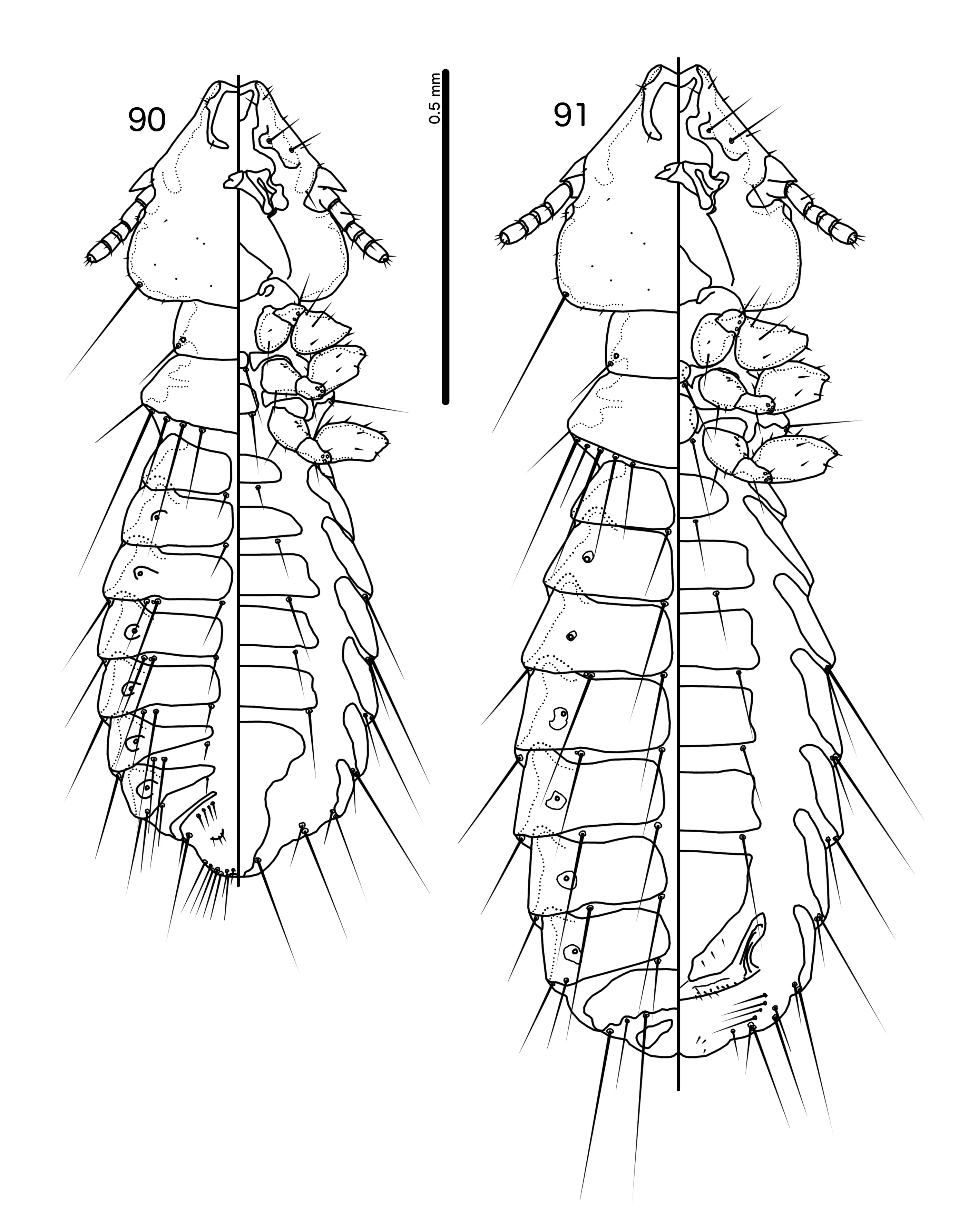
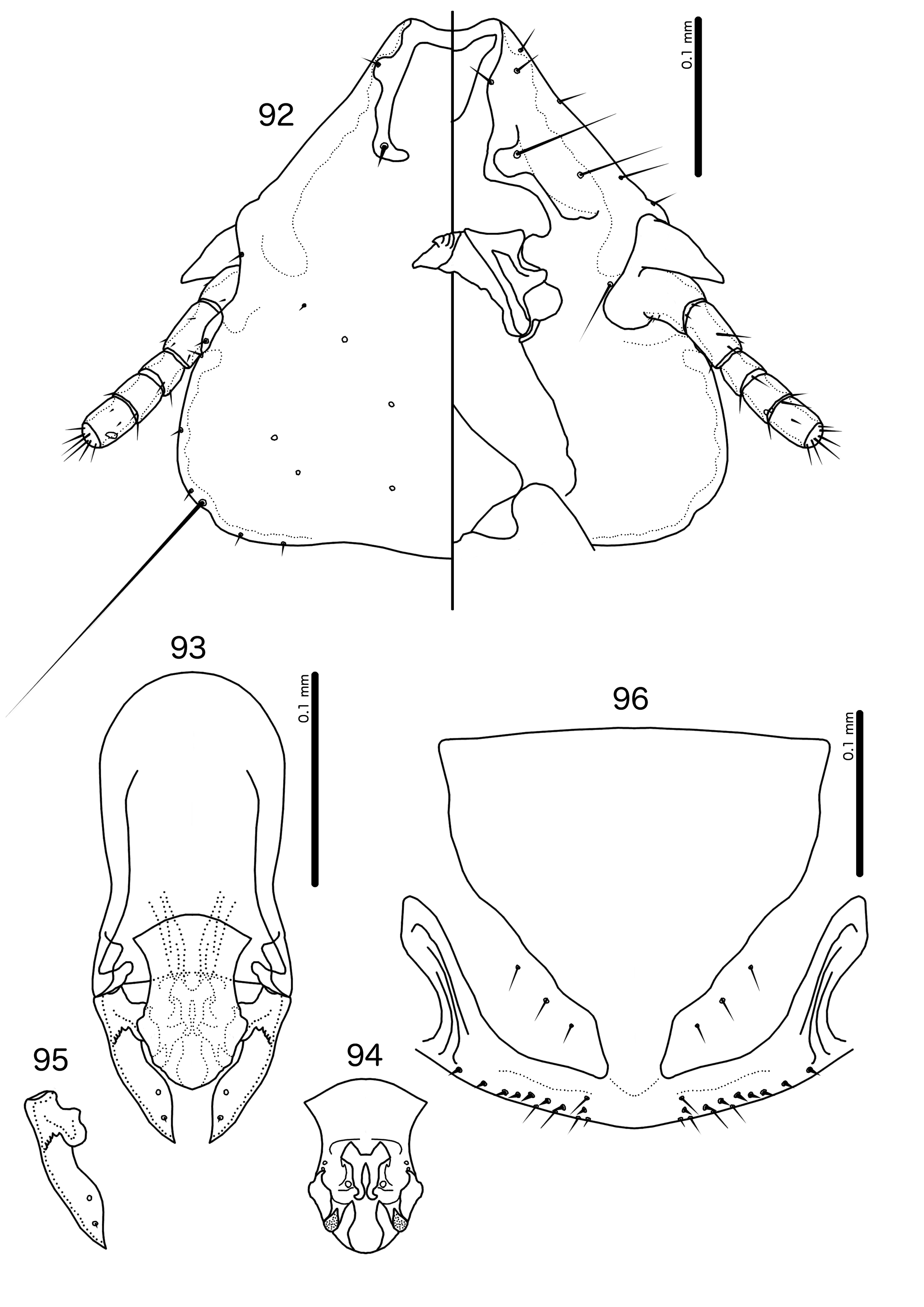
( Figs 90–96 View FIGURES 90–91 View FIGURES 92–96 )
Type host. Alcippe peracensis peracensis Sharpe, 1887 —mountain fulvetta ( Leiothrichidae ).
Type locality. Terengganu, Malaysia .
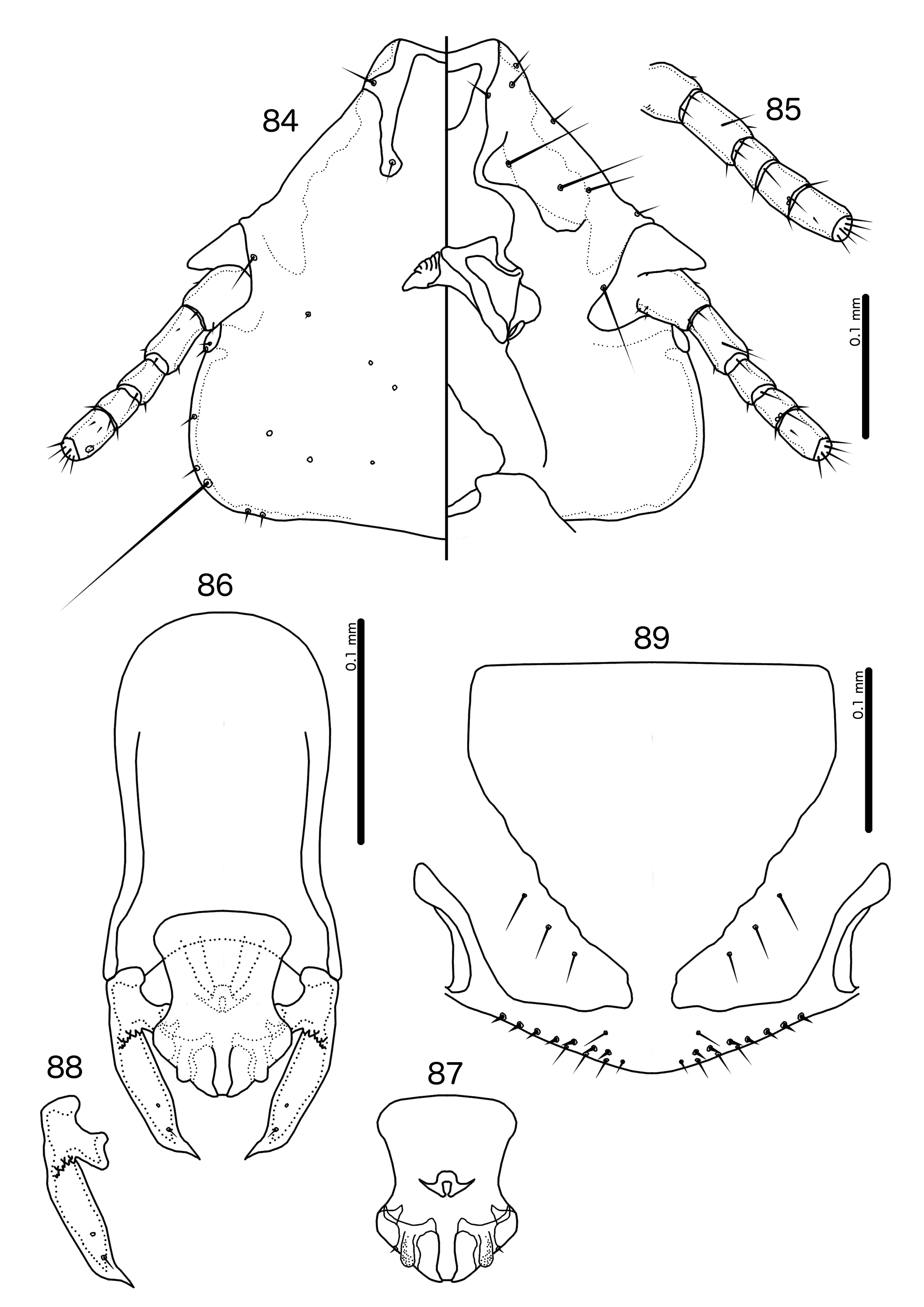
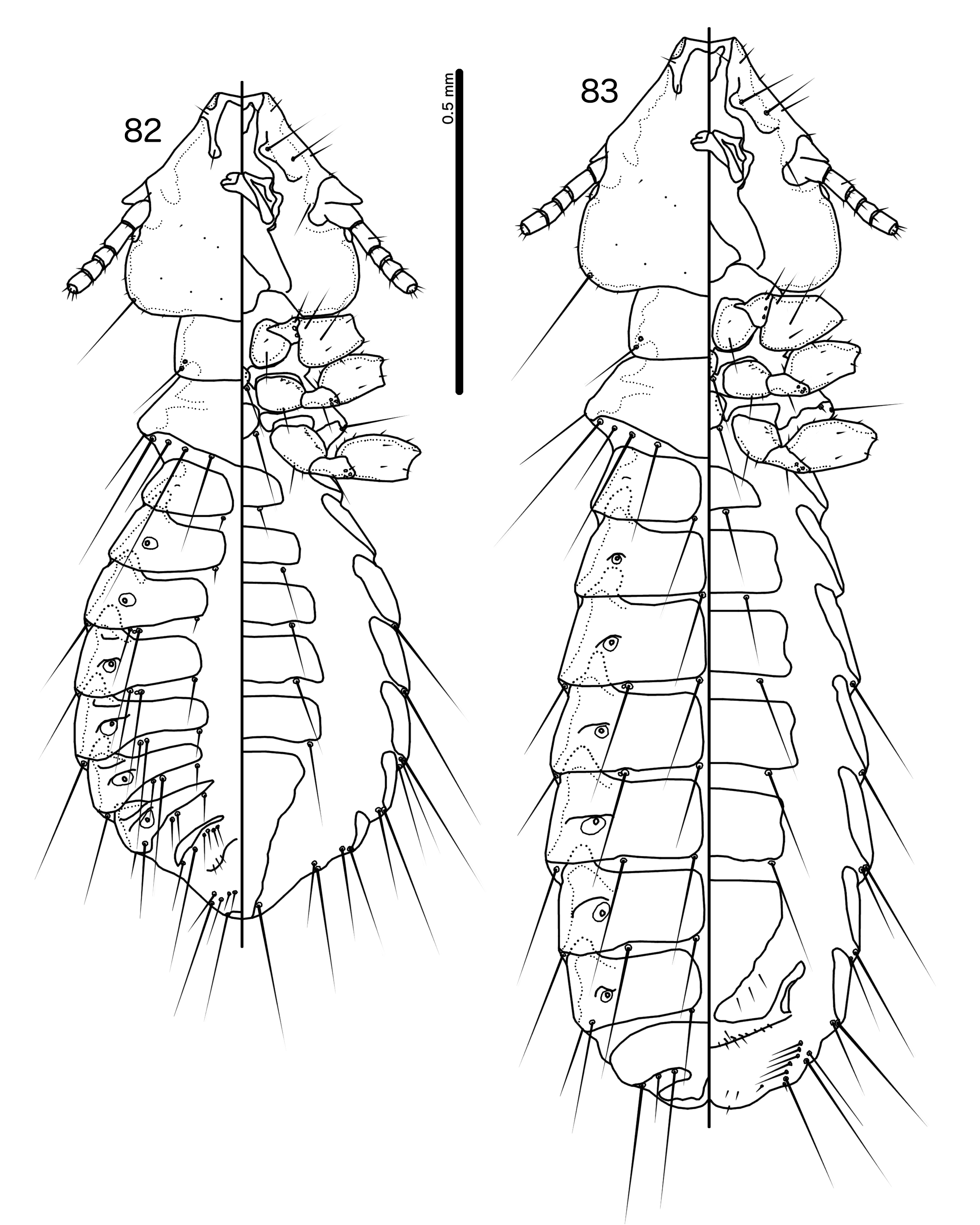
Diagnosis. Guimaraesiella (C.) iuga is most similar to G. (C.) mcgrewi , with which it shares the characters listed under the latter species, above. However, these two species can be separated by the following characters: (1) dorsal preantennal suture reaches lateral margin of head in G. (C.) mcgrewi ( Fig. 84 View FIGURES 84–89 ) but not in G. (C.) iuga ( Fig. 92 View FIGURES 92–96 ); (2) female tergopleurite XI fused with tergopleurite IX+X in G. (C.) mcgrewi ( Fig. 83 View FIGURES 82–83 ) but not in G. (C.) iuga ( Fig. 91 View FIGURES 90–91 ); (3) antennae sexually dimorphic in G. (C.) mcgrewi ( Figs 84–85 View FIGURES 84–89 ) but not in G. (C.) iuga ( Fig. 92 View FIGURES 92–96 ); (4) basal apodeme more slender in G. (C.) iuga ( Fig. 93 View FIGURES 92–96 ) than in G. (C.) mcgrewi ( Fig. 86 View FIGURES 84–89 ); (5) proximal mesosome with more pronounced convex anterior margin in G. (C.) iuga ( Fig. 94 View FIGURES 92–96 ) than in G. (C.) mcgrewi ( Fig. 87 View FIGURES 84–89 ); (6) mesosome proportionally broader in G. (C.) mcgrewi ( Fig. 87 View FIGURES 84–89 ) than in G. (C.) iuga ( Fig. 94 View FIGURES 92–96 ); (7) gonopore with quadratic antero-lateral extensions in G. (C.) iuga ( Fig. 94 View FIGURES 92–96 ) but with triangular postero-lateral extensions in G. (C.) mcgrewi ( Fig. 87 View FIGURES 84–89 ); and (8) parameral heads with prominent anterior bulge in G. (C.) mcgrewi ( Fig. 88 View FIGURES 84–89 ) but with only slight anterior bulge in G. (C.) iuga ( Fig. 95 View FIGURES 92–96 ).
Description. Both sexes. Head broadly trapezoidal ( Fig. 92 View FIGURES 92–96 ). Lateral margins of preantennal head concave. Dorsal preantennal suture does not reach lateral margins of head, and does not completely separate dorsal anterior plate from main head plate. Head chaetotaxy as in Fig. 92 View FIGURES 92–96 ; pns microsetae. Coni long and broad, reaching beyond distal margin of scape. Antennae not sexually dimorphic. Gular plate triangular. Thoracic and abdominal segments as in Figs 90–91 View FIGURES 90–91 . Reentrant heads of pleurites short, blunt, broad.
Male. Thoracic and abdominal chaetotaxy as in Fig. 90 View FIGURES 90–91 . Genitalia as in Figs 93–95 View FIGURES 92–96 . Basal apodeme comparatively slender ( Fig. 93 View FIGURES 92–96 ), rounded, with slight constrictions at about mid-length. Proximal mesosome with deeply concave lateral margins and distinctly convex anterior margin ( Fig. 94 View FIGURES 92–96 ). Ventral sclerite absent. Mesosomal lobes comparatively slender, convex. Marginal thickenings of mesosomal lobes displaced medianly in anterior end. Rugose area forms distinct nodus on each side that slightly extends beyond mesosomal margins. Elongated nodi on distal mesosome slender. Gonopore uniquely shaped, with large quadratic antero-lateral extensions and small hookshaped extensions distally; 2 ames sensilla on each side near antero-lateral corners of mesosomal lobes; 1 gpmes on each side of gonopore; lpmes not visible in examined material. Parameral heads irregular ( Fig. 95 View FIGURES 92–96 ), with slight anterior bulge. Parameral blades broad, tapering only distally; pst1–2 far apart. Measurements (n = 4): TL = 1.10– 1.20; HL = 0.32–0.34; HW = 0.32–0.34; PRW = 0.19–0.20; PTW = 0.29–0.31; AW = 0.40–0.43.
Female. Thoracic and abdominal chaetotaxy as in Fig. 91 View FIGURES 90–91 ; ss of tergopleurite VIII nearly as long as ss of tergopleurites II–VII. Subgenital plate as in Fig. 96 View FIGURES 92–96 ; cross-piece with narrow connection to subgenital plate. Crosspiece very pale, in some specimens entirely transparent. Vulval margin gently rounded ( Fig. 96 View FIGURES 92–96 ), with 3–4 slender vms on each side, and 7–9 thorn-like vss on each side; 4 vos on each side; distal vos median to vss. Measurements (n = 8): TL = 1.41–1.58 HL = 0.35–0.37; HW = 0.36–0.40; PRW = 0.21–0.23; PTW = 0.32–0.35; AW = 0.48–0.53.
Etymology. The species epithet is derived from “ iugum ”, Latin for “yoke”, referring to the large, yoke-like gonopore.
Type material. Ex Alcippe peracensis peracensis : Holotype GoogleMaps ♂, Terengganu, 102° 37’ E, 5° 25’ N, Malaysia, 4 Mar. 1974, Gunung Lawit Expedition GoogleMaps , Brit. Mus. 1974-2 (NHML). Paratypes: 2♀, same data as holotype ( NHML) GoogleMaps ; 2♂, 3♀, Terengganu, 102° 36’ E, 5° 25’ N, Malaysia, 14 Mar. 1974, Gunung Lawit Expedition , Brit. Mus. 1974-2 ( NHML) GoogleMaps ; 1♂, 3♀, Terengganu, 102° 36’ E, 5° 25’ N, Malaysia, 16 Mar. 1974, Gunung Lawit Expedition , Brit. Mus. 1974-2 ( NHML) GoogleMaps .
| NHML |
Natural History Museum, Tripoli |
No known copyright restrictions apply. See Agosti, D., Egloff, W., 2009. Taxonomic information exchange and copyright: the Plazi approach. BMC Research Notes 2009, 2:53 for further explanation.
|
Kingdom |
|
|
Phylum |
|
|
Class |
|
|
Order |
|
|
Family |
|
|
Genus |